Open Access
Research
Exercise-induced intra-ventricular gradients as a frequent potential
cause of myocardial ischemia in cardiac syndrome X patients
Carlos Cotrim*
1,3, Ana G Almeida
2and Manuel Carrageta
1,2,3Address: 1Cardiology Department, Garcia de Orta Hospital, Almada, Portugal, 2Cardiology Department, Santa Maria Hospital, Lisboa, Portugal
and 3Lisboa University Medical School, Lisboa, Portugal
Email: Carlos Cotrim* - [email protected]; Ana G Almeida - [email protected]; Manuel Carrageta - [email protected]
* Corresponding author
Abstract
Background: The development of intra-ventricular gradients (IVG) during dobutamine or exercise stress
is not infrequent, and can be associated to symptoms during stress.
The purpose of this study was to assess the occurrence of IVG during exercise stress echocardiography in cardiac syndrome X patients.
Methods: We prospectively evaluated 91 patients (pts) mean aged 51 ± 12 years (age ranged 20 to 75
years old), 44 of whom were women. All pts had angina, positive exercise ECG treadmill testing, normal rest echocardiogram and no coronary artery disease on coronary angiogram (cardiac X syndrome). After complete Doppler echocardiographic evaluation with determination of left ventricular outflow tract index (LVOTi), relative left ventricular wall thickness (RLVWT) and left ventricular end-diastolic volume index (LVDVi), all patients underwent stress echocardiography with two-dimensional and Doppler echographic evaluation during and after treadmill exercise.
Results: For analysis purpose patients were divided in 2 groups, according to the development of IVG.
Doppler evidence of IVG was found in 33 (36%) of the patients (Group A), with mean age 47 ± 14 years old (age ranged 20 to 72 years) and with a mean end-systolic peak gradient of 86 ± 34 mmHg (ranging from 30 to 165 mmHg). The IVG development was accompanied by SAM of the mitral valve in 23 pts. Three of these pts experienced symptomatic hypotension. Ten were women (30% pts). 58 pts in group B, 34 of whom were women (59%) (p = 0,01 vs group A), mean aged 53,5 ± 10,9 years old (age ranged 34 to 75 years) (p = 0,03 vs group A), did not develop IVG. LVOTi was 10,29 ± 0,9 mm/m2 in group A and 11,4 ±
1 mm/m2 in group B (p < 0,000); RLVWT was 0,36 ± 0,068 in group A and 0,33 ± 0,046 in group B (p <
0,01); LVDVi was 44,8 ± 10 ml/m2 in group A and 56 ± 11,6 ml/m2 in group B (p = 0,000).
Conclusion: 1. A significant number of patients with cardiac X syndrome developed IVG during upright
exercise in treadmill. These pts (group A) are mainly males and younger than those who did not develop IVG.
2. The development of IVG and mitral valve SAM on exertion seems to be associated with ST segment downsloping during stress testing in patients without epicardial coronary disease.
3. The development of IVG and mitral valve SAM seems to be associated with lower LVOTi, lower LVDVi and higher RLVWT.
Published: 14 January 2008
Cardiovascular Ultrasound 2008, 6:3 doi:10.1186/1476-7120-6-3
Received: 10 December 2007 Accepted: 14 January 2008 This article is available from: http://www.cardiovascularultrasound.com/content/6/1/3
© 2008 Cotrim et al; licensee BioMed Central Ltd.
This is an Open Access article distributed under the terms of the Creative Commons Attribution License (http://creativecommons.org/licenses/by/2.0), which permits unrestricted use, distribution, and reproduction in any medium, provided the original work is properly cited.
Background
The development of IVG during DSE has been largely reported and this fact is commonly associated with symp-toms during the stress study [1,2]. The occurrence of IVG during the ESE is rarely find [3]. In a group of 10 patients who developed IVG during DSE, we performed ESE and we found a small IVG in only one of them [4]. In a 23 years old male, with a positive treadmill test, a structural normal heart, normal coronary angiographies, an ESE was performed and during the study we unexpectedly detect a 102 mmHg intra-ventricular gradient [5] and systolic anterior movement of mitral valve (SAM). A similar case has been reported previously by Lau [6] and was treated successfully with β blockers.
The aim of this study was to present the results of search for intra-ventricular gradients during exercise stress echocardiography in patients with angina, positive stress electrocardiography, normal coronary arteries, and nor-mal echocardiogram (cardiac X syndrome).
Methods
This study includes 91 (pts) mean aged 51 ± 12 years (age ranged 20 to 75 years old), 44 of whom were women. All pts had angina, positive exercise ECG treadmill testing (four patients had only ischemia in a myocardial per-fusion study), normal rest echocardiogram – no left ven-tricular hypertrophy – and no coronary artery disease on coronary angiogram. Diabetes mellitus or uncontrolled hypertension in the last year were motives of exclusion. Twenty four patients (26%) are current smokers and thirty three pts (36%) had hypercholesterolemia.
At the moment of inclusion in the study, 47 (51%) patients were treated with nitrates, 10 (11%) with calcium antagonists, 18 pts (20%) on β blockers, 12 pts (13%) with angiotensin II receptor blockers or angiotensin-con-verting enzyme inhibitors, 7 pts (8%) with diuretics. All patients gave informed consent for the study.
Exercise stress echocardiography
After complete echocardiographic evaluation which also includes determination of left ventricular outflow tract index (LVOTi), relative left ventricular wall thickness (RLVWT) and left ventricular end-diastolic volume index (LVDVi), all patients underwent stress echocardiography with two-dimensional and Doppler echographic evalua-tion. We also measured the distance D1 in the end of dias-tole, in short axis view, as showed in Figure 1. Exercise stress echocardiography as performed by the authors [7] includes evaluation during all the exercise in treadmill, of contractility, and in this group of patients also pulsed, continuous and colour Doppler from apical window
(Additional Files 1 and 2). Mitral valve motion was also assessed, for the development of SAM (Additional file 3 and Figure 2). The exam was totally stored in videotape and partially in optical disk. A significant intraventricular gradient, was considered an increase in the intraventricu-lar flow velocity to or greater than 2.5 m/s at the end of systole (telesystolic peak)(Figure 3) and its occurrence separated the patients in two groups.
Statistical analysis
The results are expressed as mean ± SD for continuous var-iables, and frequency percentage for categorical variables. The variables were compared between groups with the
stu-dent T test. The X2 test was used for qualitative variables.
Results of statistic tests were considered significant if the observed p value was less than 0.05.
Results
A typical example of stress electrocardiography (Figure 4), and angiographic (Figure 5) findings is showed.
From the all group, 33 patients (36%) develop IVG (group A) and 58 pts (64%) did not develop intraventricular gra-dient (Group B) as defined by the authors. In group A the IVG at peak exercise was 86 ± 34 mmHg (ranging from 30 to 165 mmHg).
In all but 11 patients, 85% of predicted maximum theo-retical heart rate for age was reached. Clinical and
demo-A line that originates at the point where the inferior wall begins, divides the left ventricle in halfs
Figure 1
A line that originates at the point where the inferior wall begins, divides the left ventricle in halfs. The D1 distance is the distance between that line and the postero internal papil-lary muscle (arrow) at the point where it encounters the inferior wall.
graphic data is presented in Table 1, details of the exercise test are depicted in Table 2, and details of echocardiogram in Table 3, 4, and 5.
In group A 23 pts (70%) develop SAM (Figure 2, Addi-tional file 3) during exercise, associated with IVG (Figure 3, Additional file 2). No one patient developed segmental wall abnormalities.
Multivariate Analysis
A logistic regression model was constructed with the fol-lowing variables: age, sex, effort angina, left ventricular outflow tract index, left ventricular diastolic volume index, relative wall thickness, left ventricular mass index, D1 distance. From the variables included attained statisti-cal significance (p < 0.05) the contribution of effort angina, D1 distance, LVDVi, LVOTi and sex, for appear-ance of IVG as we can see in Table 6.
Discussion
Patients with a positive treadmill exercise test, and normal coronary angiography have long been recognised as an important problem in clinical practice [8-10]. These early studies identified many of the characteristics of what was subsequently characterized as syndrome X [10]. The same denomination was also applied to a syndrome, character-ized by insulin resistance, hyperinsulinemia, and diabe-tes, that is associated with dyslipidemia, hypertension, and abdominal obesity. Hence a more specific terminol-ogy comes in use: angina with normal coronary arteriog-raphy [11]. Patients with this entity, predominantly women [12], complain of pain that is frequently atypical. It may be precipitated by exertion, although the threshold for precipitating pain is highly variable [13]. Its duration may be uncharacteristically long, and it may be unusually severe and is rarely associated with symptoms such as dia-phoresis. Perfusion abnormalities have been observed commonly in patients with chest pain and normal coro-nary arteriograms, but no consistent correlation could be made among the extent of the defect, the positivity of the exercise test, and exercise tolerance [14]. Thus in many of this patients there is evidence of perfusion abnormalities that are attributed to abnormalities in the microvascula-ture [15]. However stress echocardiography allways failled to demonstrate segmental wall abnormalities even show-ing hyperdinamic ventricles[16].
The results of our study, in which 33 (36%) of 91 patients with normal coronary angiogram and positive treadmill exercise test developed intraventricular gradient, suggest that ST-segment depression may be related with the devel-opment of IVG during exercise which is possibly involved in the genesis of electrocardiographic changes. The possi-ble association between cardíac X syndrome and the development of IVG during exercise was described before [17,18] however some of the patients from these studies have arterial hypertension, and left ventricular hypertro-phy that by definition of X Syndrome [19] we have excluded and that more frequently developed IVG [3].
The flow obtained at that moment with continuous Doppler
Figure 3
The flow obtained at that moment with continuous Doppler.
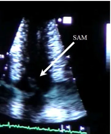
In this apical four chamber view obtained near peak exercise (before stoping) we can clearly see systolic anterior move-ment of mitral valve
Figure 2
In this apical four chamber view obtained near peak exercise (before stoping) we can clearly see systolic anterior move-ment of mitral valve.
The appearance of IVG in our study was associated with morphological determinants like reduced LVOTi, reduced left ventricular diastolic volume, a reduced distance D1, and increased relative left ventricular wall thickness. All these finding translate a proportional small heart that the multivariate model confirms.
The reduced D1 in Group A means an anterior "displace-ment" of the postero internal papillary muscle that may be involved in the development of IVG and SAM of the mitral valve [20,21] as described by other authors. We can admit that this phenomenon is eventually caused by the subtle changes in left ventricle geometric shape and dimensions with more anterior papillary muscles implan-tation [20,21], that during exercise, induce and submit the cordae and mitral valve to an abnormal systolic anterior motion and to papillary muscle ischemia. The obstruction to the outflow in left ventricle with the increase in the
intraventricular pressure that it causes may contribute, to left ventricular strain and ST-depression in this patients. The development of intraventricular gradient during exer-cise may possibly explain the ST changes in a subgroup of patients who have treadmill positive test and normal cor-onary arteries.
The patients with IVG during exercise had more angina during exercise and were predominantly male, and this may explain why these patients were submitted to coro-nary angiography much early, after the beginning of the symptons, than patients in Group B. From the all study group 42 patients (46%) reproduced symptoms during ESE, however this fact occured more frequently (22 pts from 33 in group A vs 20 from 58 pts in group B – p = 0.002) in group A, favouring the potencial participation of intraventricular gradient in the occurrence of symp-toms.
Summary of a positive exercise stress test in one patient from the study
Figure 4
In our study population, we found a great number of patients that develop SAM of the mitral valve in associa-tion with IVG contrarily to other authors [17,18]. We think that we detect SAM in a greater number of patients because we do echo during all the exercise in treadmill (Additional file 2 and 4) [7]. The magnitude of the IVG
that we have detected in our patients is also greater for the same motive (Figure 3).
Four of the 33 patients that developed intraventricular gradient are athletes [22] and we should probably study this phenomenon in this specific population and, if this Table 1: Clinical and demographic data
Group A Group B p Age, years 47,70 ± 13,36 53,53 ± 10,89 0,026 Sex, female (%) 10/33 (30%) 34/58 (59%) 0,008 BSA m2 1,8 ± 0,16 1,73 ± 0,13 0,022 Effort Angina 28/33 (85%) 33/58 (56%) 0,006 Effortless Angina 9/33 (27%) 35/58 (57%) 0,002
Duration of symptoms before cath. (months) 15 ± 10 46 ± 40 0,000
Time of FLW (months) 36,4 ± 17,9 39,1 ± 19,5 0,55 Events in FLW 6/33 (18%) 8/56 (14%) 0,31 ACS in FLW 1/33 (3%) 7/56 (13%) 0,24 β Bloq. 7/33 (21%) 11/58 (19%) 0,798 CCB 4/33 (12%) 7/58 (12%) 0,666 Nitrates 16/33 (48%) 31/58 (53%) 0,769 IECA/ARAII 5/33 (15%) 7/58 (12%) 0,680 Diuretics 2/33 (6%) 5/58 (9%) 0,663 β Bloq. FLW 20/33 (60%) 17/56 (30%) 0,003 CCB FLW 4/33(12%) 19/56 (34%) 0,530 Nitrates FLW 9/33 (27%) 33/56 (59%) 0,006 IECA/ARAII FLW 9/33 (27%) 8/56 (14%) 0,068 Diuretics FLW 4/33(12%) 4/56 (7%) 0,403
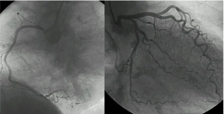
BSA – body surface area; ACS – Acute coronary Syndrome; CCB – Calcium chanell blockers; FLW – follow-up Normal angiography of coronary arteries in the same patient
Figure 5
occurs, also investigate the possible prognostic implica-tions for this event in this particular population [23]. The results of ESE have probably influenced the treatment of the patients once at the end of follow-up a greater per-centage of patients are treated with β blockers [24,25] in group A than in group B (Table 1).
The principal limitations of this study are: 1) no one patient has done a test for provocation of coronary spasm at cath. laboratory even no patient included in the study had segmental wall abnormalities with exercise 2) The
presence or absence of ischemia was only evaluated by ESE without use of scintigraphic studies. 3) We excluded all patients with left ventricular hypertrophy and uncon-troled arterial hypertension that constitutes a great number of patient in the real world of clinical practice and that should be studied in the future with the same proto-col.
Conclusion
We can conclude that a relevant number of patients with cardiac X syndrome develop significant intraventricular gradient during exercise and also that morphological var-iables are involved in is pathophysiology. The authors believe that this phenomenon may constitute a new entity that joins to the heterogeneous group of patients with angina, ST-depression during treadmill exercise test and normal coronary arteriography.
Table 6: Multivariate analysis
Variable -2 Log Likelihood Loss Function (p)
Age 114,1548 ,071918 Sex 109,0601 ,023868 LVOTi (mm/m2) 91,70272 ,000031 LVDVi ml/m2 81,35754 ,001299 RLVWT 78,80142 ,109878 LVMi g/m2 78,73733 ,800141 D1 distance 64,62039 ,000172 Effort Angina 52,25502 ,000438
LVOTi; LVDVi; RLVWT; LVMi; D1 – as previously defined
Table 4: Details of two-dimensional echocardiogram
Group A Group B p
LVOTi (mm/m2) 10,29 ± 0,9 11,4 ± 1 0,000
EF (%) 67,94 ± 5,4 66,90 ± 4,5 0,333
LVDVi ml/m2 44,8 ± 10 56 ± 11,6 0,000
D1 (mm) 10,72 ± 3,11 13,75 ± 2,98 0,000
LVOTi – left ventricular outflow tract index; EF – ejection fraction, LVDVi – left ventricular diastolic volume index; D1 – distance D1 measured as explained in figure 1.
Table 2: Exercise test data
Group A Group B p HR Baseline 70 ± 10,5 70 ± 11 0,769 HR Peak 163 ± 14 151 ± 17 0,001 Syst. BP Baseline 133 ± 13 135 ± 15 0,575 Syst. BP Peak 175 ± 21 173 ± 27 0,640 %theoretical MHR 95 ± 7 91 ± 9 0,02 Duration seconds 659 ± 159 503 ± 175 0,000 Time recovery HR 254 ± 99 260 ± 151 0,832 Double product 28760 ± 4493 26232 ± 4760 0,015 Angina during ESE 22/33 (66%) 20/58(34%) 0,002 HR – heart rate; BP – blood pressure; MHR – maxymal heart rate.
Table 3: Details of echocardiogram M Mode
Group A Group B p LVEDDi (mm/m2) 25,3 ± 2,8 28 ± 2,7 0,000 LVESDi 15,6 ± 2,4 17,4 ± 2,3 0,0002 FS (%) 38,9 ± 5,4 37,5 ± 4,9 0,219 IVSi (mm/m2) 5,2 ± 0,9 5,1 ± 0,8 0,62 PWi (mm/m2) 4,55 ± 0,7 4,59 ± 0,6 0,75 LVMi g/m2 73,9 ± 13,1 80,6 ± 13,9 0,028 LA (mm) 37,1 ± 3,2 37,8 ± 2,7 0,279 RLVWT 0,36 ± 0,068 0,33 ± 0,046 0,01
LVEDDi – left ventricle telediastolic diameter index; LVESDi – left ventricle telesystolic diameter index; FS – fraccional shortening; IVSi – interventricular septum index; PWi – posterior wall index; LVMi – left ventricular mass index; LA – left atrium; RLVWT – relative left ventricular wall thickness
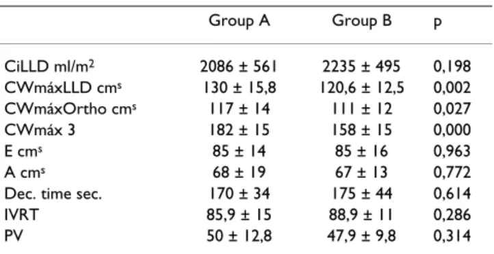
Table 5: Details of echocardiogram (Doppler)
Group A Group B p CiLLD ml/m2 2086 ± 561 2235 ± 495 0,198 CWmáxLLD cms 130 ± 15,8 120,6 ± 12,5 0,002 CWmáxOrtho cms 117 ± 14 111 ± 12 0,027 CWmáx 3 182 ± 15 158 ± 15 0,000 E cms 85 ± 14 85 ± 16 0,963 A cms 68 ± 19 67 ± 13 0,772
Dec. time sec. 170 ± 34 175 ± 44 0,614
IVRT 85,9 ± 15 88,9 ± 11 0,286
PV 50 ± 12,8 47,9 ± 9,8 0,314
CiLLD – cardiac index in left lateral decubitus before de start of the exam; CWmáxLLD – máximal velocity of flow obtained at apical five chamber view with continuous Doppler oriented through LVOT to the aorta in left lateral decubitus; CwmáxOrtho – máximal velocity of flow obtained at apical five chamber view with continuous Doppler oriented through LVOT to the aorta in orthostatic position; CWmáx3-máximal velocity of flow obtained at apical five chamber view with continuous Doppler oriented through LVOT to the aorta; E- maximal velocity of E wave of mitral flow; A – maximal velocity of E wave of mitral flow; Dec. Time sec.- deceleration time in seconds; IRVT – isovolumic relaxation time; PV propagation of velocity evaluated with M Mode color.
As as consequence of our results, exercise stress echocardi-ography should be part of new diagnostic algorithm whenever we suspect that our patients with angina may have cardiac X syndrome.
Additional material
References
1. Scandura S, Arcidiacono S, Felis S, Barbagallo G, Deste W, Drago A, Calvi V, Giuffrida G: Dynamic obstruction to left ventricular
outflow during dobutamine stress echocardiography: The probable mechanisms and clinical implications. Cardiologia
1998, 43:1201-8.
2. Pellikka PA, Oh JK, Bailey KR, Nichols BA, Monahan KH, Tajik AJ:
Dynamic intraventricular obstruction during dobutamine stress echocardiography: a new observation. Circulation 1992, 86:1429-2.
3. Peteiro J, Monserrat L, Castro-Beiras A: Labil subaortic
obstruc-tion, during exercise stress echocardiography. Am J Cardiol 84(9):1119-23. 1999 Nov 1; A10-1.
4. Cotrim C, Osório P, João I, Victor AR, Cordeiro P, Fazendas P, de Oliveira LM, Carrageta M: Do patients with intraventricular
gra-dients during dobutamine stress echocardiography have intraventricular gradients during exercise testing. Rev Port
Cardiol 2002, 21(12):1461-5.
5. Cotrim C, João I, Victor AR, Fazendas P, Cordeiro P, Sequeira A, Henriksson I, de Oliveira LM, Carrageta M: Exercise induced
ven-tricular gradient in a young patient with positive treadmill
test and normal coronary arteries. Rev Port Cardiol 2002, 21:331-5.
6. Lau TK, Navarijo J, Stainback RF: Pseudo-False-Positive exercise
treadmill testing. Tex Heart Inst J 2001, 28:308-1.
7. Cotrim C, Carrageta M: Stress-exercise echocardiography. Rev
Port Cardiol 2000, 19(3):345-0.
8. Likoff W, Segal BL, Kasparian H: Paradox of normal selective
cor-onary angiograms in patients considered to have unmistaka-ble coronary heart disease. N Engl J Med 1967, 276:1063-6.
9. Kemp HG, Elliot WC, Gorlin R: The anginal syndrome with
nor-mal coronary arteriography. Trans Assoc Am Physicians 1967, 80:59-70.
10. Kemp GH: Left ventricular function in patients with the
angi-nal syndrome and normal coronary arteriograms. Am J Cardiol
1973, 32:375-6.
11. Kaplan MN: Syndromes X: Two too many. J Am Coll Cardiol 1992,
69:1643-4.
12. João I, Cotrim C, Fazendas P, Martins C, Osório P, Henriksson I, Duarte JA, Pereira H, Oliveira LM, Carrageta M: Changes in
regional contractilty induced by effort in women with nor-mal coronary angiography – report of a clinical case. Rev Port
Cardiol 2001, 20(2):183-6.
13. Pupita G, Maseri A, Kaski JC, Galassi AR, Gavrielides S, Davies G, Crea F: Myocardial ischemia caused by distal coronary artery
constriction in stable angina pectoris. N Engl J Med 1990, 323:514-0.
14. Tweddel AC, Martin W, Hutton I: Thallium scans in syndrome X.
Br Heart J 1992, 68:48-0.
15. Epstein SE, Cannon RO: Site of increased resistance to
coro-nary flow in patients with angina pectoris and normal epicar-dial coronary arteries. J Am Coll Cardiol 1986, 8:459-1.
16. Picano E, Latanzi F, Masini M, Alessandro D, L'abbate A: Usefulness
of high-dose dipyridamole-echocardiography test for diagno-sis of syndrome X. Am J Cardiol 1987, 60:508-2.
17. Bueno FC, Bailón IR, Salguero RL, Doblas JJG, Cabeza AP, Hernández JP, Franco AD, Hidalgo LM, Galván ET: Obstrucción dinámica
intraventricuar izquierda inducida por esfuerzo. Rev Esp
Car-diol 2004, 57(12):1179-87.
18. Bueno FC, Doblas JJ, Garcia AM, Pinilla JMG, Navarro MJ, Galván ET:
Effort angina, normal coronary angiogram and dynamic left ventricular obstruction. J Am Soc Echocardiogr 2007, 20:415-0.
19. Kaski JC: Cardiac syndrome X and microvascular angina. In
Chest pain with normal coronary angiogram: Pathogenesis, Diagnosis and Management Edited by: Kaski JC. London, UK: Kluwer Academic
Pub-lishers; 1999:1-12.
20. Robert Levine A, Gus Vlahakes J, Lefebvre Xavier, Luis Guerrero J, Edward Cape G, Ajit Yoganathan P, Arthur Weyman E: Weyman.
Papillary muscle displacement causes systolic anterior motion of the mitral valve. Experimental validation and insights into the mechanism of subaortic obstruction.
Circula-tion 1995:1189-5.
21. Queiroz e Melo J, Canada M, Neves J, Ferreira MM, de Sousa JS, Rebo-cho MJ, de Santos JC, Seabra-Gomes R: Anomalous insertion of
mitral papillary muscles in obstructive hypertrophic myo-cardiopathy. Report of 2 cases. Rev Port Cardiol 1996, 15(6):499-3.
22. Cotrim C, Loureiro MJ, Simões O, Cordeiro P, Henriksson I, Vinhas H, Almeida A, Carrageta M: Intraventricular gradient during
effort in a Professional soccer player. Clinical significance.
Rev Port Cardiol 2005, 24(11):1395-1.
23. Cotrim C, Almeida AG, Carrageta M: Clinical significance of
intraventricular gradient during effort in an adolescent karate player. Cardiovascular Ulrasound 2007, 5:39.
24. Bueno FC, Pinilla JMG, Doblas JJG, Trujillo AM, Baílon IR, Galvan ET:
Beta-blocker therapy for dynamic left ventricular outflow tract obstruction induced by exercise. International Journal of
Cardiology 2007, 25:117(2):222-6.
25. Almeida S, Cotrim C, Brandão L, Miranda R, Loureiro MJ, Simões O, Lopes L, Carrageta M: Exercise-Induced left ventricular outflow
tract obstruction. A potential cause of symptoms in the eld-erly. Rev Port Cardiol 2007, 26(3):257-2.
Additional file 1
Echocardiographic images obtained during exercise. Apical four and five chamber view obtained in apical window during exercise containing two dimensional and Doppler data.
Click here for file
[http://www.biomedcentral.com/content/supplementary/1476-7120-6-3-S1.wmv]
Additional file 2
Images obtained during exercise test in the first patient with IVG. Images obtained during the exam that we repeated, after informed consent was obtained, in the first patient included in the study. IVG is easily observed during exercise echo.
Click here for file
[http://www.biomedcentral.com/content/supplementary/1476-7120-6-3-S2.MPG]
Additional file 3
Images obtained during exercise test in the first patient with IVG and SAM. Images obtained during the exam that we repeated, after informed consent was obtained, in the first patient included in the study. SAM of mitral valve is easily observed during exercise echo.
Click here for file
[http://www.biomedcentral.com/content/supplementary/1476-7120-6-3-S3.MPG]
Additional file 4
Images obtained during exercise test. Images obtained during exercise test showing the position of operator with the cubital border of the right hand attatched to the patient chest wall.
Click here for file
[http://www.biomedcentral.com/content/supplementary/1476-7120-6-3-S4.wmv]