98
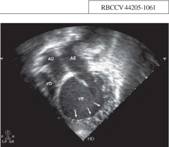
Fig. 1 - Apical four-chamber view showing dilated left ventricle with exuberant trabeculation in lateral and apical walls (arrows,) featuring noncompacted myocardium
1. São José do Rio Preto Pediatric Cardiovascular Surgery Service – Hospital de Base – São José do Rio Preto Medical School – São José do Rio Preto, SP.
Ulisses Alexandre CROTI1, Domingo Marcolino BRAILE1, Marcos Aurélio Barboza de OLIVEIRA1, Sírio
HASSEM SOBRINHO1
Rev Bras Cir Cardiovasc 2009; 24(1): 98-99
CLINICAL-SURGICAL CORRELATION
RBCCV 44205-1061
Cardioversor desfibrilador implantável em criança com miocárdio não-compactado isolado
Implantable cardioverter-defibrillator in a child
with isolated noncompacted myocardium
Correspondence address: Ulisses Alexandre Croti
Hospital de Base – FAMERP – Avenida Brigadeiro Faria Lima, 5544 – São José do Rio Preto, SP, Brasil – CEP 15090-000
Phone (Fax): (17) 3201-5025/3222-6450/ 9772-6560 E-mail: uacroti@uol.com.br
Article received on February 10th, 2009 Article accepted on March 12nd, 2008 CLINICAL DATA
9-year-old male child, 25 kg, from Barretos, São Paulo. Full-term patient presenting cardiorespiratory arrest (CRA) without defined cause and remained for 30 days in Intensive Care Unit (ICU). At 11 months presented episode of fainting, followed by cyanosis and CRA. He had undergone cardioversion due to ventricular fibrillation (sic). At the time, corticoid and digitalis were used, and the patient evolved well until the last two months, when presented new fainting episode at rest, requiring cardiac massage and new cardioversion to revert this presentation. Amiodarone, spironolactone, aspirin and suspension of digitalis were administered. After about 10 episodes of syncope and fainting, the child was referred to the ICU of our Service. On physical examination the patient was in good general condition, afebrile, acyanotic and eupneic. Precordium unchanged, regular heart rhythm, with normal and rhythmic sounds. Pulmonary auscultation was normal. Flaccid abdomen, liver at 2 cm from the right costal margin, spleen not palpable and presence of bowel sounds. Good peripheral perfusion, without edema, palpable and symmetrical pulses in all limbs, without motor sequels with only mild mental retardation, resulting from the CRA at the time of parturition (sic).
ELECTROCARDIOGRAM
Sinus rhythm, heart beat of 88 bpm, SÂP 0°, SÂQRS -30°, PR interval 0.24s. First-degree AV block, atrial and left ventricular overload, and left branch block.
RADIOGRAM
Visceral situs solitus in levocardia. Increased cardiac area with left ventricle affected, presenting a cardiothoracic index of 0.68 and second arc slightly increased. Pulmonary vascular network without apparent changes.
ECHOCARDIOGRAM
99
CROTI, UA ET AL - Implantable cardioverter-defibrillator in a child with isolated noncompacted myocardium
Rev Bras Cir Cardiovasc 2009; 24(1): 98-99
tract, left ventricular myocardium with spongiform appearance (Figure 1), left ventricular contractile dysfunction of significant degree with an ejection fraction of 33.8% and mild mitral valve insufficiency. Absence of intracavitary thrombi.
DIAGNOSIS
The patient was referred to our Service due to repeated episodes of ventricular fibrillation and recovery from sudden death [1], the treatment was quickly directed to the implantation of a cardioverter defibrillator. The diagnosis of noncompacted myocardium and not associated with other abnormalities is extremely rare, with few patients described in the literature and caused by a rare disorder of endomyocardial morphogenesis [2].
OPERATION
Patient in dorsal decubitus position, under general anesthesia, incision in the left infraclavicular region, divulsion of the pectoralis major muscle and making of the pocket with appropriate dimensions for subsequent generator implantation. Puncture of the left subclavian vein,
Fig. 2 - Induction of ventricular fibrillation
Fig. 3 - First unsuccessful ventricular defibrillation with 15J. Second ventricular defibrillation with 20J and reversion to sinus rhythm
Fig. 4 – Chest radiography in the early postoperative period with the atrial and ventricular electrodes implanted
REFERENCES
1. Andrade JC, Ávila Neto V, Braile DM, Brofman PR, Costa AR, Costa R, et al. Diretrizes para o implante de cardioversor desfibrilador implantável. Arq Bras Cardiol. 2000;74(5):481-2.
2. Chin TK, Perloff JK, Williams RG, Jue K, Mohrmann R. Isolated noncompaction of left ventricular myocardium. A study of eight cases. Circulation. 1990;82(2):507-13.

