Action of two endocrine disrupters on the sexual differenciation of
Nile tilapia
1Ação de dois desreguladores endócrinos na diferenciação sexual da tilápia do Nilo
Oscar Pacheco Passos Neto2*, André Bezerra dos Santos3, José Roberto Feitosa Silva4 and Suetônio Bastos Mota3
ABSTRACT - Endocrine disruptors are exogenous substances that can impair one or more functions of the endocrine system and cause adverse effects to the health of an intact organism. The main goal of this study was to evaluate the effect of the endocrine disruptors 17β-estradiol (E2) and 17α-ethinyl estradiol (EE2) on the sexual differentiation of Nile tilapia during spawning. Six treatments and one control with three replications, totalizing 21 experimental units, were assessed in a completely randomized design. Hormones were dosed at concentrations of 250, 500, and 1,000 ng L−1 (treatments E2–250, E2–500, E2–1,000, EE2–250, EE2–500, and EE2–1,000). Exposure to hormones occurred during the first 28 days of life. After this period, water was completely renewed and individuals kept in their respective experimental units for another 60 days, totaling 88 days. At the end of this period, animals were euthanized by ice desensitization for gonad extraction, slide preparation, and observation under an optical microscope. At the end of the experiment, the results indicated that the type of hormone and hormone concentration differentially affected the parameters of zootechnical performance. A significant difference (p<0.05) was observed in the condition factor between control and treatments. Intersex individuals were observed in treatments of both hormones, except for the control treatment. In addition, malformations were observed in individuals submitted to treatments, mainly in the head region. Thus, the presence of the chemical compounds 17β-estradiol and 17α-ethinylestradiol severely affected the development and sexual differentiation of Nile tilapia.
Key words: Emerging pollutants. Oreochromis niloticus. 17α-ethinylestradiol. 17β-estradiol.
RESUMO - Desreguladores endócrinos são substâncias exógenas que possuem a capacidade de alterar uma ou várias funções do sistema endócrino com efeitos adversos a saúde um organismo intacto. O objetivo principal do presente trabalho foi avaliar a ação dos desreguladores endócrinos 17β-estradiol (E2) e 17α-etinilestradiol (EE2) na diferenciação sexual da tilápia do Nilo durante o período de alevinagem. Foram avaliados seis tratamentos e um controle, com três repetições, totalizando 21 unidades experimentais em um delineamento inteiramente casualizado. Os hormônios foram dosados nas concentrações de 250; 500 e 1.000 ng L-1 (E2-250; E2-500; E2-1.000; EE2-250; EE2-500; EE2-1.000). A exposição aos hormônios ocorreu
durante os primeiros 28 dias de vida. Após este período a água foi totalmente renovada e os indivíduos mantidos em suas respectivas unidades experimentais por mais 60 dias, totalizando 88 dias. Ao final deste período os animais foram eutanasiados por insensibilização em gelo para extração das gônadas, preparação das lâminas e observação sob microscópio óptico. Ao final do experimento foi possível constatar que os hormônios e as concentrações hormonais avaliadas afetaram distintamente os parâmetros de desempenho zootécnico. Foi observada diferença significativa (p<0,05) no fator de condição entre o controle e os tratamentos. Indivíduos intersexo foram observados nos tratamentos de ambos os hormônios, com exceção do controle. Além disto, foram observadas malformações nos indivíduos dos tratamentos, principalmente na região da cabeça. Com os dados coletados na presente pesquisa é possível concluir que a presença dos compostos químicos 17β-estradiol e 17α-etinilestradiol afetam severamente o desenvolvimento e a diferenciação sexual da tilápia do Nilo.
Palavras-chave: Poluentes emergentes. Oreochromis niloticus. 17α-etinilestradiol. 17β-Estradiol. DOI: 10.5935/1806-6690.20190048
* Author for correspondence
Received for publication 02/07/2018; approved on 21/09/2018
1Parte da Tese de Doutorado do primeiro autor em Engenharia Civil (Recursos Hídricos) Departamento de Engenharia Hidráulica e Ambiental
-Pesquisa Financiada pelo CNPq - Edital MCTI/CNPQ Nº 14/2012 - Universal
2Departamento de Engenharia de Pesca, Centro de Ciências Agrárias, Universidade Federal do Ceará/UFC, Avenida Humberto Monte s/n
Campus do Pici, Bloco 827, Fortaleza-CE, Brasil, 60.455-760, [email protected] (ORCID ID 0000-0001-7168-2414)
3Departamento de Engenharia Hidráulica e Ambiental, Centro de Tecnologia, Universidade Federal do Ceará/UFC, Fortaleza-CE, Brasil,
[email protected] (ORCID ID 0000-0002-3395-8878), [email protected] (ORCIDI ID 0000-0001-6061-4539)
4Departamento de Biologia, Centro de Ciências, Universidade Federal do Ceará/UFC, Fortaleza-CE, Brasil, [email protected] (ORCID ID
INTRODUCTION
The first indications that substances with the potential for endocrine disrupters affected wildlife were observed in the 1980s from observations of female characteristics in male bird specimens colonizing the Great Lakes region, which occupies part of the United States and Canada (REIS FILHO; ARAÚJO; VIEIRA, 2006). Ever since, researchers around the world have observed similar effects on other animal taxa such as amphibians (PHUGE; GRAMAPUROHIT, 2015), reptiles (CZARNY et al., 2017), mollusks (BORYSKO; ROSS, 2014), and fishes (WOODS; KUMAR, 2011).
Endocrine disrupters are still poorly studied and their synergistic actions in the environment are totally unknown to most compounds since they are part of a group of compounds collectively known as emerging pollutants (NIEMUTH; KAPER, 2015). Among these endocrine disrupters, the natural estrogenic steroid 17β-estradiol (E2) and the synthetic estrogenic steroid 17α-ethinylestradiol (EE2) are the most studied since they are among the most widespread substances in the environment (CZARNY
et al. 2017). According to these authors, some of these
compounds (E2, for example) are present in the urine of animals and their concentration depends on gender, hormonal status, the existence of pregnancy, the phase of the menstrual cycle, in addition to synthetic forms used as contraceptive pills (EE2) and hormone-based medicines.
The feminizing action of endocrine disrupters of estrogenic action has been observed in fishes that inhabit rivers near large urban centers in several places of the world (DAMMANN et al., 2011; MEIJIDE et al., 2016; TETREAULT et al., 2011). In addition, laboratory tests have confirmed that considerably lower hormone doses,
in the orders of microgram (µg L-1) and nanogram per liter
(ng L-1), are likely to impair the development of various
fish species (LANGE et al., 2001; LEI et al., 2014; NIEMUTH; KLAPER, 2015).
Pessoa et al. (2012) found E2 and EE2
concentrations of the order of ng L-1 in a sewage treatment
plant (STP) located in the Metropolitan Region of Fortaleza. However, studies conducted in other Brazilian states have shown that such concentrations can reach the
order of µg L-1 in the surface water bodies close to large
urban centers (CAMPANHA et al., 2015; MACHADO
et al., 2014).
Therefore, studies that assess the effects of endocrine disrupters in a controlled environment are necessary since they allow assessing these compounds in isolation. The main goal of this study was to evaluate the effect of the endocrine disruptors 17β-estradiol (E2) and 17α-ethinyl estradiol (EE2) on the sexual differentiation of Nile tilapia during spawning. Besides that, the occurrence
of malformations and zootechnical performance (weight, length, allometric condition factor, and survival) were also evaluated.
MATERIAL AND METHODS
The present study was developed at the Laboratório de Recursos Aquáticos (LARAq) of the Departamento de Engenharia de Pesca (Laboratory of Aquatic Resources of the Department of Fisheries Engineering), the Laboratório de Saneamento (LABOSAN) of the Departamento de Engenharia Hidráulica e Ambiental (Laboratory of Sanitation of the Department of Hydraulic and Environmental Engineering), and the Laboratório de Histologia e Reprodução Animal (LAHRA) of the Departamento de Biologia (Laboratory of Histology and Animal Reproduction of the Department of Biology) of the Universidade Federal do Ceará-UFC (Federal University of Ceará).
Newly hatched larvae of Nile tilapia (Oreochromis
niloticus) were obtained from the Aquaculture Station
Professor Doctor Raimundo Saraiva da Costa affiliated to DEP/UFC and derived from the same spawning.
Six treatments and a control with three replications were assessed, totaling 21 experimental units with a useful volume of 40 L each. Ten larvae of Nile tilapia were stored in each aquarium. The experimental design was completely randomized design.
The hormones 17β-estradiol (E2) and 17α-ethinylestradiol (EE2) were purchased in a lyophilized form from the company Sigma-Aldrich. The effects of these hormones were assessed in three different concentrations:
250, 500, and 1,000 ng L-1 (treatments E2–250, E2–500,
E2–1,000, EE2–250, EE2–500, and EE2–1,000).
A stock solution with a concentration of 20 mg L-1
was prepared by diluting 10 mg of each hormone in 500 mL ethyl alcohol PA (solvent). These solutions were stored in an amber bottle and maintained in a refrigerator at 3-4 °C. The experimental aquaria (40 L) were dosed with 0.5, 1.0, and 2.0 mL of stock solution in order to obtain the experimental concentrations of 250,
500, and 1,000 ng L-1, respectively. In order to ensure
that all treatments received equal solvent volumes, 2.0, 1.5, and 0.5 mL alcohol was added to the control and
treatments of 250 and 500 ng L-1, respectively.
The individuals were exposed to hormone doses during the first 28 days of life. Weekly, the water from each tank was 100% renewed, and hormone doses replaced to the same dosages.
After 28 days of treatment, the water in each tank was completely renewed without the addition of
hormones, and the fish were kept in tanks for another 60 days, so that the mean weight of the individuals in all groups reached at least 10 g, totalizing 88 days. The water was drained from the tanks and treated with ultraviolet radiation before disposal, as described by Sornalingam, McDonagh and Zhou (2016).
Each experimental unit was covered by a glass cover and equipped with a mechanical filter driven by an air compressor under the air lift system, which remained on for 24 h. Mechanical filters were removed weekly for cleaning. Individuals were fed ad libitum four times a day at 9:00, 12:00, 14:00, and 16:00 h, with powder feed containing 45% crude protein.
Temperature (°C) and dissolved oxygen
concentration (mg L-1) were measured daily by using
a digital oximeter. In addition, pH, alkalinity (mg
CaCO3 L-1), and total ammoniacal nitrogen (mg L-1)
were measured every five days by using colorimetric and titrimetric tests according to the instructions of the manufacturer.
Two biometry assessments of the whole lot, the first 40 days after hatching (DAH) and the second 88 DAH, were carried out over the experimental period by measuring the total length (cm) and live weight (g) using a caliper (0.002 cm precision) and a digital scale (0.01 g precision), respectively.
After the second biometric evaluation, the fish were euthanized by ice desensitization, the gonads were removed, stored in histological microcassettes, labeled, and fixed in 10% formalin solution for 24 hours. Histological sections were stained with Harris hematoxylin and Gomori trichrome, as the method adapted by Tolosa
et al., (2003).
The allometric condition factor (KA) was calculated with the data of weight (W) and length
(L) by means of the equation KA = W/Lb, where b is
the allometric coefficient obtained by the regression equation of the weight-length relationship (LE CREN, 1951).
Data analysis was performed for water physical and chemical parameters (dissolved oxygen, temperature, pH, alkalinity, and total ammonia), zootechnical variables (weight, length and survival), and allometric condition factor. For this purpose, analysis of variance (ANOVA) at a level of significance of 5% was used considering the primary effect of two variables: type of hormone and hormone concentration. In cases of statistical significance, Tukey’s test was applied to compare the means using systematic component 1.
Yijk = μ + αi + βj + εijk (1)
Where: Yijk is the type of hormone i, hormonal
concentration j, and repetition k (i = 1, 2; j = 1, 2, 3; k
= 1, 2, 3); μ is the population mean; αi is the effect of
hormone type i; βj is the effect of hormone concentration
j; and εijk is the residual error.
The values expressed as percentages were transformed into arcsine for applying the statistical test. The data were subjected to the Lilliefors test to verify normality.
All statistical analyses were performed by using the Programa de Aplicações Estatísticas nas Áreas das Ciências Biomédicas (BioEstat, version 5.0) at a 5% significance level (AYRES; AYRES Jr., 2007).
Regression analysis equations of the weight-length relationship were determined by using spreadsheets for each treatment and control through the power model y =
Axb, where ln y = a + b ln x is the linearized form of this
curvilinear model.
RESULTS AND DISCUSSION
Water quality parameters assessed during the experimental period (Table 1) were within the comfort range for O. niloticus, according to Kubitza (2011). Dissolved oxygen, temperature, pH, alkalinity, and total ammoniacal nitrogen presented average values respectively between
7.37±0.42 and 6.97±0.35 mg L−1 (treatments E2–500 and
EE2–250), 26.24±0.76 and 25.93±0.81 °C (treatments EE2–250 and E2–250), 7.49±0.25 and 7.61±0.11 (treatments EE2–500 and E2–1,000), 80.55±9.80 and
68.62±7.31 mg CaCO3 L−1 (treatment E2–500 and
control), and 0.92±0.66 and 0.54±0.51 mg L−1 (treatments
E2–1,000 and EE2–500).
There was no significant difference (p>0.05) in fish weight among the groups (Table 2). However, the analysis of the other parameters of zootechnical performance indicated that the effects of the type of hormone and hormone concentration were independent. Total length and allometric condition factor were affected by the hormone concentration whereas survival was affected by the type of hormone.
There was a significant difference (p<0.05) in total length between treatments E2-250 (5.88 ± 1.56 cm) and EE2-500 (7.07 ± 1.90 cm).
For both hormone types, the allometric condition factor tended to decrease as hormone concentration was increased. There was a significant difference (p<0.05) in this variable in all cases, except in the group treated with EE2-500 when compared to the control group and the group treated with E2-1,000.
No significant difference (p>0.05) was observed for survival when comparing E2 treatment to control and to EE2 treatment, whose values were, respectively, 75.56 ± 7.26%, 80.00 ± 10.00%, and 64.44 ± 10.14%. However, a statistical difference (p<0.05) was observed between control and EE2 treatment.
As in the first biometric analysis, the effects of hormone type and hormonal concentration were different on the parameters of zootechnical performance in second biometrics (Table 3).
No significant differences (p>0.05) were found among the evaluated groups for total length and survival.
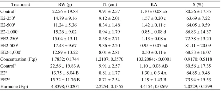
Treatment BW (g) TL (cm) KA S (%) Control1 5.59 ± 3.93 6.60 ± 1.50 ab 1.06 ± 0.08 a 80.00 ± 10.00 E2-2501 4.47 ± 4.10 5.88 ± 1.56 a 1.62 ± 0.15 c 80.00 ± 0.00 E2-5001 4.79 ± 3.18 6.23 ± 1.29 ab 1.41 ± 0.13 b 73.33 ± 5.77 E2-1,0001 5.48 ± 3.97 6.51 ± 1.37 ab 0.85 ± 0.08 e 73.33 ± 11.55 EE2-2501 5.64 ± 6.64 6.10 ± 1.70 ab 1.24 ± 0.19 f 66.67 ± 15.28 EE2-5001 7.82 ± 7.64 7.07 ± 1.90 b 0.96 ± 0.07 ae 66.67 ±11.55 EE2-1,0001 6.35 ± 4.61 6.66 ± 1.76 ab 0.49 ± 0.12 d 60.00 ± 0.00 Concentration(F;p) 1.6780; 0.1987 3.1573; 0.0356 129.2885; <0.0001 1.7091; 0.1911 Control2 5.59 ± 3.93 6.60 ± 1.50 1.06 ± 0.08 80.00 ± 10.00 A E22 4.90 ± 3.75 6.19 ± 1.42 1.30 ± 0.35 75.56 ± 7.26 AB EE22 6.57 ± 6.36 6.59 ± 1.80 1.11 ± 1.05 64.44 ± 10.14 B Hormone (F;p) 2.4135; 0.1165 1.5145; 0.2458 3.4754; 0.0518 4.6972; 0.0223
1Hormonal concentration,2Hormone, E2-250 – 17 β-estradiol, hormonal concentration 250 ng L-1; E2-500 – 17 β-estradiol, hormonal concentration 500
ng L-1; E2-1,000 – 17 β-estradiol, hormonal concentration 1,000 ng L-1. EE2-250 – 17 α-etinilestradiol, hormonal concentration 250 ng L-1; EE2-500
– 17 α-etinilestradiol, hormonal concentration 500 ng L-1; EE2-1,000 – 17 α-etinilestradiol, hormonal concentration 1,000 ng L-1
Table 2 - Zootechnical performance parameters in the first biometry of juvenile Nile tilapia (Oreochromis niloticus) under different hormonal concentration, 17 β-estradiol e 17 α- ethinylestradiol, during the first 28 days of life. Body weight (BW), total length (TL), alometric condition factor (KA) and survival (S). Mean±standard deviation. Values in the same column with different letters superscripts indicate statistical differences (p<0.05)
Treatment DO (mg L-1) Temp. (oC) pH Alk. (mg CaCO
3 L -1) TAN (mg L-1) Control 6.98 ± 0.43 26.13 ± 0.67 7.52 ± 0.22 68.62 ± 7.31 0.58 ± 0.49 E2-250 7.26 ± 0.44 25.93 ± 0.81 7.50 ± 0.14 77.57 ± 9.24 0.75 ± 0.69 E2-500 7.37 ± 0.42 26.03 ± 0.89 7.58 ± 0.08 80.55 ± 9.80 0.75 ± 0.76 E2-1,000 7.04 ± 0.38 26.09 ± 0.61 7.61 ± 0.11 71.0 ± 11.32 0.90 ± 0.74 EE2-250 6.97 ± 0.35 26.24 ± 0.76 7.60 ± 0.21 74.58 ± 7.31 0.92 ± 0.66 EE2-500 7.04 ± 0.40 26.08 ± 0.68 7.49 ± 0.25 74.58 ± 7.31 0.54 ± 0.51 EE2-1,000 7.08 ± 0.42 25.96 ± 0.71 7.53 ± 0.07 71.60 ± 2.55 0.83 ± 0.68
E2-250 – 17 β-estradiol, hormonal concentration 250 ng L-1; E2-500 – 17 β-estradiol, hormonal concentration 500 ng L-1; E2-1,000 – 17 β-estradiol,
hormonal concentration 1,000 ng L-1; EE2-250 – 17 α- ethinylestradiol, hormonal concentration 250 ng L-1; EE2-500 – 17 α- ethinylestradiol, hormonal
concentration 500 ng L-1; EE2-1,000 – 17 α- ethinylestradiol, hormonal concentration 1,000 ng L-1
Table 1 - Dissolved oxygen (DO), temperature (Temp.), pH, alkalinity (Alk.) and total ammoniacal nitrogen (TAN) in the water of juvenile Nile tilapia (Oreochromis niloticus) under different hormonal concentrations, 17β-estradiol e 17α-ethinylestradiol, during the first 28 days of life
However, when considering the effect of the type of hormone, a significant difference (p<0.05) in animal weight was found when comparing the control group (22.56 ± 19.83 g) with E2 (13.75 ± 8.04 g) and with EE2 (15.32 ± 11.76 g).
In turn, the allometric condition factor was significantly affected by hormone concentration in the two treatment groups. For hormone type, no significant difference (p>0.05) was observed between the control group and the two treatments. However, a significant difference (p<0.05) was found between the two treatments. In the group treated with EE2, the increase in hormone
concentration resulted in a significant decrease (p<0.05) in the condition factor, as observed in the first biometric evaluation.
The mean weight and body length in the control group in both biometric analyses were lower than those found by Massago et al. (2010) and Santos, Mareco and Silva (2013) for different tilapia strains.
The highest allometric condition factor values found in this study (treatment E2–250) are still considered as low when compared to those found by Tachibana et al. (2008), who studied Nile tilapia during the period of sexual reversion, and Passos-Neto et al. (2015), who studied red tilapia during the early development stages. Niemuth and Klaper (2015) found significantly lower condition factor values for Pimephales promelas exposed to the endocrine
disruptor metformin at a concentration of 40 µg L−1.
Dammann et al. (2011), on the other hand, found no significant difference in the condition factor value for P. promelas exposed to different concentrations of
estrone (10, 50, and 100 ng L−1) and 17β-estradiol (5,
25, and 50 ng L−1).
According to Vazzoler (1996), the condition factor is a quantitative indicator of the degree of healthiness or welfare of fishes, reflecting recent food conditions and/ or reserve expenditure in cyclical activities, influenced by environmental conditions and behavioral aspects of the species. According to the data obtained in our study,
Table 3 - Zootechnical performance parameters in the second biometry of juvenile Nile tilapia (Oreochromis niloticus) under different hormonal concentration, 17 β-estradiol e 17 α- ethinylestradiol, during the first 28 days of life. Body weight (BW), total length (TL), alometric condition factor (KA) and survival (S). Mean±standard deviation. Values in the same column with different letters superscripts indicate statistical differences (p<0.01)
1Hormonal concentration,2Hormone, E2-250 – 17 β-estradiol, hormonal concentration 250 ng L-1; E2-500 – 17 β-estradiol, hormonal concentration
500 ng L-1; E2-1,000 – 17 β-estradiol, hormonal concentration 1,000 ng L-1. EE2-250 – 17 α-etinilestradiol, hormonal concentration 250 ng L-1;
EE2-500 – 17 α-etinilestradiol, hormonal concentration EE2-500 ng L-1; EE2-1,000 – 17 α-etinilestradiol, hormonal concentration 1,000 ng L-1
juveniles from control and treatments presented different conditions.
Control and treatments E2–250, E2–500, E2–1,000, EE2–250, EE2–500, and EE2–1,000 presented allometric regression coefficients (b) equal to 3.2371, 3.0424, 3.1123, 3.3633, 3.2176, 3.2995, and 3.6457, respectively. Table 4 shows the equations defining the weight × length ratios
and their respective determination coefficient values (R2).
The values of b found are within the range of 2.5 to 4.0 mentioned by Le Cren (1951). Values of b higher than 3 indicate a positive allometric growth, with a higher increase in length when compared to weight, while values of b lower than 3 indicate a negative allometric growth, which means that weight is the predominant factor. In addition, values of b close to 3 characterize an isometric growth (b=3), which indicates an equal contribution of both biometric variables.
The gonads of the individuals were histologically analyzed from control (n=16) and treatments E2–250 (n=10), E2–500 (n=9), E2–1,000 (n=13), EE2–250 (n=9), EE2–500 (n=14), and EE2–1,000 (n=10). In some cases, the gonads could not be removed successfully because of their small size. Woodling et al. (2006) also demonstrated an atrophy in gonadal development in fish collected downstream from the discharge point of an STP. These authors reported difficulty in removing the gonads and in the visual identification of the individual’s sex. Dammann
et al. (2011) reported a decrease of the gonadosomatic
Treatment BW (g) TL (cm) KA S (%) Control1 22.56 ± 19.83 9.91 ± 2.57 1.10 ± 0.08 ab 80.56 ± 17.35 E2-2501 14.79 ± 9.16 9.12 ± 2.01 1.57 ± 0.20 c 63.69 ± 7.22 E2-5001 11.24 ± 5.36 8.34 ± 1.48 1.42 ± 0.11 c 64.05 ± 9.59 E2-1,0001 15.26 ± 9.02 8.94 ± 1.79 0.85 ± 0.08 d 66.83 ± 14.37 EE2-2501 15.04 ± 13.11 8.58 ± 2.71 1.13 ± 0.08 a 72.38 ± 13.20 EE2-5001 17.43 ± 9.67 9.36 ± 2.20 0.95 ± 0.07 bd 81.11 ± 20.09 EE2-1,0001 12.89 ± 13.22 8.01 ± 2.81 0.50 ± 0.11 e 68.33 ± 16.07 Concentration (F;p) 1.7832; 0.1744 1.2107; 0.3570 103.2084; <0.0001 0.9170; 0.5118 Control2 22.56 ± 19.83 A 9.91 ± 2.57 1.10 ± 0.08 AB 80.56 ± 17.35 E22 13.75 ± 8.04 B 8.81 ± 1.77 1.30 ± 0.3 4A 64.85 ± 9.48 EE22 15.32 ± 11.76 B 8.71 ± 2.54 1.19 ± 1.43 B 73.94 ± 15.53 Hormone (F;p) 4.8398; 0.0204 2.2254; 0.1355 4.4154; 0.0269 2.0229; 0.1599
index in P. promelas as estrone (10 to 100 ng L-1) and E2
concentrations (5 to 50 ng L-1) increased.
Intersex individuals were submitted to treatments with both hormones (Figure 1) at the lowest concentrations
of 250 ng L-1. Reis Filho, Araújo and Vieira (2006)
showed that endocrine disrupters are substances that although presented in small concentrations, are capable of triggering effects on the systems in which they are introduced. Campanha et al. (2015) also stated that during the degradation process, some of these compounds generated byproducts that may be more toxic than the original compound. On the other hand, laboratory studies at higher concentrations should also be conducted. In this sense, Machado et al. (2014) found E2 concentrations of
the order of µg L-1 in surface water of rivers in southern
Brazil.
The scientific interest in quantifying endocrine disrupters in the environment arose due to an increase in
the relationship between the detection of abnormalities in human health (PESSOA et al., 2012) in the presence of these micropollutants and the finding of intersex fish downstream from discharge points of STP close to large urban centers (TETREAULT et al., 2011). In addition, laboratory analyses of their effects have been observed and documented for specimens of different taxa such as fishes (Melanotaenia fluviatilis) (WOODS; KUMAR, 2011), mollusks (Nassarius burcardi and N. jonisii) (BORYSKO; ROSS, 2014), and amphibians (Euphlycts cyanophlycts) (PHUGE; GRAMAPUROHIT, 2015).
In treatment E2–500, only one intersex individual was found, while in treatments E2–250 and E2–1,000 no intersex individual was observed. In these last two treatments, however, a considerable number of situations (7 and 5 individuals, respectively) was observed in which the gonads could not be removed due to their small size and fragility. Treatments EE2–250, EE2–500, and
Treatment Equation R2 Control Y = 0.0107X3,2371 0.9940 E2-250 Y = 0.0159X3,0424 0.9862 E2-500 Y = 0.0141X3,1123 0.9884 E2-1,000 Y = 0.0085X3,3633 0.9892 EE2-250 Y = 0.0119X3,2176 0.9865 EE2-500 Y = 0.0095X3,2995 0.9943 EE2-1,000 Y = 0.0047X3,6457 0.9218
Table 4 - Equations and determination coefficient (R²) of the weight x length ratios of juvenile Nile tilapia (Oreochromis niloticus) under different hormonal concentration, 17 β-estradiol e 17 α- ethinylestradiol, during the first 28 days of life
E2-250 – 17 β-estradiol, hormonal concentration 250 ng L-1; E2-500 – 17 β-estradiol, hormonal concentration 500 ng L-1; E2-1,000 – 17 β-estradiol,
hormonal concentration 1,000 ng L-1. EE2-250 – 17 α-etinilestradiol, hormonal concentration 250 ng L-1; EE2-500 – 17 α-etinilestradiol, hormonal
concentration 500 ng L-1; EE2-1,000 – 17 α-etinilestradiol, hormonal concentration 1,000 ng L-1
Figure 1 - Photomicrography of juvenile Nile tilapia (Oreochromis niloticus) gonads under different hormonal concentration, 17 β-estradiol e 17 α-ethinylβ-estradiol, during the first 28 days of life. Testicle (A), ovary (B) e intersex (C). Harris hematoxylin and Gomori trichrome (Adapted by TOLOSA et al., 2003)
EE2–1,000 presented 2, 4, and 2 intersex individuals, respectively.
According to Czarny et al. (2017), in addition to reproductive dysfunctions such as the appearance of intersex individuals, abnormal production of vitellogenin, and a low sperm count, endocrine disrupters may still act negatively on nervous and immune systems, cause behavioral disorders, and affect the homeostasis of organisms in a way general. It is, therefore, imperative that studies in this area be developed as a basis for discussions that may result in future public policies on water quality regarding the presence of these emerging pollutants.
According to Devlin and Nagahama (2002), sex determination in fishes is controlled by a series of biochemical reactions involving different proteins. In order to change the sexual differentiation of males and females, the acting factor must cause structural and functional alterations in such proteins.
In addition to anthropogenic factors, some environmental factors can influence, on their own, sex determination in fishes. As an example is the temperature, which influences sexual differentiation in tilapias (AZAZA; DHRAÏEF; KRAÏEM, 2008; ROUGEOT et al., 2008) and in other fish species such as the rainbow trout (Oncorhynchus mykiss) (MAGERHANS; MÜLLERBELECKE; HÖRSTGEN-SCHWARK, 2009). It is not yet clear whether the influence of anthropogenic (endocrine disrupters) and natural factors (temperature) and future researches may clarify this issue.
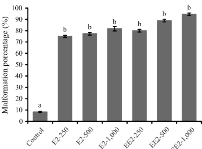
A great difference was found between the number of individuals who presented some type of malformation when comparing control (two individuals) with hormonal treatments (F=7.1771; p=0.0015). However, no significant difference (p>0.05) was observed for the number of malformed individuals when treatments were compared to each other.
The number of individuals with some type of malformation was 18 (75.00% of the total individuals that survived until the end of the experiment), 17 (77.27%), 18 (81.82%), 16 (80.00%), 16 (88.89%), and 17 (94.44%) for treatments E2–250, E2–500, E2–1,000, EE2–250, EE2–500, and EE2–1,000, respectively (Figure 2).
Lange et al. (2001) found severe malformations in P. promelas exposed to low concentrations of
17α-ethinylestradiol (16 and 64 ng L-1) during their life cycles,
such as atrophy, hemorrhage, and distended abdomen. Lei et al. (2014) observed mainly scoliosis and dilated
abdomen in Oryzias latipes exposed to estriol (E3)
concentrations ranging from 5 to 5,000 ng L-1.
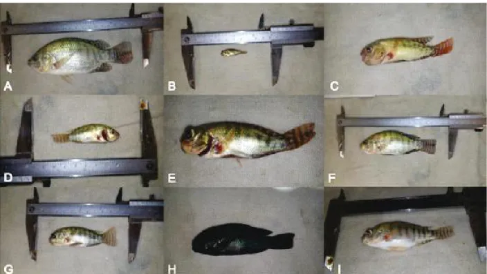
Figure 3 shows characteristic examples of malformations found in our study. The most observed deformity was the retracted belly. This condition is often associated with low-quality feed or nutritional deficiency of physiological origin. A retracted belly gives the individual low weight, resulting in an allometric coefficient higher than 3 and hence a low allometric condition factor, as shown in Figure 3 and Tables 2 and 3.
We also observed a severe developmental delay, deformities in the mouth, deformities in the shape of the head, exophthalmos (bulging eyes), distended abdomen with fluid accumulation (ascites), lowering of the mouth floor, exposed gills due to operculum deformities, change in natural color, and signs of hemorrhages in the base of the fins, especially in the pectoral fin.
In addition, we observed individuals with erratic and unbalanced swimming and buoyancy problems since they were most of the time standing at the bottom of the aquarium, which does not correspond to the natural behavior of Nile tilapia.
Figure 2 - Porcentage of malformations observed in juvenile Nile tilapia (Oreochromis niloticus) under different hormonal concentrations, 17 β-estradiol e 17 α-ethinylestradiol, during the first 28 days of life. Different letters superscripts indicate statistical differences (F=7.1771; p=0.0015). Vertrical bars indicates standard error of the mean
E2-250 – 17 β-estradiol, hormonal concentration 250 ng L-1; E2-500 – 17
β-estradiol, hormonal concentration 500 ng L-1; E2-1,000 – 17 β-estradiol,
hormonal concentration 1,000 ng L-1. EE2-250 – 17 α-ethinylestradiol,
hormonal concentration 250 ng L-1; EE2-500 – 17 α- ethinylestradiol,
hormonal concentration 500 ng L-1; EE2-1,000 – 17 α- ethinylestradiol,
Figure 3 - Types of malformations found in juveniles Nile tilapia ( Oreochromis niloticus) submitted to different hormonal concentrations of 17β-estradiol and 17α-ethinylestradiol during the first 28 days of life
CONCLUSIONS
1. Both 17β-estradiol and 17α-ethinyl estradiol are endocrine disruptors and impair the sexual differentiation of Nile tilapia (Oreochromis niloticus), causing the development of intersex fish;
2. The hormones 17β-estradiol and 17α-ethinyl estradiol cause malformations (particularly in the head), erratic swimming, problems of buoyancy, and reduce zootechnical performance;
3. The zootechnical performance parameters (weight, body length, survival, and allometric condition factor) were negatively affected by both hormones 17β-estradiol and 17α-ethinyl estradiol.
ACKNOWLEDGEMENTS
The authors would like to thank the Conselho Nacional de Desenvolvimento Científico e Tecnológico-CNPq (National Council for Scientific
and Technological Development) for financial support to perform this research (MCTI/CNPq 14/2012-Universal).
REFERENCES
AYRES, M.; AYRES JÚNIOR, M. Aplicações estatísticas nas áreas das ciências biomédicas. Manual do Programa BioEstat. Belém, 2007. 339 p.
AZAZA, M. S.; DHRAÏEF, M. N.; KRAÏEM, M. M. Effects of water temperature on growth and sex ratio of juvenile Nile tilapia Oreochromis niloticus (Linnaeus) reared in geothermal waters in southern Tunisia. Journal of the Thermal Biology, v. 33, n. 2, p. 98-105, 2008.
BORYSKO, L; ROSS, P. M. Adult expossure to the synthetic hormone 17α-ethynylestradiol affects offspring of the gastropods
Nassarius burchardi and Nassarius jonasii. Ecotoxicology
Environmental Safety, v. 103, p. 91-100, 2014.
CAMPANHA, M. B. et al. A 3-year study on occurrence of emerging contaminants in an urban stream of São Paulo State of Southeast Brazil. Environmental Science and Pollution Research International, v. 22, p. 7936-7947, 2015.
Normal individual (A); growth retardation (B); malformation in the shape of the head (C, E, G, and H); oral malformation (C, E, and G); Lowering of the mouth floor (C); malformation of the operculum (D and E); belly retraction (C, D, E, F, G, and H); distended abdomen with fluid accumulation (ascites) (I); exophthalmos (E); abnormal color (H); signs of hemorrhages in the pectoral fin (C, D, and E). All individuals are the same age
CZARNY, K. et al. The impact of estrogens on aquatic organisms and methods for their determination. Critical Reviews in Environmental Science and Technology, v. 47, n. 11, p. 1-55, 2017.
DAMMANN, A. A. et al. Comparing biological effects and potencies of estrone and 17β-estradiol in mature fathead minnows, Pimephales promelas. Aquatic Toxicology, v. 105, p. 559-568, 2011.
DEVLIN, R. H.; NAGAHAMA, Y. Sex determination and sex differentiation in fish: an overview of genetic, physiological, and environmental influences. Aquaculture, v. 208, n. 3/4, p. 191-394, 2002.
KUBITZA, F. Tilápia: tecnologia e planejamento na produção comercial. 2. ed. Jundiaí: F. Kubitza, 2011. 316 p.
LANGE, R. et al. Effects of the synthetic estrogen 17 α-ethinylestradiol on the life-cycle of the fathead minnow (Pimephales promelas). Environmental Toxicology Chemistry, v. 20, p. 1216-27, 2001.
LE CREN, E. D. The length-weight relationship and seasonal cycle in gonad weight and condition in the perch (Perca
fluviatilis). Journal of Animal Ecology, v. 20, n. 2, p. 201-219,
1951.
LEI, B. et al. Long-term exposure investigating the estrogenic potency of estriol in Japanese medaka (Orysias latipes). Comparative Biochemistry and Physiology, Part C, v. 160, p. 86-92, 2014.
MACHADO, K. S. et al. Occurrence of female sexual hormones in the Iguazu river basin, Curitiba. Acta Scientiarum. Technology, v. 36, p. 421-427, 2014.
MAGERHANS, A.; MÜLLER-BELECKE, A.; HÖRSTGENSCHWARK, G. Effect of rearing temperatures post hatching onsex ratios of rainbow trout Oncorhynchus
mykiss) populations. Aquaculture, v. 294, n. 1/2, p. 25-29,
2009.
MASSAGO, H. et al. Crescimento de quatro linhagens de tilápia
Oreochromis niloticus. Revista Acadêmica: Ciências Agrárias
e Ambientais, v. 8, n. 4, p. 397-403,2010.
MEIJIDE, F. J. et al. Effects of waterborne exposure to 17β-estradiol and 4-tert-octylphenol on early life stages of the South American cichlid fish Cichlasoma dimerus. Ecotoxicology and Environmental Safety, v. 124, p. 82-90, 2016.
NIEMUTH, N. J.; KLAPER, R. D. Emerging wastewater contaminant metformin causes intersex and reduced fecundity in fish. Chemosphere, v. 135, p. 38-45, 2015.
PASSOS NETO, O. P. et al. Reprodução e proporção sexual da tilápia vermelha, variedade Saint Peter, em diferentes
salinidades. Revista Ciência Agronômica, v. 42, n. 2, p. 310-318. 2015.
PESSOA, G. de P. et al. Desenvolvimento de metodologia para avaliar remoção de estrogênios em estações de tratamento de esgoto. Química Nova, v. 35, n. 5, p. 968-973, 2012.
PHUGE, S. K.; GRAMAPUROHIT, N. P. Sex hormones alter sex ratios in the Indian skipper frog, Euphlyctis
cyanophlyctis: determining sensitive stages for gonadal sex
reversal. General and Comparative Endocrinology, v. 220, p. 70-77, 2015.
REIS FILHO, R. W.; ARAÚJO, J. C.; VIEIRA, E. M. Hormônios sexuais estrógenos: contaminantes bioativos. Química Nova, v. 29, n. 4, p. 817-822, 2006.
ROUGEOT, C., et al. Effect of high temperature during embryogenesis on the sex differentiation process in the Nile tilapia, Oreochromis niloticus. Aquaculture, v. 276, n. 1/4, p. 205-208, 2008.
SANTOS, V. B. dos; MARECO, E. A.; SILVA, M. D. P. Growth curves of Nile tilapia (Oreochromis niloticus) strains cultivated at different temperatures. Acta Scientiarum, v. 35, n. 3, p. 235-242, 2013.
SORNALINGAM, K.; MCDONAGH, A.; ZHOU, J. L. Photodegradation of estrogenic endocrine disrupting steroidal hormones in aqueous system: process and future challenges. Science of the Total Environment, v. 550, p. 209-224, 2016.
TACHIBANA, L. et al. Densidade de estocagem de póslarvas de tilápia-do-Nilo (Oreochromis niloticus) durante a fase de reversão sexual. Boletim do Instituto de Pesca, v. 34, n. 4, p. 483-488, 2008.
TETREAULT, G. R. et al. Intersex and reproductive impairment of wild fish exposed to multiple municipal wastewater discharges. Aquatic Toxicology, v. 104, p. 278-290, 2011.
TOLOSA, E. M. C. et al. Manual de técnicas para histologia normal e patológica. São Paulo: Manole, 2003. 331 p. VAZZOLER, A. E. A. M. Biologia da reprodução de peixes teleósteos: teoria e prática. Maringá: EDUEM, 1996. 169 p. WOODLING, J. D. et al. Intersex and other reproductive disruption of fish in wastewater effluent dominated Colorado streams. Comparative Biochemistry and Physiology, Part C, v. 144, p. 10-15, 2006.
WOODS, M.; KUMAR, A. Vitellogenin induction by 17 β-estradiol and 17 α-ethynylβ-estradiol in male Murray rainbowfish (Melanotaenia fluviatilis). Environmental Toxicology and Chemistry, v. 30, p. 2620-2627, 2011.