Rev Bras Farmacogn 24(2014): 363-366
* Corresponding author.
E-mail: [email protected] (L.M.F. Lucinda).
0102-695X/$ - see front matter © 2014 Sociedade Brasileira de Farmacognosia. Published by Elsevier Editora Ltda. All rights reserved. http://dx.doi.org/ 10.1016/j.bjp.2014.07.015
Short communication
The effect of
Ginkgo
biloba
extract treatment in the Bcl-2
expression by osteoblasts in the femoral trabecular bone of
Wistar rats with glucocorticoid-induced osteoporosis
Leda M.F. Lucinda
a,b,*, Beatriz J.V. Aarestrup
a,b, Joanna S. Brandão
a,c, Vera M. Peters
a,
João E. de P. Reis
a, Martha de O. Guerra
aaCentro de Biologia da Reprodução, Universidade Federal de Juiz de Fora, Juiz de Fora, MG, Brazil bDepartamento de Morfologia, Universidade Federal de Juiz de Fora, Juiz de Fora, MG, Brazil cEscola de Medicina, Universidade Federal de Juiz de Fora, Juiz de Fora, MG, Brazil
Introduction
The bone is a dynamic tissue that is continually remodeling itself which is directly related to osteoclasts which erode cavities in bone, and osteoblasts which synthesize a new bone matrix. Most metabolic disorders such as osteoporosis are a result of the imbalance between these two types of cells (Weinstein and Manolagas, 2000). Glucocorticoids (GC) have potent
anti-inflammatory effects and their current oral use is strongly associated with serious side effects, including osteoporosis and an increase in fractures as a consequence (Lane and Yao, 2011). One of GC principal actions is to reduce osteoblast function and their number by apoptosis; (Chang et al., 2009). Apoptosis is regulated by an intrinsic process involving the activation of genes such as the Bcl-2, an anti-apoptotic protein member of the bcl-2 gene family that participates in programmed cell death (Verborgt et al., 2002).
A R T I C L E I N F O
Article history:
Received 26 March 2014 Accepted 14 May 2014
Keywords: Osteoporosis Ginkgo biloba Bcl-2 Apoptosis
A B S T R A C T
Evaluate the effect of the extract of Ginkgo biloba L., Ginkgoaceae (EGb) in the Bcl-2 expression by osteoblasts in the femoral trabecular bone of Wistar rats with glucocorticoid-induced osteoporosis. Rats were divided into five groups: osteoporosis; EGb1 (28 mg/kg); EGb2 (56 mg/ kg); alendronate (0.2 mg/animal) and control. The treatments were conducted for 20 or 30 days. The Bcl-2 expression by osteoblasts cells was evaluated in the femoral trabecular bone. The control group was compared with the osteoporosis-induced group (Student’s t-test). The other groups were analyzed by ANOVA test followed by Tukey’s test (p < 0.05). The percentage of Bcl-2 expression was reduced, when the control group (17.95 ± 3.45 20 days; 21.11 ± 3.43 30 days) was compared with the osteoporosis group (10.64 ± 3.30 20 days; 9.72 ± 2.84 30 days). Nevertheless, this percentage increased in the EGb2 group (18.58 ± 3.41 20 days; 16.51 ± 1.80 30 days) when compared to the osteoporosis group. The EGb increased the expression of the anti-apoptotic protein, suggesting a decrease in osteoblast apoptosis.
364
Leda M.F. Lucinda et al. / Rev Bras Farmacogn 24(2014): 363-366The extract of Ginkgo biloba (EGb) contains 24% of flavonoids (kaempferol, quercetin, rutin, myricetin, among others). Studies have reported that in vitro quercetin and kaempferol stimulate the osteoprogenitor cell in the bone marrow, the osteoblastic differentiation and mineralization, as well as the inhibition of the osteoclast function (Smith and Luo, 2004). In previous studies, we showed that GC reduced the percentage of the femoral trabecular bone, and the EGb was effective in increasing the bone mineral content of rats with glucocorticoid-induced osteoporosis (GIO) (Lucinda et al., 2010). Recently, we also demonstrated that in the alveolar mandibular bone, EGb reduced the Bax expression and increased the Bcl-2 expression by osteoblasts in rats with GIO suggesting a decrease of apoptosis of these cells (Lucinda et al., 2013). Therefore, not only did our study related the anti-apoptotic properties of EGb, but also Qiao et al., 2014 demonstrated that EGb exhibits a significant protective effect anti-apoptotic in rat myocardium cells, related to the down-regulation of Bax, cyt-c and caspase-3.
These studies are important to support the anti-apoptotic properties of this extract that could be responsible for the improvement in bone mineral density shown in pre-clinical studies (Lucinda et al., 2010; 2013). Based on these reports, we hypothesized that EGb could improve the expression of anti-apoptotic proteins such as Bcl-2 not only locally in the alveolar bone of the mandible but also in the trabecular femoral bone. The present study was designed to evaluate the effect of EGb in the Bcl-2 expression by osteoblast cells in the femoral trabecular bone of Wistar rats with GIO.
Materials and methods
The methodology of the present work was approved by the Ethical Committee on Animal Experimentation (protocol number 026/2009- CEEA, Federal University of Juiz de Fora, MG, Brazil), which follows the international principles in ethics for animal experimentation.
The dry extract of Ginkgo biloba L., Ginkgoaceae (Lot no. 20091112) was kindly supplied by JR Pharma (EPR-Farmácia de Manipulação e Drogaria, Co. Ltd., Juiz de Fora, Brazil), which was obtained from the Galena Pharmaceutical, Co. Ltd. (Campinas, São Paulo, Brazil) responsible for the quality test using the HPLC analytical methods. The test showed that the Ginkgo biloba extract we used was composed of 28.2% ginkgoflavonglicosides: 8.3% of terpenolactones; 15% of quercetin glycosides; 10.9% of kaempferol glycosides; 2.3% of ishorhamnetin glycosides and less than 5 ppm of ginkgolic acids.
The Ginkgo biloba dry extract was coarsely crushed and immediately suspended in distilled water at an appropriate concentration for the experiment. Afterwards, the rats of the EGb treatment group received a daily dosage of 1 ml of the suspension by oral gavage at a dose level of 28 mg/kg and 56 mg/kg of body weight.
Female Wistar rats (50 days old, weighing approximately 100-150 g) were obtained from the vivarium of the Federal University of Juiz de Fora, where they were born and bred. Groups of three animals were housed in clear plastic cages
with stainless steel wire lids and pinewood shavings as bedding, and kept in an animal room under controlled environmental conditions (12:12 light/dark cycle, temperature 22oC) on closed ventilated shelves. Animals were fed rat chow pellets (an average of 25 g daily) and received water ad libitum. Osteoporosis was induced through the intramuscular administration of dexamethasone disodium phosphate (Decadron® 4 mg/ml) at the dose level of 7 mg/kg of body weight, once a week, for five weeks in all groups, except in the control group (Lucinda et al., 2010; 2013).
After the end of the osteoporosis induction, sixty animals were selected at random and divided evenly into five groups (n = 6): Osteoporosis group (dexamethasone only); EGb1 group (Ginkgo biloba extract 28 mg/kg); EGb2 group (Ginkgo biloba
extract 56 mg/kg); Alendronate group (sodium alendronate 0.2 mg/animal/day) and Control group. The Control group was submitted to neither osteoporosis induction nor any treatment. The Alendronate, EGb1 and EGb2 groups were treated intragastrically, once a day, for 20 days (n = 30) or 30 days (n = 30), after osteoporosis induction. The choice of EGb doses was based on previous studies (Lucinda et al., 2010; 2013). The animals were euthanized on the 21st (n = 30) and 31st (n = 30) days. First they were anaesthetized intraperitoneally with Xylazine (180 mg/kg) and Ketamine (10 mg/kg), and then euthanized by total exsanguination via cardiac puncture.
The right femurs were removed and fixed in 4% phosphate-buffered formaldehyde for 24 h; decalcified in an aqueous solution of 5% nitric acid for two days, and then processed for paraffin embedding. The immunohistochemical staining was performed using mouse monoclonal Bcl-2 (C-2) antibody (Santa Cruz Biotechnology Inc., Santa Cruz, CA, USA) against amino acids 1-205 of Bcl-2 of human origin. Longitudinal 5-µm-thick sections of the femur were obtained. The paraffin embedded tissues were briefly deparaffinized, put in a solution of 0.05M citrate buffer (pH 6) to amplify the signal, placed in a water bath (96°C for 40 min), and then cooled for 20 min at room temperature. The samples were washed thrice in PBS (phosphate buffered saline) solution, 5 min each time. The endogenous peroxidase activity was blocked using a 3% hydrogen peroxidase solution for 10 min and washed in three changes of PBS for 5 min each. The samples were then incubated individually with monoclonal antibody Bax overnight at 4°C at 1:500 dilutions in 1.5% blocking serum in PBS and washed three times with PBS for 5 min each.
Leda M.F. Lucinda et al. / Rev Bras Farmacogn 24(2014): 363-366
365
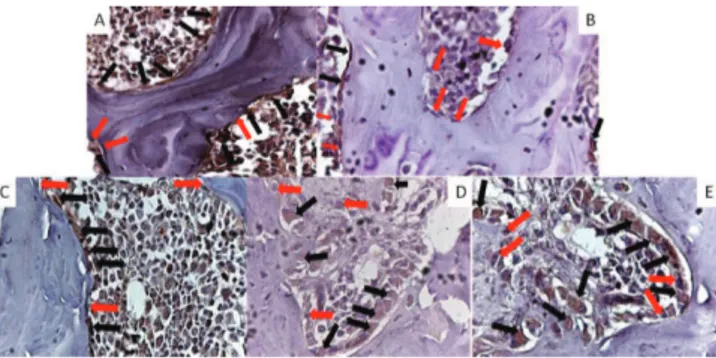
The femur samples were analyzed using an optical microscope system Zeiss (Hallbergmoos, Germany) for digitally captured microphotographs. All slides were analyzed at 250× and 400× magnifications by a pathologist. The quantitative analysis was conducted by a blind evaluator, of the experimental groups. The Bcl-2 expression was evaluated in the osteoblastic cells present in the central region of the trabecular bone tissue of the proximal epiphysis of the femur (Fig. 1). The osteoblasts were counted using the imaging software Axio vision® (version 4.5 for Windows semi-automatic), at 250× magnified images; the positive osteoblasts present a variation of brown color (Fig. 1). For the evaluation of the percentage of Bcl-2 positive cells the following formula was used: % of Bcl-2 positive osteoblasts = Bcl-2 positive osteoblast/total number of osteoblasts (where the total numbers of osteoblasts include the positive and negative cells for the Bcl-2 protein).
The data were expressed as mean ± standard deviation, and were analyzed for statistical significance using one-way analysis of variance (ANOVA), followed by Tukey’s post-hoc test, except for the control group. The control group was compared with the osteoporosis group using the Student’s t-test. A probability of p < 0.05 was considered statistically significant.
Groups (n = 6) Bcl-2 20 days (%) Bcl-2 30 days (%) % relative
Control 17.95 ± 3.45 21.11 ± 3.43 4.5 Osteoporosis 10.64 ± 3.30a 9.72 ± 2.84a 4.7
Alendronate 16.11 ± 4.70 12.06 ± 3.78 1.9 EGb1 15.17 ± 3.11 11.69 ± 1.08b 0.3
EGb2 18.58 ± 3.41c 16.51 ± 1.80c 55.8
The data are expressed by mean± standard deviation
ap < 0.05 when comparing the control group with the osteoporosis
one (Student`s t-test).
bp < 0.05 when comparing all groups, except control, with EGb2
(Tukey’s test).
cp < 0.05 when comparing all groups, except control, with
osteoporosis (Tukey’s test).
Table 1
Percentage of Bcl-2 expression in osteoblasts in the femoral trabecular bone.
In the present study, we observed that GC reduced the expression of Bcl-2, responsible of cell protection by binding to pro-apoptotic proteins, such as Bax, Bcl-xs and Bad (Gohel et al., 2009). It is known that the bone fragility from GC treatment is multi-factorial, that include the alteration of calcium and phosphorus metabolism, resulting in the elevation of secretion of parathyroid hormone and stimulation of osteoclast activity, followed by a reduction in osteogenesis and osteoblast activity (Lane and Yao, 2011).
The anti-apoptotic properties of EGb reported in this and other studies (Smith and Luo, 2004; Lucinda et al., 2013) could result from the antioxidant action of the flavonoids present in the extract, which attenuate the reactive oxygen species by chelating pro-oxidant transitional metal ions. Likewise, Brayboy et al (2001) reported that EGb was effective in protecting MC3T3-E1, an osteoblast-like cell, from death when exposed to free radicals, thus increasing their proliferation in vitro. Apart from this antioxidant action, EGb has an estrogenic effect as reported by Oh and Chung (2004), which is probably another mechanism to explain the increase in the Bcl-2 expression by osteoblasts.
Previously, it was demonstrated that EGb improves the percentage of alveolar bone in the mandible and in the trabecular bone in the femur of rats with GIO (Lucinda et al., 2010). Nevertheless, when searching for a possible mechanism to explain these results we found an important effect of the EGb, which was related directly to the expression of pro-apoptotic and anti-apoptotic proteins by osteoblasts in the mandibular alveolar bone. Nevertheless, the positive effects of EGb in bone were not limited to the mandible, as before-mentioned. The present study showed that EGb was effective in improving the expression of Bcl-2 in the femoral trabecular bone suggesting that one of the mechanisms involved in the bone increase (Lucinda et al., 2010) is to reduce the osteoblast death caused by the use of GC.
In conclusion, as the EGb increases the expression of Bcl-2 by osteoblasts in the femoral trabecular bone, this study could form the basis for further clinical studies of the potential effect of EGb in the GIO.
Results and discussion
Table 1 shows that the percentage of Bcl-2 expression of osteoblasts in the femoral trabecular bone was significantly lower in the osteoporosis group when compared with the control group (20 and 30 days). During the 20 and 30 days of treatment, the expression of Bcl-2 increased significantly in the EGb2 group, however the alendronate group didn’t show a significant increase when compared with the osteoporosis one.
Pre-clinical studies showed that EGb has a potential role in the treatment of GIO (Lucinda et al., 2010; 2013), however, to this day, the extract is not used in the clinical treatment of osteoporosis. Nevertheless, pre-clinical studies are important to clear the mechanisms that could explain the effect of EGb in osteoporosis.
366
Leda M.F. Lucinda et al. / Rev Bras Farmacogn 24(2014): 363-366Authors’ contributions
LMFL ran the laboratory work, treated the animals, analyzed the data and drafted the paper. JSB ran the laboratory work and treated the animals. BJVA contributed to the immunohistochemistry reactions and analysis. JEPR supplied and prepared the extract of Ginkgo biloba. VMP, contributed to the critical reading of the manuscript. MOG designed the study, supervised the laboratory work, drafted the paper and contributed to critical reading of the manuscript. All authors have read the final manuscript and approved the submission.
Conflicts of interest
The authors declare no conflicts of interest.
Acknowledgments
This work was financed by Fundação Mineira de Amparo à Pesquisa (APQ-00154-11 and 173/08). We thank Cassiana M Boya for the review of the English version of the manuscript.
R E F E R E N C E S
Brayboy, J.R., Chen, X.W., Lee, Y.S., Anderson, J.J.B., 2001. The protective effects of Ginkgo biloba extract (EGb 761) against free radical damage osteoblast-like bone cells (MC3T3-E1) and the proliferative effects of EGb 761 on these cells. Nutr. Res. 21, 1275-1285.
Chang, J.K., Li, C.J., Liao, H.J., Wang, C.K., Wang, G.J., Ho, M.L., 2009. Anti-inflammatory drugs suppress proliferation and induce apoptosis through altering expressions of cell cycle regulators and pro-apoptotic factors in cultured human osteoblasts. Toxicology 28, 148-156.
Gohel, A., Mccarthy, M.B., Gronowicz, G. 1999. Estrogen prevents glucocorticoid-induced apoptosis in osteoblasts in vivo and in vitro. Endocrinology 140, 5339-5347.
Lane, N.E., Yao, W., 2011. New insights into the biology of glucocorticoid-induced osteoporosis. IBMS BoneKEy 8, 229-236.
Lucinda, L.M, Aarestrup, B.J., Peters, V.M., Reis, J.E., Oliveira, R.S., Guerra, M.O., 2013. The effect of the Ginkgo biloba extract in the expression of Bax, Bcl-2 and bone mineral content of Wistar rats with glucocorticoid-induced osteoporosis. Phytother. Res. 27, 515-520.
Lucinda, L.M., Vieira, B.J., Salvador, P.A., Oliveira, T.T., Peters, V.M., Reis, J.E., Guerra M.O., 2010. Efeito do extrato de Ginkgo biloba L., Ginkgoaceae, na osteoporose induzida em ratas Wistar. Rev. Bras. Farmacogn. 20, 429-434.
Oh, S.M.; Chung, K.H., 2004. Estrogenic activities of Ginkgo biloba extracts. Life Sci. 74, 1325-1335.
Qiao Z.Y., Huang J.H., Ma J.W., Xu Y.W., Xie J., Liu H.J., Xiong S.J., Ge G.H., 2014. Ginkgo biloba extract reducing myocardium cells apoptosis by regulating apoptotic related proteins expression in myocardium tissues. Mol. Biol. Rep. 41,347-353. Smith, J.V., Luo, Y., 2004. Studies on molecular mechanisms of
Ginkgo biloba extract. Appl. Microbiol. Biot. 64, 465-472. Verborgt, O., Tatton, N.A., Majeska, R.J., Schaffler, M.B., 2002.
Spatial distribution of Bax and Bcl-2 in osteocytes after bone fatigue: Complementary roles in bone remodeling regulation. J. Bone Miner. Res. 17, 907-914.