Braz. J. of Develop.,Curitiba, v. 6, n.4,p.21791-21805 apr. 2020. ISSN 2525-8761
Antioxidant and antimicrobial activity of olive trees cultivated in the
Campanha Gaúcha region
Atividade antioxidante e antimicrobiana de folhas de oliveira cultivadas na
Região da Campanha Gaúcha
DOI:10.34117/bjdv6n4-374
Recebimento dos originais:25/03/2020 Aceitação para publicação:28/04/2020
Bruna da Fonseca Antunes
Mestra em Nutrição e Alimentos pela Universidade Federal de Pelotas
Instituição: Universidade Federal de Pelotas, Centro de Ciências Químicas, Farmacêuticas e de Alimentos
Endereço: Campus Capão do Leão - Universidade Federal de Pelotas, s/n CEP: 96010-900 - Caixa Postal 354 - Pelotas – RS, Brasil
E-mail: brunafonsecaantunes@gmail.com
Deborah Murowaniecki Otero
Pós Doutora em Ciência e Tecnologia de Alimentos pela Universidade Federal de Pelotas Instituição: Universidade Federal da Bahia, Departamento de Ciência dos Alimentos, Escola
de Nutrição
Endereço: Rua Araújo Pinho, 32 – Bairro Canela, CEP: 40110-150, Salvador – BA, Brasil E-mail: deborah.m.otero@gmail.com
Fernanda Moreira Oliveira
Mestra em Ciência e Tecnologia de Alimentos pela Universidade Federal de Pelotas Instituição: Universidade Federal de Pelotas, Departamento de Ciência e Tecnologia
Agroindustrial, Faculdade de Agronomia Eliseu Maciel
Endereço: Campus Capão do Leão - Universidade Federal de Pelotas, s/n CEP: 96010-900 - Caixa Postal 354 - Pelotas – RS, Brasil
E-mail: fer.moroli@gmail.com
Andressa Carolina Jacques
Pós Doutora em Ciência e Tecnologia de Alimentos pela Universidade Federal de Pelotas Instituição: Universidade Federal do Pampa, Cursode Engenharia de Alimentos Endereço: Avenida Maria Anunciação Gomes de Godoy, 1650 - Bairro Malafaia, CEP:
96413-172, Bagé – RS, Brasil E-mail: andressajacques@hotmail.com
Eliezer Avila Gandra
Doutor em Ciência e Tecnologia de Alimentos pela Universidade Federal de Pelotas Instituição: Universidade Federal de Pelotas, Centro de Ciências Químicas, Farmacêuticas e
Braz. J. of Develop.,Curitiba, v. 6, n.4,p.21791-21805 apr. 2020. ISSN 2525-8761 Endereço: Campus Capão do Leão - Universidade Federal de Pelotas, s/n
CEP: 96010-900 - Caixa Postal 354 - Pelotas – RS, Brasil E-mail: gandraea@hotmail.com
Rui Carlos Zambiazi
PhD em Food and Nutritional Science pela University of Manitoba
Instituição: Universidade Federal de Pelotas, Centro de Ciências Químicas, Farmacêuticas e de Alimentos
Endereço: Campus Capão do Leão - Universidade Federal de Pelotas, s/n CEP: 96010-900 - Caixa Postal 354 - Pelotas – RS, Brasil
E-mail: zambiazi@gmail.com
ABSTRACT
The aim of this study was to evaluate the antioxidant and antimicrobial potential of leaves from different olive cultivars in the Campanha Gaúcha region. The antioxidant activity was evaluated by the methods of FRAP (Ferric Reducing Antioxidant Power), ABTS (2,2'-azino-bis (3-ethylbenzthiazoline-6-sulfonic acid) and DPPH (2,2-diphenyl-1-picrylhydrazyl The antimicrobial effect of the extracts of olive leaves was evaluated by means of three phenotypic methodologies: disc diffusion, minimum inhibitory concentration and minimum bactericidal concentration, against the bacterial species Escherichia coli and Staphylococcus aureus. The three evaluated methods were efficient for the determination of antioxidant activity; however, the olive leaves showed activities that varied according to the method used, and no pattern of activity was seen among methods and cultivars. The results of the present study indicated that extracts of olive leaves presented antibacterial potential against E. coli and S. aureus. The leaves have the potential to be used in the development of new food products, and they can be an alternative to minimize the disposal of residues in the environment and to have antimicrobial activity against the pathogenic bacteria.
Keywords: Olea europaea L., by-product, biological activity. RESUMO
O objetivo deste estudo foi avaliar o potencial antioxidante e antimicrobiano de folhas de diferentes cultivares de Oliveira da região da Campanha Gaúcha. A atividade antioxidante foi avaliada através dos métodos de FRAP (Ferric Reducing Antioxidant Power), ABTS (2,2'-azino-bis (3-ethylbenzthiazoline-6-sulfonic acid) e por DPPH (2,2-difenil-1-picrilhidrazila). O efeito antimicrobiano dos extratos de folhas de oliveira foi avaliado por meio de três metodologias fenotípicas: disco difusão, concentração inibitória mínima e concentração bactericida mínima, frente às espécies bacterianas Escherichia coli e Staphylococcus aureus. Os três métodos avaliados foram eficientes para a determinação da atividade antioxidante; no entanto, as folhas de oliveira apresentaram atividades que variaram segundo o método utilizado, não apresentando um padrão de atividade entre método e cultivar. Os resultados do presente estudo indicaram que extratos de folhas de oliveira apresentaram potencial antibacteriano frente à E. coli e S. aureus. As folhas apresentam potencial para serem empregadas no desenvolvimento de novos produtos alimentícios, sendo uma alternativa para minimizar o descarte de resíduos ao meio ambiente e por possuírem atividade antimicrobiana frente às bactérias patogênicas.
Braz. J. of Develop.,Curitiba, v. 6, n.4,p.21791-21805 apr. 2020. ISSN 2525-8761 1 INTRODUCTION
The olive tree (Olea europaea L.) is native to the tropical and temperate regions of the world, it is one of the oldest cultivated plants, with the Mediterranean being the main producing region of this culture, about 98% (Vogel et al., 2014; Özcan & Matthäus, 2017). In recent years, the Campanha Gaúcha region has stood out in the cultivation of olive trees, due to the development of cultivars with different characteristics and climatic resistance, such as the cultivars Manzanilla, Koroneike, Arbosana, Arbequina and Frantoio (Cappellaro et al., 2009).
The fruits of the olive tree serve as raw material for the extraction of olive oil and for the production of preserved olives. In addition to the olives, the olive tree also produces a large number of leaves, which are discarded during the production cycle and represent about 10% of the total weight of olives destined for processing. They are considered co-products of the olive industry; however, the leaves are rich in bioactive compounds, such as phenolic compounds, considered essential for human health (Kiritsakis et al., 2010; Herrero et al., 2011). The bioactive composition of olive leaves varies according to the variety, cultivar, region of cultivation, soil and climatic conditions, among other factors. The presence of these compounds in the olive leaf makes it a complex material, including flavonoids and their glycosylated derivatives, secairidoides and their derivatives, simple phenols and acids and phenolic derivatives (Visioli et al., 2002; Farag et al., 2007; Malheiro et al., 2013).
The abiotic stresses that plants suffer from produce free radicals and consequently cause oxidative stresses in plants (Le Gall et al., 2015). Many compounds, such as phenolic compounds and flavonoids, have the function of regulating the balance of free radicals in the plant's organism and preventing its death (Nakabayashi & Saito, 2015); therefore, it is responsible for the antioxidant activity in plants, acting as regulators of the stress that plants undergo during life.
Antimicrobial activity is another very important property of the compounds present in olive leaves. Some authors have reported activity against bacteria, such as Escherichia coli, Staphylococcus aureus, Campylobacter jejuni, Listeria monocytogenes e Salmonella spp., among others (Sudjana et al., 2009; Rafiei et al., 2012; Ghomari et al., 2019 ; Bayram et al., 2020).
The interest of researchers in natural sources of compounds with specific biological properties, is increasing due to the growing suspicion of side effects and toxicity of synthetic antioxidants and antimicrobial agents (Aytul, 2010), thus, due to the possible beneficial
Braz. J. of Develop.,Curitiba, v. 6, n.4,p.21791-21805 apr. 2020. ISSN 2525-8761 properties of olive leaves, associated with the need to value its use, it is important to study its real biological potential. The industrial exploitation of the leaves may represent an option for valuing the planting of olive trees, due to the increased demand for natural products by various industrial segments, such as food and pharmaceuticals (Fernández-Bolaños et al., 2006; GUINDA, 2006; Roig et al., 2006).
Given the above, the aim of this study was to evaluate the antioxidant and antimicrobial potential of leaves of different olive cultivars grown in the region of Campanha do Rio Grande do Sul (RS), southern Brazil.
2 MATERIAL AND METHODS
Leaves of five olive cultivars (Arbequina, Koroneiki, Frontoio, Arbosana and Manzanilha) were purchased in the municipality of Pinheiro Machado / RS (31 ° 29'59.4 "S and 53 ° 30'32.7" W). About 2kg of leaves were randomly collected from about six plants of each variety.
For the determination of antioxidant activity, the extract was prepared by means of a simple extraction technique with solvent. For this, 2 g of the sample was weighed, to which 20 mL of methanol was added. The mixture was then homogenized, followed by extraction for 24 h under stirring in a water bath at 25 ° C. Next, the extract was filtered and stored in amber glass under refrigeration until the analyses were performed.
The antioxidant activity by DPPH was determined by the ability of the compounds present in the samples to scavenge the stable DPPH radical, according to the method of Brand Williams et al. (1995), with modifications. A solution of DPPH was prepared by weighing 24 mg of the reagent, dissolving this in 100 mL of methanol and storing at -20 ° C. From this, a solution of use was prepared, where 10 ml of the solution was withdrawn and diluted in 4 ml of methanol. The absorbance of this solution was adjusted to 1.1 ± 0.02, at wavelength of 517 nm. For the quantification of the antioxidant activity, 100 μL of the sample extract was added to 3.9 mL of the solution using DPPH. The solution was measured for absorbance measurement in a UV / Visible Ultrospec 2,000 spectrophotometer (Pharmacia Biotech) after 30 min of reaction, at a wavelength of 517 nm. The free radical scavenging activity was determined by comparison with a standard Trolox (5,7,8-tetramethylchromane-2-carboxylic acid) curve, and the results were expressed in μmol TEAC.g-1 sample, the antioxidant capacity
Braz. J. of Develop.,Curitiba, v. 6, n.4,p.21791-21805 apr. 2020. ISSN 2525-8761 The antioxidant activity by FRAP was determined by the iron reducing capacity, determined according to the method described by Silva et al. (2013), with few modifications. The FRAP solution was prepared, where 10 mL of acetate buffer solution, 1 mL of TPTZ (2,4,6-Tris (2-pyridyl) -s-triazine) and 1 mL of ferric chloride were added. For the quantification of the antioxidant activity, 100 μL of the sample extract was added to 300 μL of distilled water and 3 mL of FRAP solution, and then the mixture was homogenized. The solution was measured for absorbance measurement in a UV / Visible Ultrospec 2,000 spectrophotometer (Pharmacia Biotech) after 30 min of reaction (in a water bath at 37øC) at a wavelength of 595 nm. The iron reducing capacity was determined by comparison with a standard Trolox (5,7,8-tetramethylchromane-2-carboxylic acid) curve, and the results were expressed in μmol TEAC.g-1 sample, the antioxidant capacity being equivalent to relative Trolox.
The antioxidant activity by ABTS was determined by the ability of the compounds present in the samples to scavenge the ABTS radical, following the method of Re et al. (1999), with few modifications. A solution of ABTS was prepared, where 5mL of 7mM ABTS and 88mL of 140mM potassium persulfate were added; the mixture was kept in the dark at room temperature for 16 h. Then, 1 ml of the mixture was diluted in methanol until the absorbance of 1.410 ± 0.010 at 734 nm was obtained. For the quantification of the antioxidant activity, 100 μL of the sample extract was added to 3.9 mL of ABTS solution. The solution was homogenized and placed in a UV / Visible Ultrospec 2,000 (Pharmacia Biotech) spectrophotometer after 6 min of reaction to perform the absorbance measurement at a wavelength of 734 nm. The free radical scavenging activity was determined by comparison with a standard Trolox (5,7,8-tetramethylchromane-2-carboxylic acid) curve, and the results were expressed in μmol TEAC.g-1 sample, the antioxidant capacity being equivalent to relative
Trolox.
The evaluation of the antimicrobial effect of olive leaf extracts was carried out through three phenotypic methodologies: disk diffusion, minimum inhibitory concentration (MIC) and minimum bactericidal concentration (MBC). The antimicrobial effects of the extracts were tested on a gram-negative standard strain (Escherichia coli ATCC 43895) and another gram positive strain (Staphylococcus aureus ATCC 10832). The choice of strains occurred because they were considered microorganisms of reference for this type of analysis, both being considered pathogenic in foods.
Braz. J. of Develop.,Curitiba, v. 6, n.4,p.21791-21805 apr. 2020. ISSN 2525-8761 For the preparation of the plant extracts, an infusion was performed with 1.2 g of olive leaf, which was immersed in 150 ml of water at 100 ° C for 15 min at a concentration of 0.008 g.mL-1 according to the preparation of commercial teas.
The bacteria used in the experiment were kept under freezing in BHI broth (Brain Heart Infusion) and glycerol (propane-1,2,3-triol) in a ratio of 3: 1 (v: v). To perform the reactivation, an inoculum from these bacteria was transferred to Trypticasein Soy Broth (TSB) and incubated in an oven for 24 h at 37ºC. Then a part of this growth was streaked in Petri dishes with selective media, which were Eosin Methylene Blue (EMB) for E. coli and Baird-Paker agar for S. aureus, and incubated for 24 h at 37ºC for the isolation of colonies. From the bacterial growth in the Petri dishes, an inoculum was removed by loop and resuspended in saline solution (0.85% NaCl), which was standardized at concentration 0.5 on the McFarland scale
(1.5 x 10⁸ CFU. mL -1). All assays were performed in triplicate.
The disc diffusion analysis was performed according to the protocol proposed by the Clinical Manual and Laboratory Standards Institute (CLSI, 2015a), with minor modifications. The saline solution containing the inoculum was seeded with the aid of a sterile swab on the surface of plates with Muller-Hinton agar. Sterilized filter paper disks with a diameter of 6 mm were then added. Then, 10 μl of the extract were impregnated onto the paper discs and the plates were incubated for 24 h at 37 ° C. Immediately after this period, the inhibition halos were measured and the results were expressed in centimeters.
The antibacterial activity was performed by broth microdilution technique, by means of the determination of Minimum Inhibitory Concentration (MIC) and Minimum Bactericidal Concentration (CBM), according to the protocol proposed by CLSI (CLSI, 2015b), with few modifications. For this, 96-well microtiter plates were used, in which 100 μL of BHI broth, 100 μL of inoculum (80 μL of BHI broth and 20 μL of bacterial growth saline water) were added to each well, and the extract was also added to each well in three different concentrations - 1: 100 (100 μl pure extract); 1: 1000 (10 μl extract and 90 μl DMSO) and 1: 10000 (1 μl extract and 99 μl DMSO). Three controls were used: without the inoculum, without the plant extract and without the inoculum and plant extract. After preparation of the sample, the microtiter plates were read in a spectrophotometer (Biochrom EZ Read 400) between 620 nm and 630 nm. Then, incubation was performed for 24 h at 37 ° C, and a new spectrophotometer reading was performed. The MIC is considered to be the lowest concentration in which there was no bacterial growth in the culture medium. After the MIC was evaluated, 15 μL was
Braz. J. of Develop.,Curitiba, v. 6, n.4,p.21791-21805 apr. 2020. ISSN 2525-8761 removed from the wells containing samples that showed inhibition, and this was streaked on Petri dishes with tryptone soybean agar (TSA) and incubated for 24 h at 37 ° C. The minimum bactericidal concentration is considered to be the dishes where there was no bacterial growth. The results were expressed as means and standard deviation for the determinations carried out in triplicate. Statistical analysis was carried out using analysis of variance (ANOVA) and test of comparison of means (Tukey) at a significance level of 5%.
3 RESULTS AND DISCUSSION
For the antioxidant activity, it was observed that the inhibition techniques by DPPH, FRAP and ABTS showed different results in the antioxidant capacity of the extracts. By DPPH, the extracts of the leaves of the cultivars Frantoio and Arbosana were the ones that presented greater power of recovering free radicals, where as by the FRAP method, the extracts of the cultivars Koroneike and Arbosana were those that presented greatest antioxidant activity. Regarding the ABTS method, the extracts of the leaves of cultivars that presented the highest antioxidant activity were Arbequina and Arbosana (Table 1).
Table 1: Values of the antioxidant activity of leaves from olive trees of the cultivars Frantoio, Koroneike, Manzanilha, Arbosana and Arbequina, evaluated by the DPPH, FRAP and ABTS methods.
Antioxidant activity Frantoio Koroneike Manzanilha Arbosana Arbequina
DPPH (μmolTEAC.g-1) 73.44±2.67a1/ 67.63±1.07b 63.34±1.64bc 73.02±1,57a 62.92±0.86c
FRAP (μmolTEAC.g-1) 78.25±5.78b 98.19±0.92a 68.35±0.95c 91.48±0,10a 39.56±0.73d
ABTS (μmolTEAC.g-1) 66.63±1.71b 70.30±0.54ab 60.59±2.19c 71.21±1,23a 73.49±0.71a
1 / Mean (± standard deviation) followed by the same letter in the same line did not differ by Tukey's test (p≤0.05).
A study by Yancheva et al. (2016), evaluating the antioxidant activity of olive leaf cultivars, by the same methods as those used in the present study, found values for the cultivar Koroneike of 69.7; 76.2 and 51.3 μmol TEAC.g-1, by DPPH, ABTS and FRAP, respectively. These results demonstrated similar antioxidant capacity values when measured by DPPH, but were much lower when measured by ABTS and FRAP than the values of the present study. Bahmi et al. (2013) evaluated olive leaves cultivated in Tunisia, harvested in two periods, and showed that the antioxidant activity by the DPPH method of extracts of the leaves harvested in the 2nd period (January) was superior to the extracts of the leaves harvested in the first period (October). These results suggest that the antioxidant activity of leaves is influenced by the age of the leaves, since in January the leaves had already completed their growth, and by
Braz. J. of Develop.,Curitiba, v. 6, n.4,p.21791-21805 apr. 2020. ISSN 2525-8761 the higher content of polyphenols, which act as free radical scavengers. Thus, these results indicated the effectiveness of environmental factors in the efficacy of scavenging free radicals from leaves harvested in two different periods of the year. However, the antioxidant activity measured by the ABTS method showed a contradictory result, as the leaves harvested in the first period showed a higher antioxidant activity than the leaves harvested in the second period. According to the authors, this difference could be attributed to the nature of the phenolic compounds present in leaves of the first period, which can react with the ABTS radical more effectively than with DPPH.
In the present work, by the FRAP method, higher antioxidant activity was obtained for leaf extracts of the cultivars studied, except the Arbequina cultivar. However, a variation in antioxidant activity, regardless of the method used, was observed among the majority of cultivars evaluated, confirming that the activity of elimination of free radicals is influenced by the cultivar. Studies have reported that the antioxidant activity of olive leaf extracts is dependent on the phenolic profile, and since the different varieties present different profiles of phenolic compounds, different varieties are presumed to present different antioxidant activity (Bouaziz & Sayadi, 2005; Kiritsakis et al., 2010).
Each method of analysis presents particularities, owing to the various types of radicals and the different sites of action, so there is unlikely to be a single in vitro test representative of the true antioxidant activity of a compound. The type and polarity of the solvent can affect the transfer of electrons and hydrogen atoms, which are key aspects in the measurement of antioxidant activity, and the presence of other compounds in the tested solutions can also affect the results (Rufino et al., 2007).
Due to the value of the antioxidant activity presented, the olive leaves of the cultivars studied have the potential to be used in the development of food products, in order to be a natural source of bioactive compounds and to represent an alternative to minimize waste disposal in the environment.
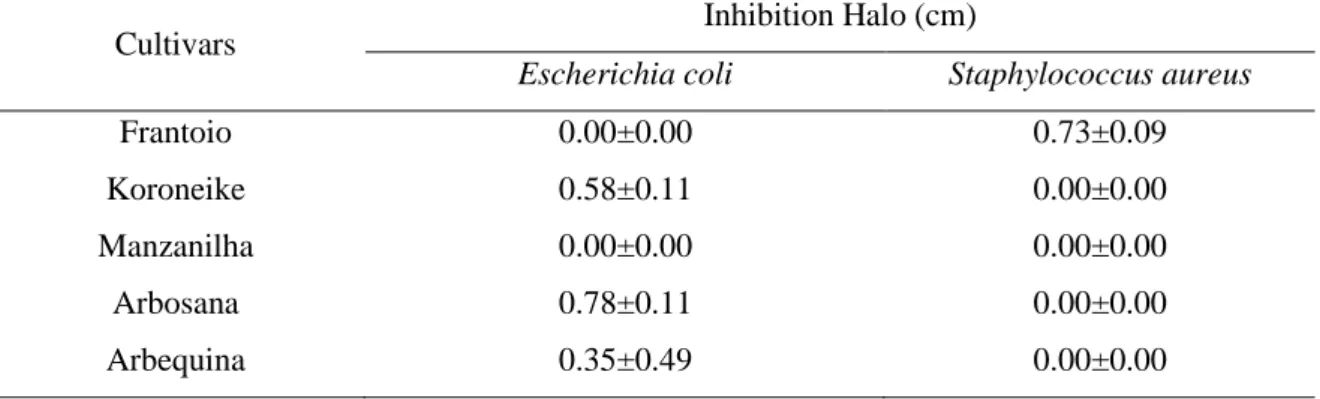
According to Arora & Kaur (1999), when halos are smaller than 0.7 cm, they are considered to be non-active against bacteria, and when they have a diameter greater than 1.2 cm they are considered to have a satisfactory inhibitory effect. Considering this criterion, it was observed that the extracts of olive leaves were active against the evaluated bacteria, but only for certain cultivars (Table 2).
Braz. J. of Develop.,Curitiba, v. 6, n.4,p.21791-21805 apr. 2020. ISSN 2525-8761
Table 2: Inhibition halos obtained by the disc diffusion method by application of extracts of olive leaves against the bacteria Escherichia coli and Staphylococcus aureus.
Cultivars Inhibition Halo (cm)
Escherichia coli Staphylococcus aureus
Frantoio 0.00±0.00 0.73±0.09
Koroneike 0.58±0.11 0.00±0.00
Manzanilha 0.00±0.00 0.00±0.00
Arbosana 0.78±0.11 0.00±0.00
Arbequina 0.35±0.49 0.00±0.00
*Mean and standard deviation.
According to the results, it was possible to observe that E. coli was inhibited by leaf extracts of the cultivars Koroneike, Arbosana and Arbequina, with halos varying from 0.35 to 0.78 cm, and it was the leaf extract of Arbequina which obtained the highest inhibition halo. However, the S. aureus bacterium was inhibited only by the leaf extract of the Frantoio cultivar, with an inhibition halo of 0.73 cm.
In a study by Lee & Lee (2010), which evaluated the antimicrobial activity of extracts of olive leaves, against the same bacteria, no halos of inhibition were evidenced. Aliabadi et al. (2012) also found that extracts of olive leaves did not inhibit the same bacteria in the same concentration of the extract evaluated in the present study; however, in higher concentrations (50 mg.mL-1) the extract obtained inhibition halos of up to 0.9 cm for S. aureus, and for E. coli bacteria it obtained inhibition halos of up to 0.82 cm, also at the concentration of 50 mg.mL-1.
Pereira et al. (2007) found the antimicrobial activity of the aqueous extract of olive leaves against Bacillus cereus, Bacillus subtilis, S. aureus, E. coli, Pseudomonas aeruginosa and Klebsiella pneumoniae bacteria and demonstrated that the extract has strong inhibitory activity against E. coli and S. aureus compared to the remaining pathogenic microorganisms. The minimum inhibitory concentration, as well as the minimal bactericidal concentration of the extracts of olive leaves of the different cultivars studied against these bacteria were also determined (Tables 3 and 4).
Braz. J. of Develop.,Curitiba, v. 6, n.4,p.21791-21805 apr. 2020. ISSN 2525-8761
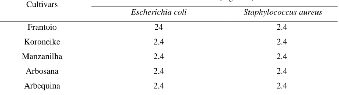
Table 3: Minimum inhibitory concentration of extracts of olive leaves from the cultivars Frantoio, Koroneike, Manzanilha, Arbosana and Arbequina, against Escherichia coli and Staphylococcus aureus.
Cultivars Concentration (mg.mL
-1) *
Escherichia coli Staphylococcus aureus
Frantoio 24 2.4
Koroneike 2.4 2.4
Manzanilha 2.4 2.4
Arbosana 2.4 2.4
Arbequina 2.4 2.4
* minimal inhibitory concentrations.
According to the results, it was observed that the extract of the leaves of the Frantoio cultivar had a minimum inhibitory concentration of 24 mg.mL-1 for E. coli and of 2.4 mg.mL
-1 for S. aureus. All other cultivars had a minimum inhibitory concentration of 2.4 mg.mL-1 for
both bacteria.
In a study by Teramoto et al. (2017), which evaluated the antimicrobial activity of the ethanolic extracts of two olive leaf cultivars, where the minimum inhibitory concentration was determined, a value of 1 and 2 mg.mL-1 was obtained for the Koroneike cultivar for the bacteria E. coli and S. aureus, respectively. For the cultivar Arbosana, the minimum inhibitory concentration was 2 mg.mL-1, for both bacteria. From the study by these authors, it was possible to observe that in all cultivars studied, microbial growth occurred, indicating that the minimum bactericidal concentration of the olive leaves was higher than the concentrations evaluated in the present study.
In the present study, the extract of the olive leaves exerted an inhibitory effect on the tested bacteria, suggesting the antimicrobial activity of the extracts, where the chemical composition of the olive leaves undoubtedly conditioned the antimicrobial effects observed. The results found in our study may be related to the phenolic compounds present in the extracts. In general, the antimicrobial capacity of the phenolic compounds is well known. In the olive leaf, the polyphenols hydroxytyrosol and oleuropein are the main phenolic compounds responsible for the antimicrobial properties (Pereira et al., 2006; Pereira et al., 2007).
Braz. J. of Develop.,Curitiba, v. 6, n.4,p.21791-21805 apr. 2020. ISSN 2525-8761
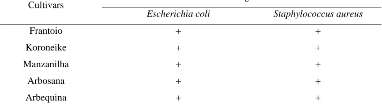
Table 4: Minimum bactericidal concentration of extracts of olive leaves from the cultivars Frantoio, Koroneike, Manzanilha, Arbosana and Arbequina, against Escherichia coli and Staphylococcus aureus.
Cultivars Microbial growth *
Escherichia coli Staphylococcus aureus
Frantoio + +
Koroneike + +
Manzanilha + +
Arbosana + +
Arbequina + +
* + microbial growth detected.
Gram-positive bacteria are more sensitive than gram-negative bacteria, and this is related to the structural characteristics of the bacteria themselves. While gram-positive bacteria have only one layer, composed mainly of peptidoglycan, gram-negative cells have an extra outer membrane composed of a double layer of lipopolysaccharides (Nazzaro et al., 2013). This layer of lipopolysaccharides in gram-negative bacteria would be more resistant to the passage of small antimicrobial molecules than the peptidoglycan layer of gram-positive bacteria (Rai et al., 2017). As a consequence, higher extracts concentrations are required to promote antimicrobial action; however, in the present study this difference was not verified in gram-positive and gram-negative bacteria.
The results of the present study indicated that extracts of olive leaves have antibacterial potential against E. coli and S. aureus. The extracts of the olive leaves had bacteriostatic effect against the strains, but did not present a bactericidal effect in the evaluated concentration. Through the techniques of Disk Diffusion and CIM, it was observed that the bacteria were inhibited by the extract of some cultivars of olive leaf, which was verified by the formation of halos and by the inhibition confirmed in the MIC technique. However, when determining the minimum bactericidal concentration, bacterial growth was detected in the same cultivars, thus verifying that there was inhibitory activity in the evaluated concentration, but there was no bactericidal activity. The absence of bactericidal activity does not necessarily mean that the extract is inactive against the microorganism tested. This fact may be related to the concentration used, where higher concentrations are required to obtain bactericidal effect or the diffusion difficulty of the extract in the cell wall due to the lipophilic characteristics of some samples or the chemical nature of isolated substances present in the extracts (Ríos et al., 1988).
Braz. J. of Develop.,Curitiba, v. 6, n.4,p.21791-21805 apr. 2020. ISSN 2525-8761 4 CONCLUSIONS
Due to the antioxidant activity presented, the olive leaves of the cultivars studied have the potential to be used in the development of food products, in order to be a natural source of bioactive compounds and to represent an alternative to minimize the disposal of residues in the environment.
Extracts of olive leaves have the potential to be used for antimicrobial control of food; however, new studies should be carried out to adjust the concentrations against antimicrobial agents.
ACKNOWLEDGMENTS
The authors are grateful to Coordenação de Aperfeiçoamento de Pessoal de Nível Superior (CAPES) and Fundação de Amparo à Pesquisa do Estado do Rio Grande do Sul (FAPERGS), for research funding.
REFERENCES
Aliabadi MA, Darsanaki RK, Rokhi ML, Nourbakhsh M & Raeisi G (2012) Antimicrobial activity of olive leaf aqueous extract. Annals of Biological Research, 3: 4189-4191.
Arora DS & Kaur J (1999) Antimicrobial activity of spices. Internation Journal of Antimicrobials Agents, 12: 257-262.
Aytul KK (2010) Antimicrobial and antioxidant activities of olive leaf extract and its food applications (Doktora Tezi), İzmir Yüksek Teknoloji Enstitüsü, İzmir-Türkiye.
Bayram M, Topuz S & Caya C (2020) Antioxidant, Antimicrobial Activity of Olive Leaf Extract and Oleuropein, Their Possibilities Usage in Foods. Turkish Journal of Agriculture - Food Science and Technology, 8(2): 337-347.
Bouaziz M & Sayadi S (2005) Isolation and evaluation of antioxidants from leaves of a Tunisian cultivar olive tree. Eur J Lipid Sci Technol, 107: 497–504.
Brahmi F, Mechri B, Dhibi M & Hammami M (2013) Variations in phenolic compounds and antiradical scavenging activity of Olea europaea leaves and fruits extracts collected in two different seasons. Industrial Crops and Products, 49: 256– 264.
Brand Williams W, Cuvelier ME & Berser C (1995) Use of a Free Radical Method to Evaluate Antioxidant Activity. Lebensm. Wiss. Technol., 28: 25-30.
Braz. J. of Develop.,Curitiba, v. 6, n.4,p.21791-21805 apr. 2020. ISSN 2525-8761 Cappellaro TH, Coutinho EF, Ribeiro FC, Araújo FA & Faria MAR (2009) Cultivares. In: Enilton Fick Coutinho, Fabrício Carlotto Ribeiro & Thaís Helena Cappellaro (Ed.) Cultivo de oliveira (Olea europaea L.). Pelotas: Embrapa Clima Temperado. p. 41-48.
CLSI, Wayne LSI (2015) M02-A11: Performance standards for antimicrobial disk susceptibility tests; Approved Standard, 11th Ed. CLSI (Clinical and Laboratory Standards Institute), vol32(1).
CLSI- Clinical and Laboratory Standards Institute (2015) LSI (2012) M07-A9: Methods for dilution antimicrobial susceptibility tests for bacteria that grow aerobically; Approved Standard,9th Ed. CLSI (Clinical and Laboratory Standards Institute), vol32(2).
Farag RS, Mahmoud EA & Basuny AM (2007) Use crude olive leaf juice as a natural antioxidant for the stability of sunflower oil during heating. International journal of food science & technology, 42: 107-115.
Fernández-Bolaños J, Rodríguez G, Rodríguez R, Guillén R & Jiménez A (2006) Potential use of olive by-products, Extraction of interesting organic compounds from olive oil waste, Grasas y Aceites, 57: 95-106.
Ghomari O, Sounni F, Massaoudi Y, Ghanam J, Drissi Kaitouni LB, Merzouki M & Benlemlih M (2019) Phenolic profile (HPLC-UV) of olive leaves according to extraction procedure and assessment of antibacterial activity. Biotechnology Reports, 23, e00347.
Guinda A (2006) Use of solid residue from the olive industry. Grasas Y Aceites, 57:107-115. Herrero M, Temirzoda TN, Segura Carretero A, Quirantes R, Plaza M & Ibañez E (2011) New possibilities for the valorization of olive oil by-products. Journal of chromatography, 42: 7511-7520.
Kiritsakis K, Kontominas MG, Kontogiorgis C, Hadjipavlou Litina D, Moustakas A & Kiritsakis A (2010) Composition and antioxidant activity of olive leaf extracts from Greek olive cultivars. Journal of the American Oil Chemists' Society, 87: 369–376.
Le Gall H, Philippe F, Domon JM, Gillet F, Pelloux J & Rayon C (2015) Cell wall metabolism in response to abiotic stress. Plants, 4: 112-166.
Lee OH & Lee BY (2010) Antioxidant and antimicrobial activities of individual and combined phenolics in Olea europaea leaf extract. Bioresource Technology, 101: 3751–3754.
Malheiro R, Casal S, Teixeira H, Bento A & Pereira JA (2013) Effect of olive leaves addition during the extraction process of overmature fruits on olive oil quality. Food and Bioprocess Technology, 6: 509-521.
Braz. J. of Develop.,Curitiba, v. 6, n.4,p.21791-21805 apr. 2020. ISSN 2525-8761 Nakabayashi R & Saito K (2015) Integrated metabolomics for abiotic stress responses in plants. Current opinion in plant biology, 24: 10-16.
Nazzaro F, Fratianni F & Martino L (2013) Effect of Essential Oils on Pathogenic Bacteria. Pharmaceuticals, 12: 1451–1474.
Özcan MM & Matthäus B (2017) A review: benefit and bioactive properties of olive (Olea europaea L.) leaves. European Food Research and Technology, 243(1): 89-99.
Pereira AP, Ferreira I, Marcelino F, Valentão P, Andrade PB, Seabra R, Estevinho L, Bento A & Pereira JA (2007) Phenolic Compounds and Antimicrobial Activity of Olive (Olea europaea L. Cv. Cobrançosa). Leaves Molecules, 12: 1153-1162.
Pereira JA, Pereira APG, Ferreira ICFR, Valentão P, Andrade PB, Seabra R, Estevinho L & Bento A (2006) Table olives from Portugal: phenolic compounds, antioxidant potential and antimicrobial activity. J. Agric. Food Chem, 54: 8425-8431.
Rafiei Z, Jafari S, Aalami M & Khomeiri M (2012) Evaluation of antimicrobial activity of olive leaf extracts by ELISA method. Iranian Journal of Medicinal and Aromatic Plants, 28: 280-292.
Rai M, Paralikar P, Jogee P, Agarkar G, Ingle AP, Derita M & Zacchino S (2017) Synergistic antimicrobial potential of essential oils in combination with nanoparticles: Emerging trends and future perspectives. International Journal of Pharmaceutics, 519: 67–78.
Re R, Pellegrini N, Proteggente A, Pannala A, Yang M & Rice Evans C (1999) Antioxidant activity applying an improved ABTS radical cation decolorizationassay. Free Radical Biology & Medicine, 26: 1231-1237.
Ríos JL, Recio MC & Villaw A (1988) Screening methods for natural products with antimicrobial activity: a review of the literature. Journal of Ethnopharmacology, 2: 127-149. Roig A, Cayuela ML & Sánchez-Monedero MA (2006) An overview on olive mill wastes and their valorisation methods, Waste Management, 26: 960–969.
Rufino MdoSM, Alves RE, Brito ESDE, Morais SMDE, Sampaio CdeG, Pérez Jiménez J & Saura Calixto FD (2007) Metodologia Científica: Determinação da Atividade Antioxidante Total em Frutas pela Captura do Radical Livre DPPH. Fortaleza-CE, Embrapa / Agroindústria Tropical. 4p. (Comunicado Técnico on line, 127).
Silva ED, Muniz MP, Nunomura RDCS, Nunomura SM & Zilse GAC (2013) Constituintes fenólicos e atividade antioxidante da geoprópolis de duas espécies de abelhas sem ferrão amazônicas. Química Nova, 36: 1-6.
Braz. J. of Develop.,Curitiba, v. 6, n.4,p.21791-21805 apr. 2020. ISSN 2525-8761 Sudjana AN, D'orazio C, Ryan V, Rasool N, Ng J, Islam N, Riley TV & Hammer KA (2009) Antimicrobial activity of commercial Olea europaea (olive) leaf extract. International journal of antimicrobial agents, 33: 461-463.
Teramoto JRS, Sachs RCC, Garcia VL, Oliveira ASS & Duarte MC (2017) Atividade antimicrobiana das folhas de duas variedades de oliveira e a contextualização deste coproduto da produção paulista e mundial de azeite de oliva. Revista Intellectus,37: 63-83.
Visioli F, Poli A & Gall C (2002) Antioxidant and other biological activities of phenols from olives and olive oil. Medicinal research reviews, 22: 65-75.
Vogel P, Kasper Machado I, Garavaglia J, Zani VT, de Souza D & Morelo SDB (2014) Polyphenols benefits of olive leaf (Olea europaea L) to human health. Nutricion Hospitalaria, 31(3): 1427-1433.
Yancheva S, Mavromatis P & Georgieva L (2016) Polyphenol profile and antioxidant activity of extracts from olive leaves. Journal of Central European Agriculture, 17: 154-163.