Henrique Luis Silva de Noronha
Outubro de 2010
Escola de Ciências
The effect of high-temperature on sugar
transport in grape cells
U M in ho |2 01 0 H en riq ue L ui s Si lv a de N or on ha T h e e ff e ct o f h ig h -t e m p e ra tu re o n s u g a r tr a n sp o rt i n g ra p e c e ll s
Mestrado em Fisiologia Molecular de Plantas
Henrique Luis Silva de Noronha
Outubro de 2010
Escola de Ciências
The effect of high-temperature on sugar
transport in grape cells
Trabalho efectuado sob a orientação do
Prof. Doutor Hernâni Gerós
e da
iii
I would like to thank Prof. Hernâni for is supervising and for helping me become a better investigator.
To all my friends in the laboratory, for providing a safe and amusing work place, a sincere “thank you”
Particularly, I thank my Family and Cecília for their constant caring and support, fundamental in these last years.
This work was funded by FCT (Fundação para a Ciência e Tecnologia), Lisboa, Portugal, as a project grant, PTDC/AGR-ALI/100636/2008
iv
Vitis vinifera is a major crop worldwide and in Portugal. Berry content in sugars,
organic acids, phenolics and aroma compounds are fundamental for fruit and wine quality. These compounds are accumulated/metabolized during the development of the berry. In particular, berry sugar content is directly related to the final alcoholic content of wine, and regulates the development of its aromatic and organoleptic properties. Massive sugar accumulation in berry mesocarp occurs after véraison due to a combined action of monosaccharide (MSTs) and disaccharide transporters (DSTs).
High-temperatures affect berry set and development and alter the normal sugar content of the fruit. Also, peaks of high temperature, nowadays more and more frequent, may stop the ripening progress. We have been exploring the mechanisms involved in sugar import and compartmentation into the berry. VvHT1 (Vitis vinifera Hexose Transporter 1) is a high affinity plasma membrane H+-dependent symporter with broad specificity for monosaccharides abundant at early stages of berry development. The expression of this transporter is tightly regulated by sugars at transcriptional and post-translational levels. In the present study we aimed at the elucidation of the effect of high temperature and temperature fluctuations on sugar transport in grape cells. Results showed that a temperature treatment of 38ºC for 12 h decreased by 40% the Vmax of 14C-glucose transport in CSB (Cabernet Sauvignon Berry) cells. Contrarily, abscisic (ABA) and salicylic acid (SA) stimulated sugar uptake by 28.7% and 62.5%, respectively. ABA and SA also stimulated 14C-glucose accumulation in intact grape berries by 88.7% and 67.8%, respectively. The down-regulation of glucose uptake mediated by high temperature corroborated the observed decrease of the VvHT1 levels in the plasma membrane. Moreover, after high-temperature treatment the intracellular ROS levels and lipid peroxidation increased by 97% and 29%, respectively. Proteomic analysis of the plasma membrane of CSB cells, allowed the identification of several proteins up-regulated in response to high temperature. It is hypothesised that intracellular ROS levels can mediate this cellular response to high-temperature. To study the recycling and turnover of VvHT1 in response to high-temperature a VvHT1-GFP expression clone was produced and a protocol for transient protoplast transfection is currently being optimized.
v
A videira (Vitis vinifera) é uma espécie agrícola de elevada importância ao nível mundial e em Portugal. O conteúdo do bago em açúcares, ácidos orgânicos, compostos fenólicos e aromáticos determina a qualidade final do fruto e do vinho. Estes compostos são acumulados/metabolizados durante o desenvolvimento do fruto. O conteúdo do bago em açúcares condiciona o teor alcoólico do vinho, além de regular o desenvolvimento das suas propriedades aromáticas e organolépticas. A acumulação massiva de açúcares que ocorre no mesocarpo após a fase de pintor (véraison) resulta da acção combinada de transportadores membranares de mono (MST) e de dissacarídeos (DST).
Temperaturas elevadas podem afectar a frutificação e o desenvolvimento do bago, bem como alterar o seu conteúdo normal em açúcares. Adicionalmente, picos de temperatura, muito frequentes no contexto das modificações climáticas em curso, podem comprometer o processo de amadurecimento. O nosso grupo de investigação tem dedicado atenção particular ao estudo dos mecanismos envolvidos no transporte e compartimentação de açúcares no bago. O transportador da membrana plasmática VvHT1 (Vitis vinifera Hexose Transporter 1), expresso nas fases iniciais do desenvolvimento do bago, medeia a incorporação da glucose e de outros monossacarídeos por um mecanismo
de simporte com H+. A expressão deste transportador é finamente regulada pelo teor em
açúcares ao nível transcricional e pós-transcricional. No presente trabalho foi estudado o efeito de temperaturas elevadas, e de flutuações de temperatura, no transporte de açúcares em células de videira. Os resultados demonstram que um pico de temperatura de
38ºC aplicado durante 12 h reduz em 40% a Vmax de transporte da 14C-glucose em culturas
celulares (CSB, Cabernet Sauvignon Berry). Contrariamente, o ácido abcísico (ABA) e o ácido salicílico (SA) estimularam o transporte de açúcar em 28,7% e 62,5%,
respectivamente. O ABA e o SA também estimularam a incorporação de 14C-glucose em
bagos intactos em 88,7% e 67,8%, respectivamente. A repressão do transporte da glucose
causada por temperaturas elevadas correlacionou-se com a detecção de níveis diminuídos do VvHT1 na membrana plasmática. Adicionalmente, a exposição a elevadas temperaturas aumentou os níveis intracelulares de ROS e de peroxidação lipídica em 97% e 29%, respectivamente. Uma análise de proteómica efectuada em membranas plasmáticas de células CSB, permitiu a identificação de diversas proteínas especificamente expressas em resposta a temperaturas elevadas. É discutido que o aumento observado dos níveis intracelulares de ROS podem mediar esta resposta celular a elevadas temperaturas. No sentido de estudar a reciclagem e turnover do transportador VvHT1 em resposta a temperaturas elevadas, foi construído um clone de expressão VvHT1-GFP e optimizado um protocolo de transformação de protoplastos.
vi
Acknowledgments ... iii
Abstract ... iv
Resumo ... v
Abbreviations and acronyms ... 1
1 Introduction ... .3
1.1 Grape berry structure and development ... 5
1.2 Hormonal control of berry development ... 8
1.3 Effect of temperature on grape berry ... 9
1.4 Long distance sugar transport ... 10
1.5 Disaccharide transporters (DST) ... 11
1.6 Monosaccharide transporters (MST) ... 13
1.7 Identification, localization and characterization of the grape monosaccharide transporter VvHT1 ... 14
2 Material and Methods ... 19
2.1 Biological material ... 20
2.1.1 CSB cultured cells ... 20
2.1.2 Intact grape berries ... 20
2.1.3 Bacterial cells ... 20
2.2 Uptake studies of radioactive sugars ... 20
2.2.1 Cell treatments ... 20
2.2.2 Determination of initial velocities of D-[14C]glucose transport ... 21
2.3 Western-Blot analysis ... 21
2.3.1 Plasma membrane isolation and purification ... 21
2.3.2 SDS-PAGE ... 22
2.3.3 Membrane transfer and immunoblotting ... 22
2.4 Study of solute incorporation in intact grape berries ... 23
2.5 Intracellular ROS quantification ... 23
2.6 Malondialdeheyde quantification ... 23
2.7 Production of a VvHT1-GFP expression clone... 24
2.7.1 Isolation and purification of total RNA from CSB cells ... 24
2.7.2 First strand cDNA synthesis ... 24
vii
2.7.3.3 Colony PCR ... 25
2.7.3.4 Isolation of plasmid DNA (miniprep) ... 25
2.7.3.5 LR recombination reaction ... 25
2.8 Protoplast isolation ... 25
2.9 Protoplast transient transfection ... 26
2.10 Protein quantification ... 26
2.11 LC MS/MS analysis ... 27
3 Results ... 28
3.1 Effect of high-temperature on glucose transport ... 29
3.2 Effect of high-temperature on ROS homeostasis ... 31
3.3 Effect of ABA and SA on glucose transport ... 34
3.4 Compartmentation studies in intact grape berries ... 35
3.5 Study of VvHT1 recycling and turnover induced by glucose repression and high-temperatures ... 37
4 Discussion ... 39
4.1 Temperature affects sugar transport/compartmentation in grape cells ... 38
4.2 High-temperature affects ROS homeostasis in grape cells ... 41
4.3 A preliminary proteomic analysis of the plasma membrane from grape cells reveals the induction of key proteins in response to high-temperature ... 42
4.4 ABA and SA stimulate sugar incorporation in grape cells ... 45
4.5 Future prospects ... 46
1
Abbreviations and acronyms
2-NBDG 2-(N-(7-nitrobenz-2-oxa-1,3-diazol-4-yl)amino)-2-deoxyglucose
ABA Abcisic acid
ASR ABA abscisic acid-, stress-, and ripening-induced
BOR1 Boron transporter 1
BSA Bovine serum albumin
cDNA Complementary DNA
CHIP Carboxylterminus of Hsc70-interacting protein
CSB Cabernet Sauvignon berry
DNA Deoxyribonucleic acid
DTT Dithiothreitol
dpm Disintegrations per minute
DST Disaccharide transporter
DW Dry weight
EDTA Ethylenediamine tetraacetic acid
FAO Food and agriculture organization
FDA Fluorescein diacetate
FW Fresh weight
GFP Green fluorescent protein
GUS β-Glucuronidase
H2DCFDA 2',7'-dichlorodihydrofluorescein diacetate
HSP Heat shock protein
kDa Kilodalton
LB Luria broth
LC MS/MS Liquid chromatography-tandem mass spectrometry
MDA Malondialdehyde
MES 2-(N-morpholino)ethanesulfonic acid
MFS Major facilitator superfamily
MS Murashige and Skoog
MST Monosaccharide transporter
NADPH Nicotinamide adenine dinucleotide phosphate
PAGE Polyacrylamide gel electrophoresis
PBS Phosphate buffered saline
PCR Polymerase chain reaction
PEG Polyethylene glycol
2
PVPP Polyvinylpyrrolidone
RNA Ribonucleic acid
ROS Reactive oxygen species
RT-PCR Reverse transcriptase-PCR
SA Salicylic acid
SDS Sodium dodecyl sulphate
SE/CC Sieve elements/companion cell
TBA Thiobarbituric acid
TCA Trichloroacetic acid
TE Tris-EDTA
Tris Tris(hydroxymethyl)aminomethane
VvHT Vitis vinifera hexose transporter
VvMSA Vitis vinifera maturation-, stress-, ABA-induced protein
VvSK1 Vitis vinifera sugar-inducible protein kinase 1
3
4
Grapes (Vitis spp.) are economically the most important fruit species in the world. More than 50 species are recognized in the grape genus Vitis, but almost all world wine is made from Vitis vinifera, native to the area south of the Caucasus Mountains and the Caspian Sea (Kunkee and Goswell 1996). In 2009 the vineyard area cultivated in the world and total fruit production was of 7,437,141 ha and 66,935,199 t, respectively. In the same year in Portugal the vineyards occupied the largest cultivated area for a fruit crop (222,700 ha) and had the greatest production (487,800 t) (FAO 2009). Approximately 71% of grape production is used for wine, 27% as fresh fruit, and 2% as dried fruit (Conde et al. 2007). Throughout antiquity the conversion of grapes into wine was considered a gift from the gods and the best wines were reserved for the elite of the society. Nowadays, wine is an integral component of the culture of many countries, a form of entertainment in others, and a libation of choice for advocates of its health benefits. Unlike many modern foods, wine’s attraction relies not on strong consistent flavors, but upon a subtle array of shifting sensations that make its charm difficult to define (Bisson et al. 2002).
Wine is composed of various constituents that include water, sugars, alcohol, phenolics, acids and mineral salts. The main constituent of wine is water accounting for 75 to 90% (v/v), and this variation is explained by the amount of the other constituents that form the wine extract that differ from wine to wine. The second largest constituent is ethyl alcohol, which, according to the type of wine, varies from 8% to 15% (v/v). Another important constituent is sugar, which is directly responsible for the final alcoholic content of the wine. A normal dry wine generally has less than 2 g sugar/L, while in a botrytized sweet wine it can reach almost 200 g sugar/L (Dominé et al. 2004).
Vineyards can be found in Europe, Northern and Southern America, Africa and Asia. Although this worldwide distribution, the most important factor for viticulture is climate and, above all, temperature. Grapes clearly prefer moderate conditions, and rarely thrive where temperatures rise above 25ºC in the summer months. In a large part of Western Europe, the location of the majority of Europe’s classic viticultural regions, average July temperatures vary between 15 and 25°C. Rainfall and drought also play an important role, and it is almost impossible to grow vines with less than 200 mm of rain a year. On the other hand, too much rain also makes it difficult to cultivate grapes. A moderate climate, with adequate to relatively high rainfall, provides ideal conditions for producing both fragrant white wines with a good structure and acidity, and well-balanced red wines with good potential for maturing (Dominé 2004). Wine quality largely depends on the vineyard and on the vine grower. Most of the wine compounds are produced by the plant itself, in the leaves (sugars and acids), and in
5
berry (acids and phenolics). Furthermore, some molecules related to aroma and taste are produced during the fruit development and ripening, being their spectrum specific to a given variety. Theses aromas, called “varietal” or primary aromas, are the grape’s signature, recognizable by the consumer during degustation. Thus, the growth and the fructification of grapevines in the vineyard are of utmost importance to wine quality (Blouin and Guimberteau 2000).
1.1 Grape berry structure and development
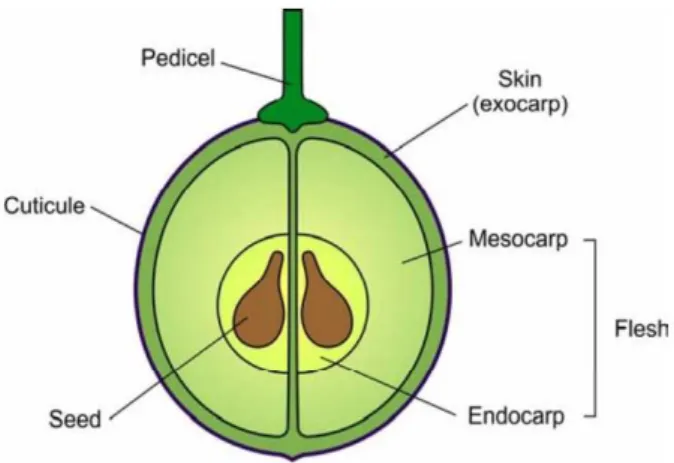
Grape berry, a non-climateric fruit, is essentially an independent biochemical factory (Gholami et al. 1995), and is mainly composed of three distinct tissues, skin, flesh and seeds (Figure 1), with the sheer bulk of wine being derived from the flesh. These tissues vary considerably in composition and contribute differently to the overall wine composition. The flesh accounts for 75-85% of total berry weight, while the skin and the seeds account for about 7% and 4%, respectively (reviewed by Conde et al. 2007; Jackson 2008). Also, the grape berry is richly supplied with vascular tissue, which after it enters the fruit via the pedicel, branches out to supply the developing seeds, the flesh and the skin (Hardie et al. 1996).
The grape berry skin contributes to the integrity of the whole berry by protecting inner tissues against mechanical damage or pathogen attack, promotes seed dispersion by providing a high contrast between background foliage and fruits as well as providing protection from UV light exposure (Grimplet et al. 2007). The vacuole of skin cells is where most of the aromas that arise from volatile compounds, such as terpenes, norisoprenoids, and thiols, are stored as sugar or amino acid conjugates. These compounds are fundamental for wine making, and the variability of skin composition plays an important role in determining the color, aroma, and other organoleptic properties of wine (Lund and Bohlmann 2006).
The primary role of the flesh is to provide a high value nutritional content for dispersal agents, including high concentrations of free amino acids and hexose sugars (mainly sucrose and fructose). During wine production, the flesh contributes with the majority of sugars, which are transformed into alcohol during the fermentation process (Grimplet et al. 2007). Also, the berry flesh accumulates organic acids (mainly tartaric and malic), mineral cations (especially K+), nitrogenous compounds (soluble proteins, ammonia and amino acids), pectic substances (cell wall structural material composed of galacturonic acid polymers) and non-flavonoid phenolics (primarily benzoic and cinnamic acid derivatives).
6
The seeds are the main source of flavan-3-ol monomers and procyanidins that have an important contribute to the organoleptic properties of wine including bitterness and astringency (Robichaud and Noble 1990).
Figure 1. Structure of a ripe grape berry, depicting the three primary tissues: skin, flesh
and seeds (Conde et al 2007)
Grape berry development, from fruit set to full maturation, is a complex series of processes requiring the coordination of a large number of events involving substantial and rapid changes in a number of tissues (Conde 2007). In grapes fruit set is defined as the stage when the berry diameter is between 1.6 and 3.2 mm. Climatic factors have a significant effect on fruit set, and, particularly, temperature can inhibit pollen tube growth and ovule fertilization. Also, fruit set is greatly reduced when temperatures fall below 18.3°C or exceed 37.8°C (Dookozlian 2000).
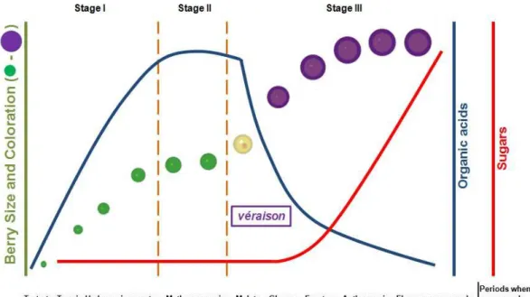
The development of the grape berry has been divided into three phases. Increases in berry weight, volume, or diameter during development are typically characterized by a double-sigmoid curve resulting from two consecutive stages of growth separated by a phase of slow or no growth (Figure 2; Coombe 1992; Dookozlian 2000). The first phase (green stage) is a period of rapid berry growth that occurs immediately after bloom. During this time, berries grow both through rapid cell division and cell enlargement. In this phase berry texture is firm, while its color is green due to the presence of chlorophyll. The sugar content of the berry remains low, while organic acids accumulate, mainly tartaric and malic acids (Figure 2). Berry growth during stage I is very sensitive to temperature. Temperatures exceeding 35°C reduce growth rate and size at harvest and light is also important for optimum berry growth. Berries subject to heavy shade immediately after berry set are significantly smaller at harvest than berries that have been well exposed to light. The second phase (lag
7
phase) is marked by decrease in the growth rate while its organic acid concentration reaches its highest level and the berries remain firm and begin to lose the chlorophyll content. This phase ends with the véraison, a French term that defines the onset of ripening, and the last phase of berry development begins (Figure 2). The third phase (ripening phase) of berry development is characterized by striking changes in fruit characteristics. The berry dramatically increases in size mainly due to cell expansion, which is caused by massive sugar accumulation, water import and also to the deposition of phenolics, among others (Reviewed by Conde et al. 2007). Also, the berries soften and lose chlorophyll, the concentration of organic acids greatly declines, and is observed a colour development and the synthesis and accumulation of aroma and flavour compounds (Figure 2). This phase of berry development is of remarkable importance to wine industry, considering that the accumulation of these compounds results in an increase of the organoleptic properties of the fruit and wine. When conditions are warm and degree-days accumulate rapidly, ripening is accelerated. Prolonged periods of excessively high temperatures following berry softening, for instance 3 to 4 consecutive days above 40.6°C, may retard berry ripening. The effects of elevated temperatures on fruit ripening are temporary and, depending upon the degree of heat stress, sugar accumulation can proceed normally once temperatures return to a normal range (Dookozlian 2000).
8
1.2 Hormonal control of berry development
Grape berry development is under tight hormonal regulation, and several hormones have been identified and characterized as regulators of fruit ripening. The pattern of ABA accumulation during berry development is well defined, with high free ABA levels in the flesh of young berries, followed by a rapid decreased to low levels that remains during most of the pre-véraison phase (Wheeler et al. 2009). After
véraison, a peak of ABA level occurs simultaneously with an increase in sugar
accumulation and color development. Following this peak in ABA levels after véraison, the levels declined as the fruit reaches full ripeness (Kondo et al. 1998; Owen et al. 2009). This pattern of accumulation provides correlative evidence for a role of ABA in ripening and perhaps in its initiation. Also, ABA is mainly found in the phloem of the berry which is consistent with a role in the unloading and uptake of photoassimilates (Kataoka et al. 1982; Shiozaki et al. 1999).
Salicylic acid is involved in signalling in plants, particularly in the induction of defense and stress responses (Bari and Jones 2009). The levels of salicylic acid during grape berry development have not yet been reported, and so it is difficult to propose a developmental role for this hormone.
Like other plant hormones cytokinins are involved in a diverse range of processes (Werner and Schmülling 2009). Cytokinins levels are high in one week old berry flesh but decreased rapidly to low levels by the time of véraison (Zhang et al. 2003). This pattern of accumulation is in agreement with the proposed roles for cytokinins in flower development and fruit set, and is also consistent with the ability of cytokinins to delay berry development (Werner and Schmülling 2009).
Brassinosteroids are generally associated with plant growth and stress response (Haubrick and Assmann 2006). Recently a role for brassinosteroids in fruit development, in particular during ripening, was described (Symons et al. 2006). High levels of brassinosteroids where found in flowers and young leaves, declining prior to
véraison. Furthermore it has been described a peak in brassinosteroids after véraison
followed by a rapid decline (Wang et al. 2001). This pattern of accumulation suggest that brassinosteroids may play a role in ripening-associated processes, for example, in the post-véraison phase of berry growth or perhaps it is part of a response to the stress resulting from the massive sugar influx that occurs after véraison.
Auxins levels are high in the early phases of berry development, after which they decline steadily to very low levels at véraison (Inaba et al. 1976). The high levels early in berry development are in agreement with the proposed role for auxin in cell division and expansion.
9
Gibberellins are regulators of many processes during plant development involving cell division and expansion (Olszewski et al. 2002). The consensus of a number of studies is that gibberellins levels in the flesh of seeded berries were high at around flowering and early in berry development after which they decreased steadily (Pérez et al. 2009). The observed pattern of gibberellins accumulation is consistent with the proposed role in cell division and expansion in the initial stages of berry development.
1.3 Effect of temperature on grape berry
High temperatures are a major threat to crop productivity. As it was already mentioned, temperature has a dramatic effect in the berry development altering normal berry set, inhibiting pollen tube growth and ovule fertilization. Also, high temperatures during the initial stages of berry development dramatically reduce the berry size and weight at harvest, and it is known that high temperature peaks may stop the ripening progress (Dookozlian 2000).
High temperature affects berry composition, especially titratable acidity, total soluble solids, and anthocyanin content (Poudel et al. 2009). It has been reported that temperature has a dramatic effect on anthocyanin accumulation in grape berry skin, a major contributor to wine organoleptic properties and quality (Mori et al. 2005; Yamane et al. 2006). It was found that a 30°C night temper ature greatly reduces the coloration of Cabernet Sauvignon grapes as compared to fruits ripened at night time temperatures of 15 and 20°C (Kliewer 1972). The lim iting factor behind this decrease in anthocyanin synthesis and accumulation under high temperature appears to be a reduced accumulation of soluble sugars in fruit (Mazza and Miniati 1993). In fact high temperatures appear to limit glucose and fructose accumulation in the berry. Poudel et al (2009) reported that vines exposed to 30°C had a lower glucose and fructose content than vines subjected to 20 and 25 °C. The e nhanced expression of various genes of the anthocyanins biosynthetic pathway in the berry skin may be correlated with the concomitant accumulation of sugars in the flesh and, concordantly, sucrose treatment promotes anthocyanin synthesis in V. vinifera cell cultures (Agasse et al 2007; Agasse et al. 2008). Although some studies have been developed to evaluate the effect of temperature on the grape berry composition, little information is available at the molecular level.
10
1.4 Long distance sugar transport
Efficient assimilation and use of nutrients by plants is of prime importance for the optimization of crop productivity. The grape berry is considered to be mainly a sink for primary metabolites essential for plant survival, and rely on the use of available carbohydrate resources produced by photosynthesis to support their growth and development. Sugars transport and allocation between the photosynthetic “source tissues” and the heterotrophic “sink tissues” is known as assimilate partitioning and is a major determinant of plant growth and productivity (Kingston-Smith 2001).
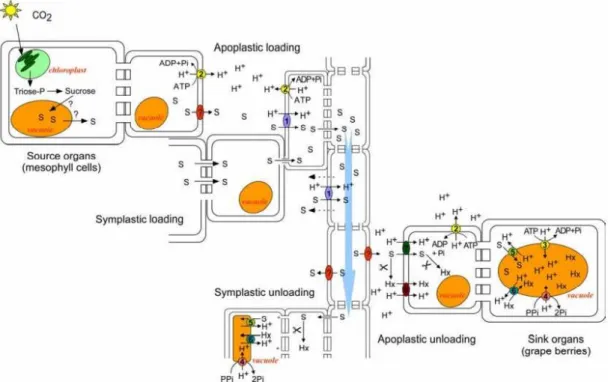
In the majority of plants sucrose is the main sugar transported via the floem, although several other solutes have been identified, including raffinose, stachiose and the sugar-alcohols mannitol and sorbitol. The main advantage for long distance transport of sucrose is its non-reducing nature and relative insensitivity to metabolism (Salisbury and Ross 1994), allowing long distance transport without the problem of metabolism easily encountered with glucose. Also, according to Munch’s mass flow hypothesis, sucrose, as the major osmotically active constituent in the phloem, also provides the driving force for translocating all other compounds in the phloem sap (Conde et al. 2006). Sucrose derived from leaf photosynthesis is exported via the phloem to the berries. From véraison and throughout ripening the berries accumulate roughly equal amounts of glucose and fructose, reaching over 1 M of each hexose, suggesting that phloem transported sucrose is hydrolyzed at some step during its transport from the leaves to the vacuole of the mesocarp cell (Figure 3; Coombe 1987; Conde et al. 2006; Conde et al. 2007; Agasse et al. 2009).
From its point of synthesis in the mesophyll sucrose may be loaded into the SE/CC complex either through plasmodesmata or via the apoplast. Apoplastic loading requires sucrose export from the mesophyll or the vascular parenchyma by a sucrose exporter that remains unidentified, and reuptake into the SE/CC complex by a sucrose/H+ symporter. When in the phloem, hydrostatic pressure drives phloem sap movement toward sink tissue. Passive leakage can take place along the path and reuptake of leaked sucrose occurs along the phloem. Phloem unloading also occurs through a symplastic (via the plasmodesmata) and apoplastic mechanism. Apoplastic phloem unloading implies the existence of a sucrose exporter that remains unidentified, at the sink tissue. In the berry tissues symplastic connections via plasmodesmata between sieve tubes and mesocarp cells remain for quite a long period during fruit development. However, several lines of evidence indicate that the apoplastic pathway play a major role at late stages of grape berry development. Indeed, there is a shift from symplastic to apoplastic phloem unloading at the onset of ripening (Zang et al.,
11
2006). The data available on invertase activity also support an apoplastic pathway of sugar unloading during grape berry development (Agasse et al. 2006; Agasse et al 2009). It appears that sugar accumulation in sink organs would rather result from the coordinated action of several mechanisms, involving various transporters and hydrolytic enzymes. Therefore, the presence of an apoplastic step requires the involvement of membrane-located sugar transporter proteins mediating the exit of sucrose from the phloem, and the uptake and compartmentation of sugars across the plasma membrane and the tonoplast of flesh cells. Concordantly, both DST and MST families have been characterized in plants (reviewed by Williams et al. 2000)
Figure 3. Long distance sugar transport through the floem. From the place of sinthesys in
the leaves mesophyll, sucrose may be loaded into the sieve elements/companion cell complex by apoplastic and symplastic mechanisms. Hydrostatic pressure drives phloem sap movement toward sink tissues. The unloading of the floem may occur via the apoplast, in coordination with cell wall invertases and monosaccharide transporters, or through plasmodesmata (Adapted from Lalonde et al. 1999).
1.5 Disaccharide transporters
It has already been mentioned that sucrose is the main carbohydrate transported through the phloem. The first plant sucrose transporter was identified in spinach and named SoSUT1 (Spinacea oleracea Sucrose Transporter 1), followed by the cloning of the potato sucrose transporter, StSUT1 (Solanum tuberosum Sucrose Transporter 1)
12
(Reismeiner et al 1992; Reismeiner et al 1993). In situ studies showed that the sucrose carrier from potato, StSUT1, is highly expressed in the phloem of the leaf minor veins, the major site of phloem loading (Reismeiner et al 1993). The cloning of several DST led to the establishment of a topological protein model. The protein structure is composed of 12 α-helices domains highly conserved and a cytoplasmatic loop with a high degree of variability.
The importance of sucrose transporters in phloem loading was further supported by studies on transgenic potato and tobacco plants. In these studies, antisense RNAs were used to reduce the level of the sucrose carrier SUT1 in the phloem (Reismer et al 1994; Kuhn et al. 1996; Lemoine et al. 1996; Burkle et al. 1998). Results correlated with the expected function of SUT1 in phloem loading, considering that the antisense plants showed a retarded growth phenotype on soil, and their source leaves were found to export fewer sugars causing an accumulation of carbohydrates in the leaves and a concomitant decrease in the sink tissues. These observations clearly showed that sucrose transporters in the phloem are essential for carbohydrate partitioning, at least in tobacco and potato, both members of the Solanaceae family (Tuernit 2001).
In Vitis vinifera three DST cDNAs were cloned (VvSUC11, also identified as
VvSUT1, VvSUC12 and VvSUC27) and characterised as proton-dependent sucrose
transporters, whereas 9 DSTs sequences are present in Arabidopsis genome (Sauer et al. 2004). VvSUC11 and VvSUC12 are intermediate affinity sucrose transporters with
Km of 0.9 mM and 1.4 mM, respectively (Ageorges et al. 2000, Manning et al. 2001), and VvSUC27 is a low affinity sucrose transporter with a Km of about 10 mM (Zhang et al. 2008). VvSUC11 is expressed in flowers and fruits whereas VvSUC12 expression is restricted to berries and young leaves. VvSUC27 expression is closely related to sink activity since its transcripts are strongly accumulated in flowers and unripe berries, roots and tendrils but poorly present in mature leaves being associated with the early stages of berry development (Davies et al. 1999). Furthermore, VvSUC11 and
VvSUC12 transcription increases with post-véraison sugar accumulation, which
suggests a direct pathway for sucrose acquisition by berry cells (Davies et al. 1999). In spite of the available information regarding grapevine sucrose transporters, the knowledge about the localization of sucrose transporters in berry flesh and the mechanisms of sucrose uptake along ripening is scarce and further investigation is needed.
13
1.6 Monosaccharide transporters (MST)
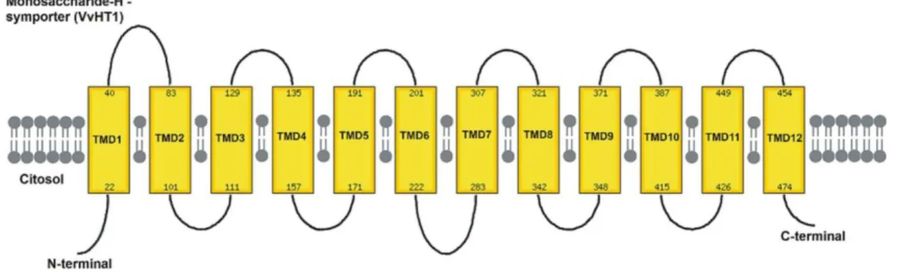
Monosaccharides represent the most important carbon and energy source for the majority of heterotrophic organisms. In the Calvin cycle and gluconeogenesis the carbon (CO2) fixated through photosynthesis is converted to monosaccharides, like glucose and fructose (Buttner and Sauer 2000). The first MST cloned was the HUP1 (Hexose Uptake Protein 1) from the unicellular algae Chlorella kessleri. These cells can switch from an autotrophic to a heterotrophic metabolism inducing hexose transporters when sugars are available. This interesting feature of the algae metabolism led to the identification of a 49 kDa protein by differential screening of cDNA libraries of induced and non-induced cells (Sauer and Tanner 1989). The functional characterization of the protein by complementation of hexose transport null-mutant yeasts demonstrated that the transport was a hexose-proton symport (Sauer et al. 1990). Since then several MST have been identified. A. thaliana genome has 53 homologous sequences encoding putative MSTs (Büttner 2007). A remarkable feature is that all MST share the same 12 transmembrane domains with N- and C- cytoplasmic termini structure, a characteristic of all sugar transporters belonging to the Major Facilitators Superfamily (Figure 4; Williams et al. 2000, Delrot et al. 2001).
In V. vinifera 59 putative hexose transporters encoding genes have been identified based on protein motif recognition (Samson et al. 2004, Jaillon et al. 2007). Six full length cDNAs encoding for MST and named VvHT1-6 (V. vinifera Hexose Transporter 1-6) were previously cloned from various grape cultivars such as Pinot noir, Ugni blanc, Chardonnay, Cabernet Sauvignon and Syrah (Fillion et al. 1999; Vignault et al. 2005; Hayes et al. 2007). The predicted peptides share about 60% identity to each other (Büttner and Sauer 2000, Büttner 2007). Interestingly, VvHT6 is related to AtTMT2 (Arabidopsis thaliana Tonoplast Monosaccharide Transporter 2), a member of the Tonoplast Monosaccharide Transporter subfamily of the Major Facilitators Superfamily. AtTMTs are tonoplastic hexose-proton antiporters induced by abiotic stresses such as cold or drought and were suggested to play a role as sensors (Wormit et al. 2006).
Uptake activities of VvHT1, VvHT4 and VvHT5 have been demonstrated by heterologous expression in the hxt-null mutant yeast EBY VW 4000 (Wieczorke et al. 1999). All three VvHTs are high affinity, H+-dependent transporters mediating the uptake of radiolabelled D-[U-14C]glucose according to saturable Michaelis-Menten kinetics. VvHT1 exhibits the highest affinity for glucose (Km of 70 µM) compared to VvHT4 and VvHT5 (Km about 150 µM and 100 µM, respectively) and is the only one able to restore the growth of the complemented yeast on glucose. VvHT3 was not able
14
to transport any of the tested radiolabelled sugars in the deficient yeast model (Vignault et al. 2005; Hayes et al. 2007). Up to date, attempts to confirm the transport activity of both VvHT2 and VvHT6 in yeast has had little success.
Figure 5. Topological model of VvHT1, a typical MST, with 12 trans-membrane domains
with N- and C- cytoplasmic termini structure characteristic of the Major Facilitators Superfamily.
1.7 Identification, localization and characterization of the grape berry monosaccharide transporter VvHT1
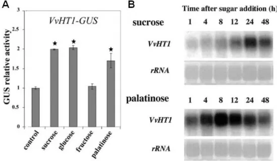
VvHT1 was firstly identified and cloned by Fillion et al. (1999) from two different grape cultivars, Pinot Noir and Ugni-Blanc. Preliminary studies by reverse transcription-PCR suggested that VvHT1 was mainly expressed in the berries, with a first peak of expression at anthesis, and a second peak about 5 weeks after véraison. Although it has been reported that VvHT1 expression slightly increases after véraison in Ugni-Blanc berries (Fillion et al. 1999), detailed microarray analysis suggested that the second peak of expression does not occur in berries from Chardonnay, Shiraz, and Cabernet Sauvignon varieties (Terrier et al. 2005). VvHT1 was characterized as an MST by heterologous expression in both Nicotiana tabacum and yeast (Leterrier et al. 2003; Vignault et al. 2005). Studies performed on tobacco Bright-Yellow cells transformed with different lengths of the VvHT1 promoter transcriptionally fused to β -glucuronidase reporter gene (VvHT1-GUS) suggested that this MST is regulated by sucrose, the non-transported sucrose isomer palatinose and glucose, whereas fructose did not affect it. Furthermore, the authors demonstrated that in grape cell suspension sucrose and palatinose increase the expression of VvHT1 (Figure 5; Atanassova et al
15
2003). These results provided the first example of a putative sugar transporter, which is induced by both glucose and sucrose in higher plants (Atanassova et al 2003).
Figure 5. Studies of the regulation of VvHT1 expression. A) Regulation by different
sugars studied with a fusion of the VvHT1 promoter with the reporter gene GUS. B) Analysis of the VvHT1 transcripts enhanced by sucrose and sucrose analog palatinose (Adapted from Atanassova et al. 2003).
Following this line of work, Cakir et al. (2003) identified an ASR protein in grape, VvMSA (Vitis vinifera Maturation-, Stress-, ABA-induced protein), by means of a yeast one-hybrid approach using as target the proximal promoter of the VvHT1, that contains two sugar boxes and is induced by sucrose and glucose, as it was previously mentioned. VvHT1 and VvMSA are both inducible by sucrose in grape berry cell culture, and sugar induction of VvMSA is enhanced strongly by ABA. Gel-shift assays demonstrated a specific binding of VvMSA to the VvHT1, suggesting that this ASR protein may be part of the transcriptional complex mediating the sugar-inducible expression of VvHT1. Also, the positive regulation of VvHT1 promoter activity by VvMSA in planta was confirmed by coexpression experiments (Cakir et al. 2003). VvHT1 localization was elucidated by Vignault et al. (2005) using in situ hybridization, immunofluorescence and immunogold labelling experiments. In situ hybridization showed that VvHT1 transcripts are primarily found in the phloem region of the conducting bundles, and immunofluorescence and immunogold labelling experiments localized VvHT1 in the plasma membrane of the sieve element/companion cell interface and of the flesh cells. These studies suggested that VvHT1 is involved in the retrieving of the monosaccharides needed to provide the energy necessary for cell division and cell growth at an early stage of berry development (Vignault et al. 2005).
16
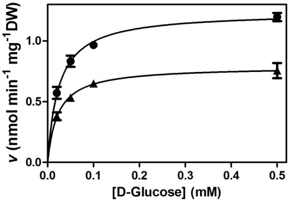
Figure 6. Kinetics and specificity of the monosaccharide transport system of V. vinifera
cultured cells (CSB, Cabernet Sauvignon Berry). A) Initial uptake rates of 0.02 - 0.5 mM D-[14C]glucose. B) Eadie-Hofstee plots of the initial uptake rates of D-[14C]glucose in the absence of other sugars (closed squares) and in the presence of 5 mM xylitol (open circles), 5 mM galactose (lozenge), 5 mM mannitol (open squares), and 5 mM arabinose (inverted triangles). C) Eadie-Hofstee plots of the initial uptake rates of D-[14C]glucose in the absence of other sugars (closed squares) and in the presence of the glucose analogs L-glucose (8 mM; open triangle), 2-deoxy-D-glucose (0.5 mM; open lozenge) and 3-ortho-metil-glucose (0.5 mM; closed circles). Adapted from Conde et al. (2006).
In grape cell suspensions Conde et al. (2006) characterized VvHT1 as a high-affinity (Km= 0.05 mM glucose), broad-specificity (Figure 6) monosaccharide-proton co- transport system. The high affinity measured for the H+-dependent monosaccharide transport may be important for cell growth in media with limiting sugar supply.
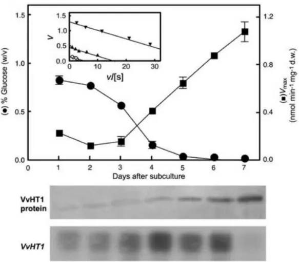
As shown in Figure 7, VvHT1 transcription, VvHT1 protein amount and glucose transport are tightly regulated by sugar availability in the culture medium. Additional evidences supported the involvement of hexokinase as a sugar sensor. When high levels of glucose are present, energy-independent, diffusional uptake is the preferred mode of sugar absorption. Under these conditions, VvHT1 expression is maintained at basal levels due to the balance between a positive induction signal generated by the presence of glucose and a repression signal due to high glucose levels sensed by
17
hexokinase. Additionally, high glucose levels seem to repress glucose transport activity at the protein level, triggering inactivation, mistargeting, and/or proteolysis of VvHT1. The absence of the repression signal generated by hexokinase allows the increase of
VvHT1 transcripts and, in accordance, the number of high-affinity monosaccharide/H+
symporters in the plasma membrane (increase of Vmax), ensuring a high-transport capacity at limiting glucose conditions (Figure 8). These findings correlate well with the expression of the VvHT1 in the early stages of berry development, when the sugar levels in the berry are low.
Figure 7. Regulation of VvHT1 transcripts and protein levels in CSB cells by glucose
18
Figure 8. Model of the glucose regulation of VvHT1 expression and glucose transport
activity. Glucose (closed circles), proton (opened circles), hexokinase (HXK). (Conde et al. 2006)
19
20
2.1 Biological Material 2.1.1 CSB cultured cells
CSB (Cabernet Sauvignon Berry) cells were maintained in 250 mL Erlenmyers with MS medium (Murashige and Skoog 1962) supplemented with 2% (w/v) sucrose, in an orbital shaker at 100 rpm and 23°C in the dark. The cells were sub-cultured after 7 days, at the end of the exponential growth phase.
2.1.2 Intact grape berries
Grape berries from the Alvarinho cultivar were used to study sugar incorporation. Berries were randomly collected from the bunches 8 weeks after fruit set (10 to 12 mm; 0.75 to 0.85 g) and maintained at 4ºC before use.
2.1.3 Bacterial cells
The E. coli cells used to amplify the plasmid vectors were from the One Shot MAX Efficiency DH5α-T1R (Invitrogen), and were transformed with a chemical method, accordingly to the manufacturer’s instructions. For long term storage, glycerol stocks prepared by adding 400 µL 45% (v/v) sterile glycerol to 200 µL bacterial culture were maintained at -80°C.
2.2 Uptake studies of radioactive sugars 2.2.1 Cell treatments
Cell suspensions were collected at day 6 after subculture by centrifugation at 3000 g for 3 min, washed twice in MS medium without carbon source and resuspended in the same medium. The cells were placed for 12 h in the orbital shaker, in the same conditions to overexpress VvHT1 (Conde et al. 2006). In the following day cells were centrifuged at 3000 g for 3 min and resuspended at approximately 5 mg FW/mL in MS medium without carbon source, pH 5.0. This pH provides the necessary proton gradient to energize the monosaccharide/H+ transport system (Conde et al. 2006). To study the influence of high temperature on sugar transport, the cells were incubated for 12 h at 38ºC prior uptake studies. To study the influence of SA and ABA on sugar transport the cells were incubated with 150 µM of both hormones for 24 h at 23ºC prior uptake studies.
21
2.2.2 Determination of initial velocities of D-[14C]glucose transport
One mL of cell suspension was added to 10 mL flasks under constant agitation (100 rpm). After 5 min incubation at room temperature, the reaction was started with 40 µL of a radioactive solution with a specific activity of 500 dpm/nmol glucose. Different solutions were used according to the desired final concentrations (0.02-0.5 mM). After 3 min incubation the reaction was stopped by adding 5 mL of cold MS medium without carbon source. This step stops the transport reaction by both decreasing the temperature and diluting the reaction mixture. The cells were then separated from the culture medium by filtration under vacuum through a GF/C membrane (Whatman), and washed in 10 mL of cold MS medium. The membranes were placed in 20 mL scintillation vials containing 5 mL of scintillation liquid (OptiPhase HiSafe II, LKB) and the radioactivity incorporated in the cells was measured in a Packard Tri-Carb 2200 CA (Packard Instruments Co., Inc., Rockville, Md) scintillation counter. D-[14C]glucose was obtained from the Radiochemical Center (Amersham).
2.3 Western Blot analysis
2.3.1 Plasma membrane isolation and purification
Plasma membrane enriched vesicles from both control cells and temperature treated cells were isolated and purified as previously described (Conde et al. 2006). Approximately 200 mL of suspension cells were centrifuged at 3000 g for 3 min at 4°C, washed twice in ice-cold MS medium without carbon source, and resuspended in 250 mL of ice-cold extraction buffer [250 mM sucrose, 2 mM EDTA (pH 8.0) 2 mM DTT, 1 mM PMSF, 70 mM Tris-HCl (ph 8.0), 3 mM MgCl2, 100 mM KCl, 0.1% (w/v) BSA and 0.2% (w/v) PVPP], and all subsequent procedures were carried out at 4°C. The cells were homogenized with an ultra-turrax T25 (IKA WERKE, Janke and Kumkel IKA, Germany) for 5 min with approximately 20 s pulses, and centrifuged at 10000 g for 10 min. The pellet was discarded and the supernatant was filtered through 3 layers of cheese cloth and centrifuged at 100000 g for 60 min to obtain the microsomal fraction. After discarding the supernatant, the pellet was aspirated and gently homogenized in 8 mL of resuspension buffer [20 mM Tris-HCl (pH 7.5), 1 mM EDTA (pH 7.5), 1 mM DTT, 1 mM PMSF and 15% (v/v) glycerol]. The microsomal fraction was layered over a 32% and 46 % (w/v) discontinuous sucrose gradient [32/46% sucrose (w/v), 20 mM Tris-HCl (pH 7.5), 1mM EDTA (pH 7.5), 1mM DTT and 1 mM PMSF] and centrifuged at 80000 g for 3 h in a Beckman SW 28 rotor. The plasma membrane-enriched fraction was
22
collected from the 32/46% sucrose interface with the help of a pipette, diluted 4 times in resuspension buffer and centrifuged at 100000 g for 30 minutes. The supernatant was discarded and the pellet resuspended in 600 µL of resuspension buffer, aliquoted and quickly stored at -80°C.
2.3.2 SDS-PAGE
SDS-PAGE of membrane proteins was performed according to Laemmli (1970). A stacking gel [4% (w/v) polyacrylamide, 250 mM Tris-HCl (pH 6.8) and 0.1% (w/v) sodium dodecyl sulphate (SDS)] was placed on the top of the resolving gel [10% polyacrylamide (w/v), 375 mM Tris-HCl (pH 8.8) and 0.1% (w/v) SDS] and submerged in TRIS-Glycine running buffer [25 mM Tris-HCl, 250 mM glycine and 0.05% (w/v) SDS]. The samples were heated at 70°C for 10 min pr ior to separation in Laemmli buffer [50 mM TRIS-HCl (pH 6.8), 2% (w/v) SDS, 5% (v/v) β-mercaptoethanol and 10% (v/v) glycerol] and 15 µg of protein from each sample was loaded into the wells.
2.3.3 Membrane transfer and immunoblotting
The proteins separated by the SDS-PAGE were electro-transferred to a 0.45 µm thick Immobilion-PSQ nitrocellulose membrane (Millipore) in transfer buffer [50 mM Tris-HCl, 380 mM glycine, 0.02% (w/v) SDS and 20% (v/v) methanol] in a TE Series transfer electrophoresis unit (Hoeffer Scientific Instruments). The nitrocellulose membrane was incubated 1 h in the blocking solution [5% (w/v) fat-free milk powder, 1% (w/v) BSA in PBS (137 mM NaCl, 2.7 mM KCl, 10 mM Na2HPO4, 1.76 mM KH2PO4, pH 7.4), containing 0.1% (v/v) Tween-20] at room temperature in the dark with small agitation. All the following steps were performed at room temperature in the dark. The membrane was then placed 15 min in the washing solution [1% (w/v) fat-free milk powder, 1% (w/v) BSA in PBS, containing 0.1% (v/v) Tween-20] for 15 min, followed by 2 washes in the same solution for 5 min. The membrane was then incubated with the primary antibodies against the C-terminus of VvHT1 diluted 1:1000 in PBS containing 01% (v/v) Tween-20 and 0.3% (w/v) BSA for 1 h, followed by 3 washes, of 5 min each, with the washing solution. A 1 h incubation of the secondary antibodies, anti-rabbit immunoglobulin G (IgG) conjugated to goat peroxidase, diluted 1:3000 in PBS containing 0.1% (v/v) Tween 20 and 0.3% (w/v) BSA was performed and followed by 3 washes, 5 min each, with the washing solution. The immunodetection was accomplished using the chemiluminescent ECL detection substrate (Amersham). Relative levels of antigens on the nitrocellulose membranes were analyzed by the KEMIDOK and quantified in the Quantity One Software (Bio-Rad).
23
2.4 Study of solute incorporation in intact grape berries
The method used to study substrate compartmentation into intact grape berries was based in an approach reported several years ago (Kriedmann 1968). The berries were placed in 126-wells ELISA plates in contact with the desired radioactive solution via the pedicel. After incubation with the radioactive substrates (30 mM of D-[14C]glucose or [14C]sucrose at 250 dpm/nmole) at 23ºC during 12 h, the pedicel was cut and each berry was placed in 6 mL scintillation liquid and homogenized with a pestle. The radioactivity entrapped in the mesocarp tissue was measured in a Packard Tri-Carb 2200 CA (Packard Instruments Co., Inc., Rockville, Md) scintillation counter, and the results expressed in disintegrations per minute (dpm). SA or ABA (150 µM) were added to the radioactive solution to study their effect on glucose internalization by grape berries. To study the effect of high temperature on the incorporation of D-[14C]glucose the berries were placed in a green-house at 38ºC 12 h. The incorporation of the fluorescent glucose analog 2-NBDG was also tested. The berries were incubated overnight with 1.35 mM of 2-NBDG and the results visualized under a epifluorescent microscope.
2.5 Intracellular ROS quantification
ROS production was measured using the H2DCFDA probe (Molecular Probes, Invitrogen). Cultured cells (1 mL) were incubated with 2 µM H2DCFDA for 15 min in the dark. After a quick centrifugation, 0.5 mL of the supernatant was collected and diluted in 2.5 mL of sterile water. The fluorescence was measured in a Perkin Elmer LS50 spectrofluorimeter at 488 nm excitation and 525 nm emission with 1 s integration (Franklin et al. 2008).
2.6 Malondialdehyde quantification
MDA was determined according to Heath and Packer (1968). Briefly, 0.5 g of filtered cells were grounded in liquid nitrogen and homogenized in 0.5 mL of 10% (w/v) TCA and centrifuged at 18000 g for 10 min at 25ºC. 250 µL of the supernatant was mixed with 250 µL of reaction buffer [10% (w/v) TCA and 0.6% (w/v) TBA], incubated for 30 min at 95°C and then quickly cooled on ice. The absorbance of the supernatant was measured at 532 nm, and the value for non specific absorption at 600 nm was
24
subtracted. The amount of MDA complex was calculated from the extinction coefficient 155 mM-1cm-1.
2.7 Production of a VvHT1-GFP expression clone
2.7.1 Isolation and purification of total RNA from CSB cells
For the isolation of total RNA approximately 2 mL from CSB cultured cells were filtered and grounded in liquid nitrogen with a mortar and pestle. The TRI Reagent (Ambion) was used to isolate the total RNA, according to the manufacturer’s instructions. Total RNA was purified with the RNeasy purification kit (QIAGEN), according to the manufacturer’s instructions. RNA concentration was measured in a Nanodrop ND-1000, and the 260/280 nm and 230/260 nm ratios were used to estimate the quality of the isolated RNA. Also, the integrity of the isolated RNA was checked by a 1% agarose gel electrophoresis.
2.7.2 First strand cDNA synthesis
First strand cDNA synthesis was performed with the LongRange 2Step RT-PCR (QIAGEN) following the manufacturer’s instructions.
2.7.3 Gateway technology
2.7.3.1 Production of attB-PCR products
To insert VvHT1 into the donor vector by site specific recombination the attB regions were added to both ends of the VvHT1 cDNA. Primers were designed, accordingly to the manufacturer’s instructions, to incorporate the attB regions by PCR. The primers included a gene specific template sequence and the 25 bp attB1 and attb2 sequences (VvHT1 forward: ggg acaa gttt gta caa aaa agc agg ctt caa tat gcc ggc tgt cgg agg ctt tga taa g; VvHT1 reverse: ggg gac cac ttt gta caa gaa agc tgg gtt tac att ctt aac agg gta gtt ttc ctt gac cag ttc gac). The PCR was performed with the Phusion kit (FINNZYMES) according to the manufacturer’s instructions. The DNA polymerase of the Phusion kit is a PFU (isolated from the hyperthermophilic Pyrococcus furiosus) and as a superior thermostability and proofreading properties compared to other thermostable polymerases.
25
2.5.3.2 BP recombination reaction
The VvHT1 entry clone, pDONR-VvHT1, was generated by a BP recombination reaction between the attB-PCR product and the donor vector pDONR221, witch has the gene that confers resistance to kanamycin. The reaction mixture was composed of 75 ng VvHT1 attB-PCR product, 75 ng pDONR221, 1 µL BP clonase enzyme and TE buffer [10 mM TRIS, pH 8.0 and 1 mM EDTA, pH 8.0] to a final volume of 5 µL. After overnight incubation bacterial cells were transformed and plated on solid LB medium supplemented with 50 µg/mL kanamycin to allow the selection of transformed bacteria. Positive clones were identified by colony PCR.
2.7.3.3 Colony PCR
To identify positive clones a colony PCR was performed with the Hot Star Taq DNA Polymerase kit (QIAGEN), accordingly to the manufacturer’s instructions. The primers are those used for the VvHT1 and a sample of the selected colonies was used as template. The results were checked by 1% agarose gel electrophoresis. The positive clones were inoculated in liquid LB with the appropriate antibiotic and growth overnight at 37°C with vigorous shacking. In the fo llowing day an aliquot of the cells was used to make glycerol stocks, and rest to isolate plasmid DNA.
2.7.3.4 Isolation of plasmid DNA (miniprep)
The Easy Spin kit (Citomed) was used to isolate plasmid DNA according to the manufacturer’s instructions.
2.7.3.5 LR recombination reaction
The VvHT1 expression clone, VvHT1-pH7FWG2, was generated by a LR recombination reaction between pDONR-VvHT1 and the entry vector pH7FWG2 which confers resistance to spectinomycin. The reaction mixture was composed of 75 ng pDONR-VvHT1, 75 ng pH7FWG2, 1 µL of LR clonase and TE buffer to a final volume of 5 µL, and incubated overnight. In the following day, bacteria was transformed and plated on solid LB medium supplemented with 100 µg/mL spectinomycin to allow the selection of transformed cells. Positive clones were identified by colony PCR.
2.8 Protoplast isolation
The suspension cells were collected at day 7 after subculture, centrifuged 4 min at 3000 g, and resuspended in digestion buffer [Gamborg B5 medium supplemented
26
with 0.4 mM sucrose, 1.15% (v/v) Y-C cellulase and 0.15 % Y-23 pectoliase], and incubated overnight at 25 rpm and 22°C in the dark. In the following day, the digestion mixture was centrifuged 8 min at 750 g and the protoplasts remain in the upper fraction that is collected, diluted 1:1 with digestion buffer without enzymes, to remove all the remaining enzymes from the protoplasts, and centrifuged again in the same conditions. A discontinuous gradient was performed by adding W5 medium [5 mM glucose, 154 mM NaCl, 125 mM CaCl2 and 5 mM KCl (pH 5.8)] to the protoplasts suspension and centrifuged 8 minutes at 750 g. The protoplasts were collected from the interface of the discontinuous gradient, diluted 1:1 in W5 medium and centrifuged 8 minutes at 750 g. The supernatant was discarded and the pellet washed and resuspended in MMM medium [400 mM mannitol, 15 mM MgCl2 and 5 mM MES (pH 8.0)] (Papadakis et al. 2009; Fontes et al. 2010).
2.9 Protoplast transient transformation
Transient protoplast transfection protocol was adapted from Yoo et al. (2007). 50,000 protoplasts were centrifuged for 3 min at 90 g, the supernatant was discarded and 100 µL of transfection buffer [600 mM mannitol, 15 mM CaCl2 and 5 mM MES, pH 5.7] was added. Following, 15 µg of plasmid DNA was added, incubated 15 min at room temperature, 110 µL of PEG [40% (w/v) PEG 4000, 300 mM mannitol and 100 mM Ca(NO3)2] was added and incubated 2 min at room temperature. After this, 440 µL of W5 medium [154 mM NaCl, 162 mM CaCl2, 2.5 mM KCl and 2 mM MES, pH 5.7] was added, centrifuged 1 min at 110 g and the supernatant was discarded, except ~ 50 µL. Finally, 100 µL of culture buffer [600 mM mannitol, 4 mM MES and 4 mM KCl, pH 5.7] was added and incubated overnigh at 20ºC. The transformation was confirmed under an epifluorescence microscope.
2.10 Protein quantification
Protein concentration was determined by the method of Lowry (1951), using BSA as the standard.
27
2.11 LC MS/MS analysis
Plasma membrane proteins were separated by SDS-PAGE (Laemmli 1970), in 10% acrylamide gel, and the proteins stained with colloidal Blue G-250. A specific section of the gel was sliced (~ 31-65 kDa)and immediately subjected to in-gel tryptic digestion (Shevchenko et al. 1996). Tryptic peptides were further fractionated by reverse-phase chromatography coupled online to an LCQ Deca XP ion trap mass spectrometer (Thermo Finnigan). The peptides were analyzed by MS full scan and MS/MS scans ofthe three most intense parent ions. MS/MS data sets were searched by SEQUEST through Bioworks 3.2 interface (Thermo Finnigan) against a subset of the NCBI protein database downloaded on 08/12/2008 consisting of Vitis vinifera sequences.Vitis vinifera homologs were identified using the BLAST search of the NCBI
28
29
3.1 Effect of high-temperature on glucose transport
A detailed kinetic characterization of glucose transport by CSB cells was performed in a previous paper (Conde et al., 2006). In the present study the effect of high temperature was evaluated. For this purpose, D-[14C]glucose uptake in derepressed cells ([glucose]medium << 0.02%) incubated for 12 h at 38°C and 25ºC (control cells) was measured (Figure 9).
Figure 9. Glucose transport by CSB cells cultivated with 2% sucrose up to the
mid-exponential growth phase and transferred to a sugar-depleted medium before treatment at different temperatures (derepressed conditions). Initial uptake rates at pH 5.0 by control cells cultivated at 25ºC (circles) and by cells exposed for 12 h to high temperature (38°C) (triangles). Inset, Eadie-Hofstee plot of the initial glucose uptake rates. Uptake rates are mean values ± SE; n = 3.
The kinetic parameters exhibited by control cells were as follows: Km, 0.024 ± 0.003 mM glucose and Vmax, 1.24 ± 0.04 mmol glucose min-1 mg-1 DW, similar to those reported before (Conde et al., 2006). In high-temperature treated cells, the Km was similar (0.024 ± 0.003 mM) but the Vmax decreased by 36%, to 0.79 ± 0.04 mmol glucose min-1 mg-1 DW. To study if high temperature affects sugar transport at protein activity level or by altering protein amount a Western-Blot analysis with a polyclonal
30
antibody targeting to the C-terminal end of the VvHT1 protein was performed (Figure 10). Plasma membranes from control and temperature-treated cells were purified, as described in Material and Methods.
Figure 10. Effect of high-temperature on VvHT1 levels at the plasma membrane of CSB
cells. The western-blot analysis was performed on purified plasma membrane fractions from control cells (incubated at 23ºC for 12 h) and high temperature treated cells (incubated at 38ºC for 12 h).
The analysis of the results by the Quantity One software (Biorad) showed that the overnight incubation at 38°C reduces the amount of the VvHT1 protein in the plasma membrane by 55%, which correlated to the decrease of the Vmax of glucose uptake.
Changes induced by high-temperature in the plasma membrane polypeptide pattern were assessed by SDS-PAGE analysis. As shown in Figure 11, a group of polypeptides with a molecular mass of ~ 31-65 kDa was up-regulated by high-temperature. LC MS/MS analysis (Pole Proteomic; Bordeaux, France) allowed the identification and characterization of these proteins (Table I).
31
Figure 11. Polypeptide pattern of plasma membrane proteins from CSB cultured cells
after SDS-PAGE. Purified plasma membranes were isolated from control cells (cultivated at 23°C) and high-temperature treated cells (cultivate d at 38°C).
3.2 Effect of high temperature on ROS homeostasis
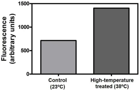
High-temperature is commonly associated with ROS production (Dat et al. 2000). In the present study the influence of a 12 h incubation of CSB cells at 38ºC in the redox state was evaluated. The estimation of the intracellular ROS levels with the H2DCFDA fluorescent probe showed that CSB cells exposed to 38ºC increased the levels of fluorescence by 97%, when compared to the control (Figure 12). Following this result, the effect of the increased intracellular ROS levels on membrane damage was evaluated by measuring lipid peroxidation with the MDA method (Figure 13). Results showed a 28.7% increase in MDA amount, when the cells were exposed to high-temperature.
32
Table I. Characterization of the proteins up-regulated by temperature identified by LC MS/MS
analysis of the excised band depicted in Figure 14.
Acc. No.1 Protein Function Taxonomy MW
(kDa)2
gi|147828051 Plasminogen activator inhibitor 1 RNA-binding protein, putative
Ricinus communis 39.17
gi|147834848 Hsp70-interacting protein 1 Vitis labrusca 41.02 gi|147845028 Chaperone protein dnaJ, putative Ricinus communis 45.49 gi|147769068 Chaperone protein dnaJ 15 Arabidopsis thaliana 45.88 gi|147818771 Methylthioribose kinase, putative Ricinus communis 47.93 gi|147783001 Eukaryotic peptide chain release
factor subunit, putative
Ricinus communis 49.10
gi|147780810 Eukaryotic peptide chain release factor subunit, putative
Ricinus communis 49.10
gi|147780179 Glycosyl hydrolase family-like
protein
Salvia miltiorrhiza 49.59
gi|147856780 Serine/threonine-protein kinase PBS1, putative
Ricinus communis 50.06
gi|147790061 Leucine rich repeat-containing protein, putative
Ricinus communis 58.47
gi|147784740
Chaperonin containing t-complex protein 1, gamma subunit, tcpg, putative
Ricinus communis 60.44
gi|147790061 Leucine rich repeat-containing protein Ricinus communis 58.47 gi|147784740 Chaperonin containing t-complex
protein 1, gamma subunit, tcpg, putative
Ricinus communis 60.44
gi|147805226 UBQ10 (Polyubiquitin 10) Arabidopsis thaliana 51.16
gi|147834511 Polyubiquitin Zea mays 60.25
1
Accession number in the NCBI database
2
33
Figure 12. Intracelular ROS quantification with the fluorescent probe H2DCFDA in CSB
cells incubated for 12 h at 38ºC (high temperature treated cells) and in control cells (cultivated at 23ºC). Mean of two independent experiments are shown.
Figure 13. Quantification of MDA in CSB cells incubated for 12 h at 38ºC (high
temperature treated cells) and in control cells (cultivated at 23ºC). Mean of two independent experiments are shown.
34
3.3 Effect of ABA and SA on glucose transport
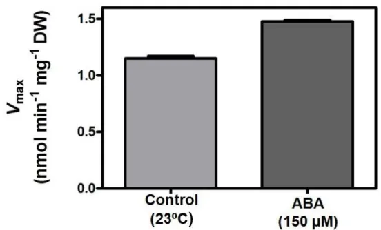
As previously reported (Cakir et al. 2003) VvHT1 is regulated by an ASR protein, VvMSA, which specifically binds to its promoter region. Also, VvMSA expression is strongly enhanced by ABA and glucose, suggesting that ABA can regulate VvHT1. Furthermore, ABA levels increase in parallel with sugar accumulation in the grape berry and color development and it is mainly found in the phloem of the berry suggesting a role on the unloading and uptake of sugars (Kataoka et al. 1982; Shiozaki et al. 1999). In the present study the incubation of CSB cells with 150 µM ABA for 12 h promoted a consistent increase of the Vmax of the glucose transport system from 1.15 ± 0.02 nmol glucose min-1 mg-1 DW (control cells) to 1.48 ± 0.01 nmol glucose min-1 mg-1 DW (Figure 14).
Figure 14. Effect ABA (150 µM; 12 h incubation) on the activity of glucose transport in
CSB cells. Mean of two independent experiments are shown.
SA has been involved in signaling in plants, particularly in the induction of defense and stress responses (Bari and Jones 2009), but its role during grape berry development remains unclear. In the present study the incubation of CSB cells with 150 µM SA for 12 h increased the Vmax of the glucose transport system from 0.80 ± 0.06 nmol glucose min-1 mg-1 DW (control cells) to 1.30 ± 0.04 nmol glucose min-1 mg-1 DW (Figure 15).
35
Figure 15. Effect SA (150 µM; 12 h incubation) on the activity of glucose transport in CSB
cells. Mean of two independent experiments are shown.
3.4 Compartmentation studies in intact grape berries
In a paper published several years ago (Kriedmann 1978) the pattern of glucose incorporation into intact grape berries incubated via the pedicel with radioactive substrates was evaluated by autoradiography. That work was the inspiring basis of this study where intact grape berries were incubated in ELISA plates with radioactive sugars and sugar analogs to evaluate the effect of high temperature, ABA and SA on sugar incorporation (Figure 16). As shown in figure 17, after the incubation of intact grape berries with 1.35 mM 2-NBDG for 12 h the fluorescent sugar distributed throughout the mesocarp tissue. Figure 18 shows that high temperature (38ºC), 150 µM ABA and 150 µM SA increased D-[14C]glucose uptake by grape berries, after 12 h incubation as follows: control, 2.55 ± 0.47 nmol glucose h-1 g-1 FW; 38ºC treatment, 4.86 ± 1.30 nmol glucose h-1 g-1 FW; ABA treatment, 4.81 ± 0.72 nmol glucose h-1 g-1 FW and SA treatment, 4.28 ± 0.85 glucose h-1 g-1 FW.