BASIC RESEARCH
Institute of Orthopedics and Traumatology, Hospital das Clinicas da Fac-uldade de Medicina da Universidade de São Paulo - São Paulo/SP, Brazil. Email: [email protected]
Tel.: 55 11 3069.6776 / 8101
Received for publicaiton on November 23, 2009 Accepted for publication on December 03, 2009
HISTOLOGICAL STUDY OF FRESH VERSUS FROZEN
SEMITENDINOUS MUSCLE TENDON ALLOGRAFTS
Alexandre Carneiro Bitar, Luiz Augusto Ubirajara Santos, Alberto Tesconi Croci, João Alberto Ramos Maradei Pereira, Edgard N. França Bisneto, Arlete Mazzini Miranda Giovani, Claudia Regina G. C. M. Oliveira
doi: 10.1590/S1807-59322010000300010
Bitar AC, Santos LAU, Croci AT, Pereira JARM, França-Bisneto EN, Giovani AMM, Oliveira CRGCM. Histological study of fresh versus frozen semitendinous muscle tendon allografts. Clinics. 2010;65(3):297-303.
OBJECTIVE: The purpose of this study was to histologically analyze allografts from cadaveric semitendinous muscle after cryo-preservation at -80°C in comparison to a control group kept at only -4°C to test the hypothesis that the histological characteristics of the tissue are maintained when the tendons are kept at lower temperatures.
METHODS: In a tissue bank, 10 semitendinous tendons from 10 cadavers were frozen at -80ºC as a storage method for tissue preservation. They were kept frozen for 40 days, and then a histological study was carried out. Another 10 tendon samples were analyzed while still “fresh”.
RESULTS: There was no histological difference between the fresh and frozen samples in relation to seven variables.
CONCLUSIONS: Semitendinous muscle tendon allografts can be submitted to cryopreservation at -80ºC without suffering his-tological modiications.
KEYWORDS: Cryopreservation; Tissue banks; Histology; Tendons; Tissue transplantation.
INTRODUCTION
The use of allogenic tissues is growing in orthopedic practice, as is the number of studies on methods for processing, sterilization and cryopreservation that interfere as little as possible with the original physiological properties of the tissues.1
The literature compares allografts with autologous grafts in knee ligament reconstruction surgery, with little difference in clinical results.2,3 However, compared with
autologous transplants, allografts do have some advantages. For example, they do not increase morbidity for the donor, they require a shorter surgery time, and they are available without restriction on size and morphology. They are best indicated for multiple ligament reconstructions, revision
surgeries, and patella baja.4-7 In ligament reconstruction
surgeries, the possibility exists of an immune response from the recipient tunnel enlargement, and delayed incorporation of the allograft.3 The risk of disease transmission and the
potential for immunogenicity are the major disadvantages of allografts,8 but these complications can be controlled.9,10
Grafts can be processed and stored in different ways, and the preparation techniques have the potential to change the initial resistance and mechanical properties of the graft prior to implantation.11 Theoretically, any method that promotes
cell debridement of the collagenous matrix should reduce the antigenicity of the tissue. However, it is essential that the extraction does not destroy the collagen matrix, which could result in alteration of the biomechanical properties of the graft.11,12
Although there are studies on the biomechanical behavior of cryopreserved tendons,13 the literature does not address the
histological changes of the tissue at -80oC.
kept at only -4 °C to test the hypothesis that the histological characteristics of the tissue are maintained when the tendons are kept at lower temperatures.
METHODS
Twenty semitendinous muscle tendons (10 from the left limb and 10 from the right) from 10 cadavers were used in this study. The cadavers, all aged 20 to 40 years old, were obtained from the local mortuary service and were kept for up to 48 hours at -4oC. None of the cadavers
demonstrated neoplasias, infections, previous surgeries, collagen diseases, or degenerative diseases, and none had received corticosteroid therapy or chemotherapy that might have altered the tendinous structures. The causes of death are listed in Table 1.
The tendons of the semitendinous muscle were removed after dissection of the skin and subcutaneous tissue with a tendon extractor commonly used in surgeries for knee ligament reconstruction. The technique was standardized: 5-cm portions of tendon were removed from an area 3 cm away from the insertion site, a region where there is no transition of muscle or bone, only tendon tissue.
The samples were soaked in 0.9% sodium chloride (saline) solution to prevent dehydration or alteration of the tissue architecture. Of the 20 tendons obtained, 10 were prepared for microscopic analysis the next day (Group A), and the remaining 10 (Group B) were sent to the tissue bank for cryopreservation for 40 days. Each cadaver had a tendon in Group A and a tendon in Group B. The determination of groups according to the sides (right and left) was random.
The 10 tendons in Group A were ixed for 24 hours in a 10% formalin solution. The 10 tendons for the tissue
bank (Group B) were packaged in sealed 0.5 micrometer polyethylene and nylon bags, with triple protection, in accordance with international standards of the European Association of Tissue Banks (EATB) and the American Association of Tissue Banks (AATB),14 and were kept
cryopreserved at -80oC in an electric vertical ultrafreezer
(model SANYO MDF U3086S, Sanyo Electric Co. Ltd.) equipped with a CO2 backup, a warning system and temperature monitoring via satellite, producing a printed report of temperature levels every six hours.
After 40 days of cryopreservation, the tendons in Group B were thawed by immersion in 0.9% NaCl isotonic saline solution for 30 minutes at room temperature and ixed in formalin solution for a further 24 hours. As with Group A, which was already ixed in formalin, these samples were stained using hematoxylin and eosin and cut longitudinally and transversely to produce 2 mm-thick pieces. The analysis of the histological slides was always done by the same pathologist and in random order.
The cellular and structural components of each piece were analyzed using an optical microscope. The cellular components were investigated for the presence of inflammatory reaction (cell infiltration and edema) and ibroblast proliferation. The histological architecture, orientation of the ibers, quality of the matrix and presence or absence of angiogenesis were structurally evaluated.
The Fischer exact and chi-square tests were used for statistical analysis. Differences with p ≤ 0.05 were considered signiicant.
RESULTS
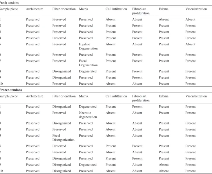
The data from the histological study of the tendons ixed in formalin and the cryopreserved tendons are listed in Table 2. Table 3 lists the histological features for the total samples of fresh and frozen tendons, some of which are illustrated in Figures 1 to 4. The Fischer exact and chi square tests did not show statistical differences (p > 0.3 for all variables); this shows that for all variables, there was no tissue modiication due to freezing.
DISCUSSION
Grafts are used in various procedures in different branches of orthopedics, including ligament reconstruction. The literature shows the importance of the use of allografts in knee surgery, especially in revision surgeries, multiple ligament reconstructions and, more recently, surgery for primary reconstruction of the anterior cruciate ligament (ACL) in active3,15 patients and in those aged over 40 years.16
The irst report of the use of allografts in humans dates Table 1- Characteristics and cause of death among the
ca-davers studied
No. Sex Age (years) Cause of death
1 Male 38 Heart failure
2 Female 34 Obstructive acute abdomen
3 Male 39 Stroke
4 Male 33 Cirrhosis
5 Male 27 Heart failure
6 Male 40 Pulmonary embolism
7 Male 31 Heart failure
8 Male 36 Heart failure
9 Female 29 Cirrhosis
10 Male 38 Pulmonary embolism
-back to 1881.17 The irst tissue bank of bone grafts was
created in 1940 in the United States, and the initial clinical results were published in 1942 by Inclan.18 Since then, a
series of regulations and studies has emerged relating to the use of grafts in orthopedic practice.19 In our institute,
the Tissue Bank has been in operation since 1999 and is governed by local legislation.20
There have been at least 11 clinical studies comparing the use of auto and allografts in the reconstruction of the ACL.2,3 Most of these show little difference between the
two techniques with respect to long-term results. However, there has been no prospective randomized study, and the comparison methods (scores), types of graft, as well as methods of preparing and ixing the graft are highly variable. Furthermore, most studies use the patellar tendon graft; therefore, it may not be possible to generalize the conclusions of these studies to the lexor tendon.
Macey21 was the irst to use the semitendinosus tendon
for reconstruction of the intra-articular knee. The practice of using this free graft has increased since the 1990s and is currently compared with the patellar tendon as the irst option for autografting in primary surgeries for ACL reconstruction.15
Some authors, such as Lawhorn and Howell22, suggest the
use of allografts without a bone plug because of the potential for slower incorporation of the bone due to immunogenicity and smaller cross-sectional area of transplants with bone plugs. Hadjicostas et al.,23 compared the histological features
of the patellar tendon and semitendinosus and gracilis tendons in cadavers. Differences were found in the relationship between ibrils/interstitium (20 to 30% higher in the lexor) and density of ibroblasts (35 to 50%). It was noted that there is an advantage of these grafts with regard to the renovation and integration of the patellar tendon.
Table 2 - Histological analysis of fresh and frozen semitendinous tendons
Fresh tendons
Sample piece Architecture Fiber orientation Matrix Cell iniltration Fibroblast proliferation
Edema Vascularization
1 Preserved Preserved Preserved Absent Absent Absent Absent
2 Preserved Preserved Preserved Present Present Present Present
3 Preserved Preserved Preserved Present Present Present Present
4 Preserved Preserved Preserved Present Present Present Present
5 Preserved Preserved Hyaline
Degeneration
Absent Absent Present Absent
6 Preserved Preserved Preserved Present Present Present Present
7 Preserved Preserved Focal
Degeneration
Present Present Present Present
8 Preserved Disorganized Degenerated Present Present Present Present
9 Preserved Disorganized Preserved Present Present Present Present
10 Preserved Preserved Preserved Absent Absent Present Present
Frozen tendons
Sample piece Architecture Fiber orientation Matrix Cell iniltration Fibroblast proliferation
Edema Vascularization
1 Preserved Disorganized Degenerated Present Present Present Present
2 Preserved Preserved Necrotic
degeneration
Absent Absent Present Present
3 Preserved Disorganized Preserved Absent Absent Present Present
4 Preserved Preserved Preserved Absent Absent Present Present
5 Preserved Focal
Disorganization
Preserved Absent Absent Present Present
6 Preserved Preserved Preserved Present Present Present Present
7 Preserved Preserved Preserved Absent Absent Present Present
8 Preserved Disorganized Preserved Present Present Present Present
9 Preserved Disorganized Degenerated Present Absent Absent Present
Figure 1 - Histological aspect of sample #3, a fresh tendon (hematoxylin-eosin, 40 X).
Figure 2 - Histological aspect of sample #8, a frozen tendon: degenerated
matrix and intact vascularization (hematoxylin-eosin, 40 X).
Figure 3 - Histological aspect of sample #1, a frozen tendon: ibroblastic
proliferation and degenerated matrix (hematoxylin-eosin, 100 X).
Figure 4 - Histological aspect of sample #2, a frozen tendon: focal necrosis
and degenerated matrix (hematoxylin-eosin, 100 X). Table 3 - Statistical analysis of the histological results
Fresh Frozen Total
Histological architecture p = 1.0 (Fisher exact test)
Preserved 10 10 20
Not preserved 0 0 0
Total 10 10 20
Fiber orientation p = 0.35 (Fisher exact test)
Preserved 8 5 13
Disorganized 2 5 7
Total 10 10 20
Matrix p = 1 (Fisher exact test)
Preserved 7 7 14
Degenerated 3 3 6
Total 10 10 20
Cell iniltration p = 0.37 (Fisher exact test)
Present 7 4 11
Absent 3 6 9
Total 10 10 20
Fibroblast proliferation p = 0.18 (Chi squared test)
Present 7 3 11
Absent 3 6 9
Total 10 10 20
Edema p = 1.0 (Fisher exact test)
Present 8 8 16
Absent 2 2 4
Total 10 10 20
Vascularization p = 0.47 (Fisher exact test)
Present 8 10 18
Absent 2 0 2
Total 10 10 20
Currently, tendon allografts are used in knee surgeries, in elbow ligament reconstructions, and for revisions of the acromioclavicular joint.24 In our medical service, allografts
are used mainly in knee surgeries, ACL reconstruction, multiple ligament reconstructions, ligament surgery in skeletally immature patients and with double bundle
reconstruction. Other studies with patellar tendon grafts and Achilles tendon grafts have been carried out by our institute, analyzing the biomechanical aspects and comparing the same methods of preparation, with no statistically signiicant differences.25,26
selection of the cadavers included in our sample. Like the work of Hadjicostas et al.,23 we included bodies of both
sexes. Limiting the age of the cadavers in this study served to remove the degenerative effects that can be seen after age 40 as a result of tissue aging, and also to reduce the risk of injuries and disruptions to which aging individuals are naturally predisposed. The patients included in the study by Hadjicostas et al.,23 had a higher mean age (56.6 years) than
our sample. In fact, age and sex appear to have little effect on the composition of samples for tissue donation.27
The increased frequency of the use of allografts in traumato-orthopedics requires the adoption of storage techniques that interfere as little as possible in the quality of the parts.17,19 Allografts can be stored in different ways;
they can be chilled in residential mechanical freezers at temperatures of +2oC to -4oC for up to ive days. In freezers
with temperatures of -20oC to -40oC, they can be stored
for up to six months.14 At these temperatures, the enzymes
present in the tissue are still active and can destroy the tissue. Therefore, storage periods of longer than a few months are not recommended. The methods of sterilization used at low temperatures are effective against fungi and do not seem to change the mechanical characteristics of the grafts. The period of 40 days chosen in this study coincides with the period of incubation necessary for microbiological investigations for bacteria and fungi.19 The deep-freezing
process enables storage for up to ive years19, and this is the
method we use in our service.
Many services prefer to carry out the manipulation of tissues under aseptic conditions from acquisition through clinical use, and the samples are discarded when microbiological assays show positive bacterial cultures (20 to 30%).28 Sterilization methods, therefore, are
not completely safe. They can alter the biomechanical characteristics of tissues or fail to penetrate tissue layers, resulting in the protection of microorganisms rather than their destruction. Irradiation with gamma rays is the most common method of sterilization.12, 29 However, to achieve
safer sterilization in frozen tissues, high-dose irradiation is necessary, which can alter the biomechanical properties of the tissue. Also, the sterilization effectiveness against viruses is low.19 Ethylene oxide sterilization requires strict control of
the levels of waste gas in contact with the allograft and is no longer used by tissue banks, due to the possibility of toxic effects for the recipient (dissolution of the graft and articular inlammatory reactions).19
The processing techniques used in the preparation and preservation of grafts have been questioned as potentially altering the initial resistance and mechanical properties of the graft prior to implantation.11 Two studies carried out in
Brazil address the biomechanical properties of patellar25 and
calcaneus26 tendons of cadavers with the same preparation
method as that used in our study, comparing fresh and cryopreserved allografts. They found no differences. A study on metric measurements and attachment levels of the medial patellofemoral ligament shows this to be a distinct structure.30
Although there have been studies on the biomechanical behavior of tendons,13 the literature does not address
histological changes of tendons cryopreserved at -80oC under
aseptic conditions. During cryopreservation at -80oC, the
destruction of the allograft enzyme appears to be minimal and at least one enzyme, collagenase, which can destroy the tissue, is inactive.31 Furthermore, with cryopreservation
there is no intracellular free water, which is thought to be necessary for enzymatic activity, bacterial proliferation and lipid oxidation.32,33 Lipid oxidation inside the tissues induces
apoptosis and inhibits cell differentiation; such oxidation can be minimized or avoided with cryopreservation at temperatures of at least -70oC.33
The literature refers to histological changes due to cryopreservation only in cartilage34 (one of the most
commonly used grafts in surgical practice),34 concluding
that during freezing, the vitality of the cells is threatened. Other injuries may also occur, such as the formation of extracellular ice crystals, intracellular ice nucleation, collapse of the matrix, and breakage of intercellular bridges. In our study, the histological study of one tendon (not cartilage) was carried out, and none of these histological phenomena were observed with cryopreservation at -80oC.
Freezing with liquid nitrogen at -179oC has also been
used as a storage method with similar results but higher cost.28 Another widespread storage method is lyophilization.
Cryopreservation and lyophilization have been related to a reduction in allograft antigenicity.6 The use of chilled
saline solution is not a guaranteed method because the stock can only be kept safely for short periods.28 Treatment with
paraformaldehyde and fixation with glutaraldehyde are no longer recommended because of the toxicity of these solutions to the recipient tissue.
Fibroblasts play a key role in the regeneration of tendinous tissues, regardless of the location. Papandrea et al.,.35 reported evidence of regeneration of the medial lexor
Ferretti et al.,36 carried out a histological study with three
patients undergoing review surgery (after 6, 24 and 26 months), using the medial lexors as the initial graft. Biopsy specimens taken after the six-month period still showed histological immaturity, with ibroblast proliferation but irregularity in the organization of the collagen fibers. However, after two years, both tendons showed well-oriented ibers, similar to the tendons of patients who had not been operated on. Pufe et al.,37 noted that the density of
ibroblasts negatively inluences the mechanical properties of the tendons. Hadjicostas et al.,.23 speculated that a high
concentration of ibroblasts in the medial lexor tendons could lead to a better healing response of the tendons. In our sample, the freezing process did not interfere with ibroblast proliferation or the structure of the tendon.
Vasculature also appeared to be preserved after freezing of the tendons in the present work. Hadjicostas et al.,23
showed a higher concentration of vessels in the flexor tendons when compared to the patellar tendons, but no correlation was observed with the other variables. There is some controversy, however, concerning the inluence of angiogenesis in the process of tendon healing, as previous studies show that the density of blood vessels adversely affects the biomechanical properties of the tendons.38
Graf et al.,39 studied the effects of freezing the tendon
through histological, biochemical and biomechanical
analyses of the patellar tendon after reconstruction of the ACL in rabbits, and did not observe any signiicant difference in comparison with the control group. The only investigation in the literature comparing the histological features of fresh versus frozen tendons in cadavers, to our knowledge, is the present study.
We believe that other variables, such as storage time11,
should be examined in future works. The quantiication of histological variables through cell counts and histomorphometry could make the analysis of the effect of freezing more objective. Special staining for collagen and glycogen or even other variables could also deepen the histological analysis. In the future, we believe that these data will serve to improve the use of these tissues for transplant allografts, with the possibility of lower tissue reaction, lower inlammation rates and perhaps lesser effects of rejection, increasing the resistance of allografts and making their use in surgical practice more feasible.
CONCLUSION
Tendons of the semitendinosus muscle of human cadavers, when subjected to cryopreservation at -80oC, retain their
histological architecture, orientation of ibers, matrix, cellular iniltration, ibroblast proliferation, swelling and blood, with no histological changes when compared to fresh tendons.
REFERENCES
1. Nutton RW, McLean I, Melville E. Tendon allografts in knee ligament surgery. J R Coll Surg Edinb. 1999;44:236-40.
2. Chang SK, Egami DK, Shaieb MD, Kan DM, Richardson AB. Anterior cruciate ligament reconstruction: allograft versus autograft. Arthroscopy. 2003;19:453-62.
3. Marrale J, Morrissey MC, Haddad FS. A literature review of autograft and allograft anterior cruciate ligament reconstruction. Knee Surg Sports Traumatol Arthrosc. 2007;15:690-704.
4. Flahiff CM, Brooks AT, Hollis JM, Vander Schilden JL, Nicholas RW. Biomechanical analysis of patellar tendon allografts as a function of donor age. Am J Sports Med. 1995;23:354-8.
5. Jackson DW, Corsetti J, Simon TM. Biologic incorporation of allograft anterior cruciate ligament replacements. Clin Orthop. 1996;324:126-33. 6. Jackson DW, Windler GE, Simon TM. Intraarticular reaction associated with the use of freeze-dried, ethylene oxide-sterilized bone-patella tendon-bone allografts in the reconstruction of the anterior cruciate ligament. Am J Sports Med. 1990;18:1-10; discussion 10-1. 7. Levitt RL, Malinin T, Posada A, Michalow A. Reconstruction of anterior
cruciate ligaments with bone-patellar tendon-bone and achilles tendon allografts. Clin Orthop Relat Res. 1994;303:67-78.
8. Barrios RH, Leyes M, Amillo S, Oteiza C. Bacterial contamination of allografts. Acta Orthop Belg. 1994;60:293-5.
9. Albert A, Leemrijse T, Druez V, Delloye C, Cornu O. Are bone autografts still necessary in 2006? A three-year retrospective study of bone grafting. Acta Orthop Belg. 2006;72:734-40.
10. Urabe K, Itoman M, Toyama Y, Yanase Y, Iwamoto Y, Ohgushi H, et al. Current trends in bone grafting and the issue of banked bone allografts based on the fourth nationwide survey of bone grafting status from 2000 to 2004. J Orthop Sci. 2007;12:520-5.
11. Sterling JC, Meyers MC, Calvo RD. Allograft failure in cruciate ligament reconstruction. Follow-up evaluation of eighteen patients. Am J Sports Med. 1995;23:173-8.
12. Salamon A, Hámori J. Development of collagenous ibres in autologous and preserved homologous tendon grafts. Acta Morphol Acad Sci Hung. 1976;24:11-22.
13. Pearsall AW 4th, Hollis JM, Russell GV Jr, Scheer Z. A biomechanical comparison of three lower extremity tendons for ligamentous reconstruction about the knee. Arthroscopy. 2003;19:1091-6. 14. American Association of Tissue Banks. Standards for Tissue Banking.
15. Sherman OH, Banffy MB. Anterior cruciate ligament reconstruction: which graft is best? Arthroscopy. 2004;20:974-80.
16. Barrett G, Stokes D, White M. Anterior cruciate ligament reconstruction in patients older than 40 years: allograft versus autograft patellar tendon. Am J Sports Med. 2005;33:1505-12.
17. Malinin TI. Allografts for the reconstruction of the cruciate ligaments of the knee: procurement, sterilization and storage. Sports Med Arthroscopy. 1993;1:31-41.
18. Inclan A. The use of preserved bone graft in orthopaedic surgery. J Bone Joint Surg Am. 1942;24:81-96. Disponível em: http://www.ejbjs.org/cgi/ reprint/24/1/81. Acessado em 2008 (09 out).
19. Vangsness CT Jr, Garcia IA, Mills CR, Kainer MA, Roberts MR, Moore TM. Allograft transplantation in the knee: tissue regulation, procurement, processing, and sterilization. Am J Sports Med. 2003;31:474-81. 20. Amatuzzi MM, Croci AT, Giovani AMM, Santos LAU. Banco de tecidos:
estruturação e normatização. [Tissue bank: structure and organization]. Rev Bras Ortop. 2000;35:165-72.
21. Macey HB. A new operative procedure for the repair of ruptured cruciate ligament of the knee joint. Surg Gynecol Obstet. 1939;69:108-9. 22. Lawhorn KW, Howell SM. Scientiic justiication and technique for
anterior cruciate ligament reconstruction using autogenous and allogenic soft-tissue grafts. Orthop Clin North Am. 2003;34:19-30.
23. Hadjicostas PT, Soucacos PN, Paessler HH, Koleganova N, Berger I. Morphologic and histologic comparison between the patella and hamstring tendons grafts: a descriptive and anatomic study. Arthroscopy. 2007;23:751-6.
24. Costic RS, Labriola JE, Rodosky MW, Debski RE. Biomechanical rationale for development of anatomical reconstructions of coracoclavicular ligaments after complete acromioclavicular joint dislocations. Am J Sports Med. 2004;32:1929-36.
25. Giovani AM, Croci AT, Oliveira CR, Filippi RZ, Santos LA, Maragni GG, et al. Comparative study of cryopreserved bone tissue and tissue preserved in a 98% glycerol solution. Clinics. 2006;61:565-70. 26. Reiff RBM, Croci AT, Bolliger Neto R, Pereira CAM. Estudo
comparativo de propriedades biomecânicas da porção central do tendão calcâneo congelado e a fresco. [Comparative study on biomechanical properties of the central portion of frozen and fresh calcaneus tendon]. Acta Ortop Bras. 2007;15:6-8.
27. Pietrzak WS, Woodell-May J. The composition of human cortical allograft bone derived from FDA/AATB-screened donors. J Craniofac Surg. 2005;16:579-85.
28. Zimmerman MC, Contiliano JH, Parsons JR, Prewett A, Billotti J. The biomechanics and histopatology of chemically processed patellar tendon allografts for anterior cruciate ligament replacement. Am J Sports Med. 1994;22:378-86.
29. Jackson DW, Grood ES, Wilcox P, Butler DL, Simon TM, Holden JP. The effects of processing techniques on the mechanical properties of bone-anterior cruciate ligament-bone allografts. An experimental study in goats. Am J Sports Med. 1988;16:101-5.
30. Aragão JA, Reis FP, de Vasconcelos DP, Feitosa VL, Nunes MA. Metric measurements and attachment levels of the medial patellofemoral ligament: an anatomical study in cadavers. Clinics. 2008;63:541-4. 31. Tomford W. Transmission of disease through musculoskeletal
transplantation. Portland Bone Symposium. Portland: Oregon Health Sciences University; 1997.
32. Galea G, Kearney JN. Clinical effectiveness of processed and unprocessed bone. Transfus Med. 2005;15:165-74.
33. Laitinen M, Kivikari R, Hirn M. Lipid oxidation may reduce the quality of a fresh-frozen bone allograft. Is the approved storage temperature too high? Acta Orthop. 2006;77:418-21.
34. Schachar NS, McGann LE. Investigations of low-temperature storage of articular cartilage for transplantation. Clin Orthop Relat Res. 1986;146-50.
35. Papandrea P, Vulpiani MC, Ferretti A, Conteduca F. Regeneration of the semitendinosus tendon harvested for anterior cruciate ligament reconstruction. Evaluation using ultrasonography. Am J Sports Med. 2000;28:556-61.
36. Ferretti A, Conteduca F, Morelli F, Masi V. Regeneration of the semitendinosus tendon after its use in anterior cruciate ligament reconstruction: a histologic study of three cases. Am J Sports Med. 2002;30:204-7.
37. Pufe T, Petersen WJ, Mentlein R, Tillman BN. The role of vasculature and angiogenesis for the pathogenesis of degenerative tendons disease. Scand J Med Sci Sports. 2005;15:211-22.
38. Yoshikawa T, Tohyama H, Katsura T, Kondo E, Kotani Y, Matsumoto H, et al. Effects of local administration of vascular endothelial growth factor on mechanical characteristics of the semitendinosus tendon graft after anterior cruciate ligament reconstruction in sheep. Am J Sports Med. 2006;34:1918-25.