Horizontal and vertical maxillary
osteotomy stability, in cleft lip and palate patients,
using allogeneic bone graft
Kelston Ulbricht Gomes1, Wilson Denis Benato Martins2, Marina de Oliveira Ribas2
How to cite this article: Gomes KU, Martins WDB, Ribas MO. Horizontal
and vertical maxillary osteotomy stability, in cleft lip and palate patients, using al-logeneic bone graft. Dental Press J Orthod. 2013 Sept-Oct;18(5):84-90.
Submitted: January 16, 2012 - Revised and accepted: August 17, 2012
» The authors report no commercial, proprietary or inancial interest in the prod-ucts or companies described in this article.
Contact address: Kelston Ulbricht Gomes
Av. Silva Jardim, 2042 – Sala 1205 – Curitiba/PR, Brazil CEP: 80250-200 – E-mail: [email protected]
1 PhD in Oral and Maxillofacial Surgery, Pontifical Catholic University of Paraná
(PUC-PR).
2 Professor of Dentistry at PUC-PR.
Objective:This study was carried out to evaluate maxillary stability after orthodontic-surgical treatment of patients with cleft lip and palate. Cephalometric analysis was applied to two different groups, with and without allogeneic bone graft. Methods: The sample comprised 48 patients with cleft lip and palate. The test group comprised 25 patients who, after correction of maxillary position, received allogeneic bone graft at the gap created by Le Fort I osteotomy. The control group comprised 23 patients and its surgical procedures were similar to those applied to the test group, except for the use of bone graft. Manual cephalometric analysis and comparison between lateral teleradiographs, obtained at the preopera-tive phase, immediate postoperapreopera-tive phase and after a minimum period of six months, were carried out. Results: An higher horizontal relapse was observed in the control group (p<0.05). There were no statistically significant differences in vertical relapses between test and control groups (p>0.05). Conclusion: The use of allogeneic bone graft in cleft lip and palate patients submitted to Le Fort I osteotomy contributed to increase postoperative stability when compared to surgeries without bone graft.
Keywords:Orthognathic surgery. Oral surgery. Bone grafting.
Objetivo:avaliar a estabilidade maxilar após o tratamento ortodôntico-cirúrgico de pacientes portadores de fissura la-biopalatal, por meio de análise cefalométrica, em dois diferentes grupos, com e sem utilização de enxerto ósseo alógeno. Métodos: a amostra foi constituída de 48 pacientes portadores de fissura labiopalatal. O grupo teste foi composto de 25 pacientes que, após a correção da posição da maxila, receberam enxertia óssea alógena para preenchimento dos espaços gerados pelas osteotomias maxilares do tipo Le Fort I. O grupo controle foi composto por 23 pacientes, com os procedimentos cirúrgicos similares aos do grupo teste, com a diferença de que não se utilizou enxertia óssea. Reali-zaram-se traçados cefalométricos manuais e comparação entre as telerradiografias em perfil, obtidas no pré-operatório, no pós-operatório imediato e após o período mínimo de seis meses. Resultados: ocorreu maior recidiva horizontal no grupo controle (p < 0,05). Não houve diferenças estatisticamente significativas nas recidivas verticais entre os grupos teste e controle (p > 0,05). Conclusão: a utilização de enxertos alógenos em pacientes portadores de fissura labiopala-tal submetidos a osteotomias maxilares Le Fort I contribuiu para o aumento da estabilidade horizonlabiopala-tal pós-operatória, quando comparada com cirurgias sem enxertia óssea.
INTRODUCTION
Cleft lip and palate deformities are amongst the most
common congenital anomalies of the face.1
Cleft lip and palate surgery as well as orthodontic treatment are amongst the therapeutic possibilities for re-covering patients’ esthetics and function. In patients with cleft lip and palate, some occlusal deleterious situations such as teeth crowding and unilateral crossbite with seg-ments collapse, open bite on the affected side and
retru-sion of the maxilla, are identified.2-6
After the growth spurt, orthognathic surgery is indi-cated to correct skeletal and dental discrepancies in pa-tients who present dentofacial deformity.
Hirano and Suzuki 7 described potential aspects
which are responsible for maxillary retrusion in adult cleft patients: Unfavorable muscular action due to scars caused by early surgeries in lip and palate, pharyngeal flaps and absence of teeth, which reduces occlusal stability.
The stability of orthognathic surgeries depends on the type and the extent of movements performed by the maxilla. Stability is considered difficult especially in patients with cleft lip and palate. Usually, these pa-tients have undergone surgery in the soft and hard pal-ate, which normally results in fibrosis, limiting the ex-tent of both transverse and anteroposterior movements
of the maxilla.8-12
In order to avoid relapses when treating dentofacial deformities in cleft patients, some authors suggest in-creasing the time of intermaxillary fixation during the postoperative phase, performing bimaxillary surgeries, using face masks with reverse traction of the maxilla and interpositioning bone grafts between the gaps created by
maxillary advancement.13,14,15
A successful correction of dentofacial deformities depends on effective stabilization and prompt union of the repositioned bone segments. When there is a large area of contact between the segments, safe and satisfac-tory bone union is expected. When the contact area is small, there may be instability, relapse or fibrous union (pseudoarthrosis) between segments. In such cases,
grafts are recommended. Some authors16,17 suggest
al-logeneic bone graft in orthognathic surgery. However, in the aforementioned studies, allogeneic bone graft was performed in patients without cleft lip and palate.
The study of Precious,18 in 2007, concluded that
scars on the upper lip and on the palate interfere with nose, lips, soft adjacent tissues and skeletal development.
When intervention is performed with bone graft and correction of nasolabial musculature at the age of five or six years old, a symmetric function is established, which improves facial development. The primary muscle sur-gery improves growth and decreases the chances of
un-dergoing orthognathic surgery.19
Nique et al 2 have studied the use of allograft bone
for alveolar reconstruction in unilateral cleft patients. The receptor area was radiographically observed for a period of 3 to 6 months. The allograft bone is an ex-cellent alternative to repair alveolar cleft, its use brings significant benefits for the patient, eliminating the morbidity of a second surgical site.
Garrison et al19 evaluated twenty patients who
were simultaneously submitted to both alveolar bone graft and Le Fort I osteotomy. The research-ers evaluated the extent of maxilla relapse at the an-teroposterior and vertical direction through lateral teleradiographs. They concluded that there was no significant change in the horizontal plan, however, in the vertical direction there was a great tendency to relapse. The intermaxillary fixation time lasted for eight weeks and mandibular fixation was used at the orbital rim and zygomatic crest. For the evaluation, cephalometry was adopted, the SN plan was traced and a perpendicular line was drawn from the Nasion. The researchers measured the distance from this line to point A in order to evaluate potential changes in the horizontal direction (anteroposterior). To deter-mine the vertical movement, a line was drawn per-pendicular to the SN up to the point A.
Another research, carried out by Heliovaara et al,20
examined the causes of relapse through a retrospective analysis of 71 patients, 58 of which had unilateral and bi-lateral clefts. The mean advancement of the maxilla was 6.9 mm. Grafts were harvested from calvaria or man-dible and there were used four miniplates for containing the maxilla as well as intermaxillary fixation which was kept during 6 weeks and maintained after releasing fixa-tion with class III elastics. The researchers concluded that the type of cleft (unilateral or bilateral), the scars in the soft palate, muscle tension, adaptation and stability of bone segments are amongst the main causes of relapse in maxillary osteotomies. The occlusal stability is im-portant to prevent relapses.
Hirano and Suzuki7 evaluated one group comprised of
of 11 patients with bilateral cleft lip and palate. The gaps cre-ated by Le Fort I osteotomy were filled with autogenous bone without applying intermaxillary fixation or surgical guide. Patients were evaluated through lateral teleradio-graphs and point A was used as the reference point. Patients were evaluated at pre- and immediate postoperative phases as well as one year after the surgery. The average relapse in the group with cleft palate only was 8.5% in the horizontal direction and 16.7% in the vertical direction. In the group with bilateral cleft, relapse was 9.4% horizontal, and 17.8% vertical. The authors suggest that the main factors for relaps-es are: the method used for fixating the osteotomized seg-ments, neuromuscular adaptation, the extent of movement of the maxilla and previous orthodontic preparation.
Ianetti et al21 evaluated the use of bimaxillary
surger-ies for minimizing potential relapses in patients with cleft lip and palate. They highlight intense scarring and soft tissue tension as being responsible for relapse. To reduce the relapse, authors suggest overcorrection of the maxilla; however, they warn that major advances of the maxilla can result in velopharynx incompetence. These conclu-sions were based on the evaluation of 15 patients who underwent combined bimaxillary surgery. In order to improve the stability of the maxilla, the authors suggest performing bone graft in the space created by Le Fort I osteotomy, with the indicated use of intermaxillary elas-tics for three weeks and the surgical guide being removed after six weeks. The stability evaluation was carried out by means of lateral teleradiographs, taken at the preop-erative phase, six weeks, a year and two years after the surgery. The references were point A, the posterior nasal spine and point B. For cases in which only the maxilla was operated, relapse was of 25%, and in cases of
maxil-lary and mandibular osteotomy, relapse was of 8%.21
In another study, conducted by Erbe et al,22
cepha-lometric analysis was performed during immediate and late postoperative phases (39-110 months) for patients simultaneously undergoing both Le Fort I osteotomies for advancement and autogenous alveolar bone graft. Op-erative changes in the position of the maxilla were evalu-ated in vertical and horizontal directions. All parameters used in the cephalometric measurements were manually measured by one single examiner as an attempt to elimi-nate observer bias. Some reference points were difficult to identify; however, the careful observation of a series of lateral films of the head increased accuracy and the iden-tification of reference points was made possible.
Even with surgical correction of the maxilla, some degree of relapse is expected due to the aforementioned shortcomings and peculiarities (previous surgery on the palate and lack of occlusal stability by the absence of teeth). Bone grafting performed in the space created by both osteotomy and correction of the position of the maxilla can reduce the occurrence of relapse.
Thus, the objective of this study was to evaluate hori-zontal and vertical stability of maxillary osteotomy using allogeneic bone graft in patients with cleft lip and palate.
MATERIAL AND METHODS
The stability of orthodontic-surgical treatment of patients with cleft lip and palate was evaluated through cephalometric analysis in two different groups, one with and another without the use of allogeneic bone graft.
The study was approved by the local Institutional Re-view Board under the number 0003716/10.
Material
The sample consisted of 48 patients with cleft lip and palate, submitted to surgery at the Assistance Center for Cleft Lip and Palate (CAIF) in Curitiba, Paraná, Brazil, from January 2006 to March 2009.
All patients underwent orthognathic maxillary surgery, performed with the Le Fort I technique, with rigid inter-nal fixation and intermaxillary fixation lasting for an aver-age of 4 (four) weeks. The test group (TG) consisted of 25 patients of both genders with unilateral and bilateral clefts, with an average age of 23.16 years. The surgeries were isolated in the maxilla or combined with mandibular sur-geries. After the maxilla had been repositioned, allogeneic bone graft, from the Bank of Muscle and Bone Tissue of the Clinical Hospital from Federal University of Paraná, was inserted to fill the gaps created by the osteotomies.
e) With a new radiograph obtained afterwards, at least 6 months after the first one, the process of com-parison was repeated by means of superimposing the tracings. At this time, the immediate postoperative ra-diograph was used and the values for assessing the oc-currence of relapse were obtained.
f) The purpose of these measurements was to lin-early measure possible vertical and horizontal postop-erative changes, over time, and relate them to the use of bone grafts.
RESULTS
Both groups (CG and TG) presented normal distri-bution with regard to the following variables: horizontal advancement, horizontal relapse, vertical movement and vertical relapse. The average horizontal advancement was similar in both groups. The average vertical movement was higher in TG than in CG (Table 1).
Horizontal relapse was higher in CG (p <0.05). There were no statistically significant differences in vertical re-lapses between TG and CG (p> 0.05) (Tables 2 and 3).
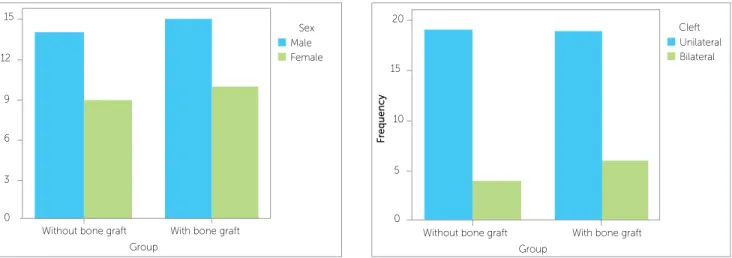
Variables such as gender, type of procedure and type of cleft did not influence the stability of the sur-gery in any group (p>0.05) (Figs 2, 3 and 4).
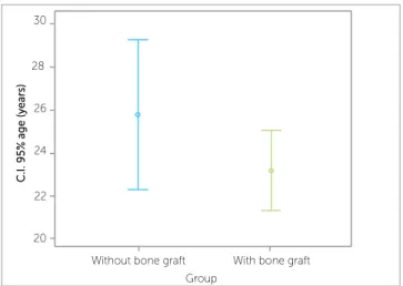
The CG had a follow-up period longer than the TG. However, despite this difference, there is no correla-tion between this variable and horizontal or vertical relapses (Pearson Correlation Coefficient p>0.05). (Table 4; Figs 5 and 6).
By using Pearson Correlation Coefficient it was obtained a p-value> 0.05, indicating that there is no correlation between the two variables. Therefore, de-spite the follow-up time of the group with graft was smaller than in the group with no graft, there was no correlation between this variable and relapse, both horizontal and vertical.
Methods
a) A blind study in which manual cephalometric anal-ysis of the lateral teleradiographs was carried out by one single examiner. Radiographs were obtained at the pre-operative phase, immediate postpre-operative phase and after a minimum period of six months.
b) The records as well as the cephalometric analysis were carried out using advocated parameters and
mea-sures.19,23 The anteroposterior position of the maxilla
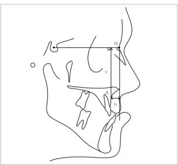
was determined by drawing the SN plan and a perpen-dicular line in relation to it, from the Nasion (Na) point. The distance from this line to point A was measured, de-termining the anteroposterior preoperative maxilla posi-tion which was compared to the postoperative posiposi-tion, over time (h) (Fig 1).
c) A perpendicular line was drawn from the SN plane towards point A in order to determine the preoperative vertical position of the maxilla which was compared to the postoperative position, over time (v) (Fig 1).
d) Having such reference points as guides, the max-illary tracing in the preoperative radiograph was super-imposed over the first postoperative (immediate) radio-graph. Tracings were repeated, resulting in horizontal and vertical linear values which correspond to the amount of movement obtained with surgery.
Table 1 - Descriptive statistics of variables according to each group.
Variable Group n Mean ± SD Median
Horizontal advancement (mm)
Without bone graft 23 5.52 ± 2.25 6.00 With bone graft 25 5.44 ± 2.36 6.00 Horizontal
relapse (mm)
Without bone graft 23 -1.09 ± 1.12 -1.00 With bone graft 25 -0.36 ± 0.95 0.00 Vertical
movement (mm)
Without bone graft 23 1.61 ± 2.76 2.00 With bone graft 25 4.00 ± 3.54 3.00 Vertical
relapse (mm)
Without bone graft 23 -0.30 ± 1.29 0.00 With bone graft 25 -0.88 ± 1.48 0.00 Figure 1 - Cephalometric tracings: reference lines and points used for
evalu-ating postoperative results. S= Sella; N= Nasion; A= Point A; h= horizontal measurement; v= vertical measurement (Adapted from: Garrison et al19).
N
Table 2 - Student’s t-test carried out in order to assess whether the mean horizontal and vertical relapses are different from zero in the group with-out bone graft.
Group without bone graft
Variable n Mean ± SD
Horizontal relapse (mm) 23 -1.087 ± 1.124 Vertical relapse (mm) 23 -0.304 ± 1.294
One-sample test Test value = 0
Variable T D.F. p-value
Horizontal relapse (mm) -4.635 22 0.0001 Vertical relapse (mm) -1.127 22 0.271
P-value< 0.05 indicates that the variable mean is diferent from zero.
Table 3 - Student’s t-test carried out in order to assess whether the mean horizontal and vertical relapses are different from zero in the group with bone graft.
P-value< 0.05 indicates that the variable mean is diferent from zero.
Group with bone graft
Variable n Mean ± SD
Horizontal relapse (mm) 25 -0.360 ± 0.952 Vertical relapse (mm) 25 -0.880 ± 1.481
One-sample test Test value = 0
Variable T D.F. p-value
Horizontal relapse (mm) -1.890 24 0.070 Vertical relapse (mm) -2.970 24 0.006
Without bone graft Group
F
requency
15
12
9
6
3
0
With bone graft
Sex Male Female
Figure 2 - Distribution frequency according to the variable sex. Figure 3 - Distribution frequency according to the variable type of cleft. Without bone graft
Group
F
requency
20
15
10
5
0
With bone graft
Cleft Unilateral Bilateral
Figure 4 - Distribution frequency according to the variable type of procedure. Procedure
Maxillary surgery Bimaxillary surgery
F
requency
20
15
10
5
0
Without bone graft Group
With bone graft
DISCUSSION
The authors agree with the literature regarding the instability of orthognathic surgery in patients with cleft lip and palate. The cause of instability is attribut-ed to some variables such as several previous surger-ies, fibrous tissue resulted from previous procedures, changes in dentition and muscle balance. At the same time, for non-cleft patients, stability and predictabil-ity in orthognathic surgery usually vary depending on the direction and magnitude of the surgical
proce-dures, generally in that order of importance.9,10
The literature indicates a significant trend towards a higher number of postoperative relapses in cleft patients than in patients with non-cleft maxillary hypoplasia who
Figure 5 - Confidence interval with regard to age in each group. Without bone graft
Group
With bone graft
C.
I. 95% age (years)
30
28
26
24
22
20
Figure 6 - Confidence interval with regard to postoperative follow-up period in each group.
Without bone graft Group
With bone graft
C.
I. 95% Follow-up time between immedia
te and
la
te postopera
tive phases in each gr
oup (months)
30
28
26
24
22
20
Some authors suggested that to improve stability, a bet-ter, more effective and rapid healing should be provided by means of performing bone grafts adapted in the gaps
creat-ed by the correction of the maxilla.16,17 To evaluate the
ef-fectiveness of the grafting procedure, the authors proposed a study carried out by means of cephalometric analysis of patients undergoing orthognathic surgery.
Additionally, taking into account the benefits ob-served with the use of allogenic bone graft, the authors included in the study patients who had allogeneic type only, since it is known that allogenic bone graft-ing offers several advantages such as easy handlgraft-ing, great amount of available material, cost reduction and, espe-cially, decrease in patient’s postoperative morbidity.
Nique et al2 studied the use of allograft in patients with
alveolar defects and cleft lip and palate, obtaining good results for bone integration. Other authors have also had good results concerning allograft bone grafting in
or-thognathic surgery for non-cleft patients.16,17
As for the methods, the authors used those already de-scribed in the literature, for instance, radiographic evalua-tion by means of cephalometric analysis performed at three different stages (preoperative, immediate postoperative and
late postoperative).7,19-22 As shown in the studies of Erbe
et al22 and Iannetti et al,21 these methods demonstrated to
be efficient, since they were manually performed by one single and trained examiner.
After applying the methodology, the results showed more horizontal relapse in the CG (without graft) than the observed in the TG (with grafting), i.e., more stability was obtained with the use of grafts. This fact was also observed
by Hirano and Suzuki;7 however, in their study, relapses
occurred both horizontally and vertically and the only dif-ferent approach was the use of autogenous bone graft to fill the gaps created by osteotomies, which may suggest the formation of an autograft mechanical barrier that is less ef-ficient to restrict the movements of relapse.
At last, the present results corroborate the studies of
Heliovaara et al20 and Iannetti et al,21 demonstrating the
positive effects of performing bone grafting in order to minimize relapses in orthognathic surgery for cleft patients.
CONCLUSIONS
The use of allogeneic grafts in cleft patients under-going Le Fort I maxillary osteotomy contributes to increase postoperative stability when compared to sur-geries without bone grafting.
Table 4 - Descriptive statistics of variables.
Variable Group n Mean ± SD Median
Age (years) Without bone graft 23 25.78 ± 8.11 23.00 With bone graft 25 23.16 ± 4.66 23.00 Follow-up
time between immediate and late postoperative
phases
Without bone graft 23 24.87 ± 11.37 23.00
1. Ankola AV, Nagesh L, Hegde P, Karibasappa GN. Primary dentition status and treatment needs of children with cleft lip and/or palate. J Indian Soc Pedod Prev Dent. 2005;23(2):80-82.
2. Nique T, Fonseca RJ, Upton LG, Scott R. Particulate allogeneic bone grafts into maxillary alveolar clefts in humans: a preliminary report. J Oral Maxillofac Surg. 1987;45(5):386-92.
3. Spina Vea. Classiicação das issuras lábio-palatinas. Sugestão de modiicação. Rev Hosp Clin Fac Med São Paulo. 1972;27(1):5-7. 4. Bill J, Prof P, Bayerlein T, Weingaertner J, Fanghanel J, Reuther
J. Treatment of patients with cleft lip, alveolus and palate: a short outline of history and current interdisciplinary treatment approaches. J Craniomaxillofac Surg. 2006;34 Suppl 2:17-21.
5. Schultes G, Gaggl A, Karcher H. Comparison of periodontal disease in patients with clefts of palate and patients with unilateral clefts of lip, palate, and alveolus. Cleft Palate Craniofac J. 1999;36(4):322-7. 6. Dahllof G, Ussisoo-Joandi R, Ideberg M, Modeer T. Caries, gingivitis, and
dental abnormalities in preschool children with cleft lip and/or palate. Cleft Palate J. 1989;26(3):233-7; discussion 37-8.
7. Hirano A, Suzuki H. Factors related to relapse after Le Fort I maxillary advancement osteotomy in patients with cleft lip and palate. Cleft Palate Craniofac J. 2001;38(1):1-10.
8. Bailey LJ, Cevidanes LH, Proffit WR. Stability and predictability of orthognathic surgery. Am J Orthod Dentofacial Orthop. 2004;126(3):273-7.
9. Kerawala CJ, Stassen LF, Shaw IA. Inluence of routine bone grafting on the stability of the non-cleft Le Fort 1 osteotomy. Br J Oral Maxillofac Surg. 2001;39(6):434-8.
10. Aylife PR, Banks P, Martin IC. Stability of the Le Fort I osteotomy in patients with cleft lip and palate. Int J Oral Maxillofac Surg. 1995;24(3):201-7.
11. Bishara SE, Chu GW, Jakobsen JR. Stability of the LeFort I one-piece maxillary osteotomy. Am J Orthod Dentofacial Orthop. 1988;94(3): 184-200.
REFERENCES
12. Proit WR, Phillips C, Turvey TA. Stability following superior repositioning of the maxilla by LeFort I osteotomy. Am J Orthod Dentofacial Orthop. 1987;92(2):151-61.
13. Steinberg B, Padwa BL, Boyne P, Kaban L. State of the art in oral and maxillofacial surgery: treatment of maxillary hypoplasia and anterior palatal and alveolar clefts. Cleft Palate Craniofac J. 1999;36(4):283-91. 14. De Riu G, Meloni SM, Raho MT, Gobbi R, Tullio A. Delayed iliac abscess as
an unusual complication of an iliac bone graft in an orthognathic case. Int J Oral Maxillofac Surg. 2008;37(12):1156-8.
15. Bothur S, Blomqvist JE, Isaksson S. Stability of Le Fort I osteotomy with advancement: a comparison of single maxillary surgery and a two-jaw procedure. J Oral Maxillofac Surg. 1998;56(9):1029-33; discussion 33-4. 16. Christian JM, Peterson LJ. Frozen femoral head allogeneic bone grafts
for orthognathic surgery. J Oral Maxillofac Surg. 1982;40(10):635-9. 17. Schefer P, Blanchard P, Attar A, Assa A. Cryopreserved allografts in
orthognathic surgery]. Rev Stomatol Chir Maxillofac. 1988;89(4):220-8. 18. Precious DS. Treatment of retruded maxilla in cleft lip and palate:
orthognathic surgery versus distraction osteogenesis: the case for orthognathic surgery. J Oral Maxillofac Surg. 2007;65(4):758-61. 19. Garrison BT, Lapp TH, Bussard DA. The stability of Le Fort I maxillary
osteotomies in patients with simultaneous alveolar cleft bone grafts. J Oral Maxillofac Surg. 1987;45(9):761-65.
20. Heliovaara A, Ranta R, Hukki J, Rintala A. Skeletal stability of Le Fort I osteotomy in patients with isolated cleft palate and bilateral cleft lip and palate. Int J Oral Maxillofac Surg. 2002;31(4):358-63.
21. Iannetti G, Cascone P, Saltarel A, Ettaro G. Le Fort I in cleft patients: 20 years’ experience. J Craniofac Surg. 2004;15(4):662-9.
22. Erbe M, Stoelinga PJ, Leenen RJ. Long-term results of segmental repositioning of the maxilla in cleft palate patients without previously grafted alveolo-palatal clefts. J Craniomaxillofac Surg. 1996;24(2):109-17. 23. Louis PJ, Waite PD, Austin RB. Long-term skeletal stability after rigid