Interview with
Stephen Yen
Dr. Stephen Yen was born in Boston and lived in different parts of Asia during his childhood. His mother, Chin-Ho Yu Yen, was a physician and his father, Peter Kai-Jen Yen, an orthodontist who taught at Harvard for twenty five years. His parents taught Dr. Yen about faith, family and work as they served two terms as Christian missionaries in Taiwan and Hong Kong. Dr. Yen’s father was a pioneer in the field of orthodontics and founded departments in Taipei, Hong Kong, Xian, Chengdu, Shanghai, Beijing and Guangzhou. Dr. Yen graduated from the Harvard School of Dental Medicine before completing his Orthodontic Residency at the University of Southern California. While completing his PhD at the Center for Craniofacial Molecular Biology where he currently conducts research, Dr. Yen worked for two years with Bill Shaw at Children’s Hospital Los Angeles, and later took over the care of the craniofacial patients at the hospital. At USC, he teaches orthognathic surgery to the oral and maxillofacial surgery residents and takes part on the joint seminars between the orthodontic and oral surgery departments. He also works one day per week at the USC–Los Angeles County Hospital treating adult patients who need reconstructive surgery due to trauma. He directs a post-residency fellowship in craniofacial orthodontics. His research interests include surgical-orthodontic treatments for cleft lip and palate patients and molecular determinants of facial overgrowth. He is married to Christine Kuida and has three children: Leia, Daniel and Lauren. They add humor, affection and unpredictability to his daily life.
Luciane Macedo de Menezes • B.A. in Biology from Harvard College.
• D.M.D. from Harvard School of Dental Medicine.
• Orthodontic Specialty Training from University of Southern California.
• PhD in Craniofacial Biology from University of Southern California.
• Associate Professor in Orthodontics, Oral and Maxillofacial Surgery and Basic Sciences, Ostrow School of Dentistry, University of Southern California.
• Researcher at the Center for Craniofacial Molecular Biology.
• Director of Craniofacial Orthodontic Fellowship, Children’s Hospital Los Angeles.
• Staff orthodontist, USC–Los Angeles County Hospital.
• Diplomate, American Board of Orthodontists.
First of all I would like to congratulate you for the abnegation and brilliance with which you are engaged in the research of new therapeutic techniques and in assist-ing patients with severe congenital defor-mities. The closing of the alveolar cleft with autogenous donor sites such as the iliac crest or the chin region, among others, is well established. Taking that into account, what would be your choice of treatment in the presence of large oronasal communica-tion in the hard palate region, considering patients in mixed or permanent dentition? Roberto Rocha
I work with a craniofacial team that has surgeons from different disciplines — speech pathologists, pediatricians, geneticists, nurses, child psychologists, pediatric dentists and au-diologists — who help to determine what type of treatment is feasible for each patient. Dur-ing early mixed dentition, we may obturate a large anterior palatal fistula to improve speech. Currently, we are testing the use of Nance ap-pliances in order to provide patients with an appliance that does not have to be remade every few months. Our surgeons may opt to first try to close a fistula using soft tissue flaps from the cheek, tongue or free tissue grafts from other parts of the body with blood supply. However, one potential problem with soft tissue flaps is post-treatment expansion that can re-open the fistula during expansion. A narrow arch form with a large anterior palatal fistula is very chal-lenging to treat. In the past, we have collapsed cleft segments in order to graft the segments so that they could be expanded by distraction osteogenesis to provide additional bone and soft tissue. We have also distracted segments of palatal bone across a palatal opening by using a tooth or a palatal microimplant (TAD) as a
handle for osseous transport. This requires mak-ing custom transpalatal wires for the tooth or TAD to travel across a palatal opening.
Distraction osteogenesis to lengthen the mandible in patients with congenital defor-mities can lead to poor occlusion and open bite. In order to compensate for problems that occur during lengthening, orthodontic intermaxillary elastics can be used to guide the distracted mandibular segment. It is re-ported that some clinicians remove the dis-tractor before total bone consolidation to take advantage of the phenomenon called “callus molding”. Is it necessary to remove the distractor before bone consolidation to
better mold the regenerated bone?
Eduar-do Franzotti Sant’Anna
The “floating bone technique” describes the early removal of distractors so that the distrac-tion segments can be guided into occlusion with orthodontic elastics.
A
D
B
E
C
F
Do you have a speciic protocol (time to start, with or without the distractor in place, type of maxillary archwire, elastic force used and time to stop when the open bite does not close) for callus molding? Eduardo Franzotti Sant’Anna
If you do not remove the distractor, then orthodontic guidance can begin as early as the distraction period. If the distractors are to be removed for callus molding, then the distractors should be left in place at least for two weeks of bone consolidation. Our animal studies suggest that callus molding can occur after two weeks, but the rate of correcting an opening will slow down and may result in only a partial correction while the distraction site is mineralizing. We are balancing opposite needs: Stabilizing the distrac-tion site for bone formadistrac-tion vs. maintaining some elastic properties of the distraction site for open
bite correction. Heavy stainless steel rectangular archwires are used with heavy elastics in order to guide a distraction procedure.
What could be the deleterious effects of re-directing and manipulating the distraction site (callus molding) with orthodontic elastic
forces? Eduardo Franzotti Sant’Anna
and surgical relapse that we are investigating. In order to produce skeletal movements and limit skeletal relapse, we add microimplants (TAD) to the orthodontic guidance protocol so that wire loops will connect the brackets and wire to the bone. When orthodontic elastics are applied to the archwire, the force will also be applied against the microimplant in bone.
What is your experience with distraction
os-teogenesis in grafted areas? Carlos Alberto
Estevanell Tavares
It is possible to distract a bone graft, but the quality of the bone graft may not be ideal for distraction. For example, if there are voids or dips in the level of the bone graft at the distraction site, then these irregularities will be stretched out during the distraction process. The ideal bone for distraction is dense, has ideal height and width and does not contain sutures.
Do you believe that BMP grafts represent a promising future for the cleft palate pa-tients? Carlos Alberto Estevanell Tavares
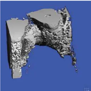
Bone morphogenetic proteins will have a place in craniofacial surgery in the future but the long-term complications need to be identi-fied and understood. We are moving away from alveolar bone grafts from the iliac crests to a combination of BMP2 in demineralized bone matrices. This bone substitute can eliminate the morbidity of harvesting bone from the iliac crest. In a study reported at IADR and ACPA this year (2011), we compared autogenous bone grafts and MP2/demineralized bone matrices. BMP2, as sold in the original collagen sponge, can be compressed in the cleft site and produce only limited amounts of volumes of bone. In order to maintain the space and volume, the BMP2 was placed inside a roll of demineralized bone. We studied the graft outcomes with the Kodak 3000 which has the highest resolution for a cone beam CT and a limited field of three teeth (Fig 2).
We found that neither type of graft completely filled the cleft site but BMP2 with demineral-ized bone matrices produced almost twice as much bone. Interestingly, both types of bone grafts can show 100% bone fill in the vertical and mesial-distal dimensions as seen in an oc-clusal radiograph but only 20-60% bone fill in the missing transverse dimension. We need to do better in the future.
In your opinion, what are the main indica-tions for skeletal anchorage in cleft palate
patients? Luciane Macedo de Menezes
I use microimplants differently than most orthodontists because I am not trying to elimi-nate the surgery. Most craniofacial patients will need surgery to improve their function and appearance. The microimplants are used to sup-port surgeries and limit surgical complications. Skeletal anchorage can help to protract a max-illa, widen a fused maxmax-illa, set up a wire system for osseous transport and provide anchorage in edentulous spaces.
A B C
A B
Do you think skeletal anchorage can reduce
the use of corticotomies? Carlos Alberto
Es-tevanell Tavares
I think corticotomies and skeletal anchorage can be used together. In terms of anchorage for tooth movement, corticotomies are a method for reducing resistance to tooth movement whereas microimplants (TAD) can increase resistance. Both methods can target specific teeth. The combination of techniques provides a way to alter the bone biology of tooth movement. This is an area of active research for us, as well as several other laboratories.
Basically, in which situations would you recom-mend:
- Orthodontic tooth movement associ-ated to corticotomy?
- Surgically assisted block displacement? Roberto Rocha
I am a little afraid of losing bone during a corticotomy procedure which is why, I believe,
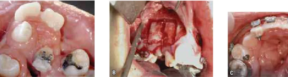
periodontists place a bone graft over the dental roots to hold the space for bone remineraliza-tion. Most of the time, I use osteotomy-assisted tooth movement for reshaping the arch form in craniofacial patients (Figs 3 and 4). I ligate the segments against the host bone for three days to ensure a good distraction callus before distracting the segments into position. Corticotomy-assisted tooth movement is used by some orthdontists to accelerate tooth movement. In the future, there may be less invasive ways to produce the bone response needed to accelerate tooth movement.
What are the main challenges in treating children with congenital malformations? Luciane Macedo de Menezes
I think the main challenges for the future are financial and educational.
One challenge is to make the medical and orthodontic care affordable to patients with congenital malformations through private and government medical insurance programs.
FIGURE 3 - Osteotomy-assisted tooth movement facilitates difficult tooth movements.
Another challenge is to help orthodontists to take care of patients with specialized needs through a post-residency fellowship such as the one we have in craniofacial orthodontics at Chil-dren’s Hospital Los Angeles.
Since the 50’s the treatment protocol of cleft lip and palate patients has evolved and several paradigms have changed. From your point of view what new boundaries are to be unfolded in orthodontics and surgery? Roberto Rocha
Certain innovations such as distraction osteo-genesis and bone morphogenetic proteins have provided new strategies for dealing with osseous deformities. However, as an orthodontist, one para-digm that has changed for me is my approach to the Class III malocclusion. I used to be afraid that any procedure that might worsen a Class III maloc-clusion would automatically lead to orthognathic surgery later in the life of the patient. Currently, I don’t worry about Class III malocclusions as much because we now use several maxillary protraction protocols during adolescence to achieve Class III correction even in large skeletal Class III cases. These protraction techniques are supported by alternating expansion and constriction to loosen the sutures, SARPE/LeFort I surgeries in cases of fused sutures and microimplants to limit side effects of treatment. The benefits for early interventions used to be weighed against the post-treatment effects on
maxillary growth. The timing of an alveolar bone graft is such an example. In the past five years, the calculation of risks and benefits for different procedures has changed for me because I have a way to deal with Class III malocclusions without orthognathic surgery. The goal in my research is to re-create the bone response for distraction os-teogenesis and rapid tooth movement in order to eliminate or limit the need for surgery. I welcome collaborations with colleagues in Brazil to help studying these new areas of research.
During the period that I had the opportuni-ty to accompany you at Children’s Hospital, the affection and dedication provided not only to the cleft children but also to their parents called my attention. I would like to know what was the most important lesson taken from your contact with these children and their parents?
Luciane Macedo de Menezes
1. S Yen. Protocols for late maxillary protraction in cleft lip and palate patients at Children’s Hospital Los Angeles. Semin Orthod. 2011;17(2):138-48.
2. Lypka M, Hammoudeh J, Yen S. Correcting vector problems with bilateral internal maxillary distractors by using rapid palatal expanders. J Craniofac Surg. 2011. In press. 3. Lee W, Karapetyan G, Moats R, Yamashita DD, Moon HB,
Ferguson DJ, et al. Osteotomy/corticotomy-assisted tooth movement microCts Differ. J Dent Res. 2008;87(9):861-7. 4. Wang L, Lee W, Lei DL, Liu YP, Yamashita DD, Yen S. Tissue
responses in osteotomy and corticotomy-assisted tooth movement in rats: histology and immunostaining. Am J Orthod Dentofacial Orthop. 2009;136(6):770.e1-11. 5. Yen S, Gross J, Wang P, Yamashita DD. Closure of a large
alveolar cleft by bony transport of a posterior segment using orthodontic archwires attached to bone. J Oral Maxillofac Surg. 2001;59(6):688-91.
6. Yen S, Yamashita DD, Kim TH, Baek SH, Yen S. Closure of an
unusually large anterior palatal istula by bony transport and
corticotomy-assisted tooth movement. J Oral Maxillofac Surg. 2001;61:1346.
7. Yen S, Yamashita DD, Gross J, Meara J, Yamazaki K, Kim TH, et al. Combining orthodontic tooth movement with distraction osteogenesis to close spaces and improve arch form in cleft lip and palate patients. Am J Orthod Dentofacial Orthop. 2005;127(2):224-32.
REFERENCES
Carlos Alberto Estevanell Tavares
- Dentistry Graduate, Federal University of Rio Grande do Sul State.
- MSc and PhD in Dentistry (Orthodontics), Rio de Janeiro Federal University.
- Professor of the Specialization Course in Orthodontics at the Brazilian Association of Dentistry of Rio Grande do Sul State.
- Diplomate of the Brazilian Board of Orthodontics and Facial Orthopedics (BBO).
Eduardo Franzotti Sant’Anna
- Dentistry Graduate, Federal Fluminense University. - MSc and PhD in Dentistry (Orthodontics), Rio de Janeiro
Federal University.
- Was a clinician and visiting professor at Rush Craniofacial Center, Chicago/USA.
- Adjunct Professor of Orthodontics at the School of Dentistry, Bahia State Federal University.
Roberto Rocha
- Dentistry Graduate, Federal University of Santa Catarina. - Residency in Preventive and Interceptive Orthodontics at
HPRLLP USP-Bauru.
- MSc and PhD in Dentistry (Orthodontics), Rio de Janeiro Federal University.
- Diplomate of the Brazilian Board of Orthodontics and Facial Orthopedics (BBO).
- Associate Professor at the Federal University of Santa Catarina.
Luciane Macedo de Menezes
- Dentistry Graduate, Federal University of Rio Grande do Sul State.
- MSc and PhD in Dentistry (Orthodontics), Rio de Janeiro Federal University.
- Professor of Orthodontics, School of Dentistry, Pontifical Catholic University of Rio Grande do Sul State.
- Coordinator of the Specialization Course in Orthodontics at the Brazilian Association of Dentistry, Rio Grande do Sul State.
8. Yen S, Gross J, Yamashita DD, Kim TH, McAndrew J, Shuler C, et al. Correcting an open bite side effect during distraction with spring forces. Plast Reconstr Surg. 2002;110(6):1476-84. 9. Yen S, Shang W, Shuler C, Yamashita DD. Orthodontic spring guidance in bilateral mandibular distraction in rabbits. Am J Orthod Dentofacial Orthop. 2001;120(4):435-42.
10. Yen S, Shang W, Shuler C, Yamashita DD. Bending the distraction site during mandibular distraction osteogenesis: a model for studying segment control and side effects. J Oral Maxillofac Surg. 2001;59(7):779-88.
11. Vachiramon A, Urata M, Kyung HM, Yamashita DD, Yen S. Clinical applications of microimplants in craniofacial patients. Cleft Palate Craniofac J. 2009;46(2):136-46. Epub 2008 Jun 3. 12. Lypka M, Afshar A, Pham D, Fortman K, Yamashita D, Yen S.
Implant-supported distraction osteogenesis: a technique to advance anterior maxilla. J Craniofac Surg. 2009;20(2):525-7. 13. Vachiramon A, Yen S, Lypka M, Bindignavale VJ, Hammoudeh
J, Reinisch J, et al. A novel model surgery method for LeFort III Advancement. J Craniofac Surg. 2007;18(5):1230-5.
14. Shang W, Scadeng M, Yamashita DD, Pollack H, Faridi O, Tran B, et al. Manipulating the mandibular distraction site at different stages of consolidation. J Oral Maxillofac Surg. 2007;65(5):840-6.
Contact address Stephen Yen E-mail: [email protected]
Submitted: June 10, 2011