Healing of the abdominal wall after parcial hepatectomy
Healing of the abdominal wall after parcial hepatectomy
Healing of the abdominal wall after parcial hepatectomy
Healing of the abdominal wall after parcial hepatectomy
Healing of the abdominal wall after parcial hepatectomy
Cicatrização da parede abdominal após hepatectomia parcial
Cicatrização da parede abdominal após hepatectomia parcial
Cicatrização da parede abdominal após hepatectomia parcial
Cicatrização da parede abdominal após hepatectomia parcial
Cicatrização da parede abdominal após hepatectomia parcial
MARIADE LOURDES PESSOLE BIONDO-SIMÕES – TCBC-PR1; FLÁVIA THAIANA BONATO2; ALINE MORAES MENACHO2; MARIANA DRECHMER2;
TEREZA CRISTINA SANTOS CAVALCANTI3 ; SAULO JOSÉ ALVES FELIZOLA4
A B S T R A C T A B S T R A C T A B S T R A C T A B S T R A C T A B S T R A C T
Objective Objective Objective Objective
Objective: To evaluate the wound healing of the abdominal wall incision in hepatectomized rats as for the concentration of collagen, inflammatory reaction and angiogenesis. MethodsMethodsMethodsMethods: We used 48 rats randomly assigned to laparotomy with orMethods without hepatectomy. The scars were studied in the 3rd, 7th and 14th postoperative days. We analyzed the density of collagen by the histochemical method and angiogenesis, by immunohistochemistry. ResultsResultsResultsResultsResults: The analysis showed a lower total collagen concentration in skin and subcutaneous tissue in the abdominal scars of the Experiment group (p3 = 0.011, p7 = 0.004 and p14 = 0.008). The density of collagen I was lower in the hepatectomy group, especially in the third day, in the skin, subcutaneous tissue (p = 0.038) and in the aponeurotic plane (p = 0.026). There was a lower concentration of collagen III in the two abdominal wall layers studied, although not statistically significant. The inflammatory response was similar at all times in both groups. It was found that angiogenesis was developed earlier in the Control group (p3 = 0.005 and p7 = 0.012) and later in the Experimental group (p14 = 0.048). ConclusionConclusionConclusionConclusionConclusion: Hepatectomy leads to a delay in the healing process, interfering with collagen
synthesis and angiogenesis.
Key words Key words Key words Key words
Key words: Liver. Regeneration. Hepatectomy. Wound healing.
Work conducted in the discipline of Surgical Technique and Experimental Surgery of the Faculty of Medicine of the Paraná Federal University – UFPR, Curitiba, Paraná – PR, Brazil.
1. Associate Professor, Department of Surgery, Faculty of Medicine, Paraná Federal University – UFPR, Curitiba, Paraná – PR, Brazil; 2. Graduates, Scientific Initiation Program, UFPR-PR-BR; 3. Assistant Professor, Pathological Anatomy Department, UFPR-PR-BR; 4. Resident, Pathological Anatomy Department, Erasto Gaertner Hospital – PR – BR.
INTRODUCTION
INTRODUCTION
INTRODUCTION
INTRODUCTION
INTRODUCTION
F
ailure in healing of the abdominal wall remains a problem for surgeons. Despite technological advances, problems such as dehiscence and incisional hernias conti-nue highly incident. In the United States 200,000 surgical repairs of incisional hernias are held each year1. The typeand magnitude of the operative act are among the known risk factors for suture dehiscence of abdominal wall. Emergency operations, perioperative periods of hemodynamic instability, procedures involving the biliary tree, liver disease and surgical treatment of aneurysm of the aorta are associated with increased incidence of failed of acute wound healing2.
Gómez et al.3 reported incisional hernias in
11.6% of patients undergoing liver transplantation, Müller et al.4 in 12%, Piazzese et al.5 in 4.9% and Vardanian et
al.6 in 4.6%. Similar situation is described for patients
undergoing partial hepatectomies. Rudow et al.7 reported
an incidence of 20%, while D’Angelica et al.8, 9.8%.
According to Van’t et al.9, most severe situations with
evisceration led 25% of patients to death within 60 days. Among those who survived, 69% of them have developed incisional hernias.
Living donor hepatic transplant, when the donor cedes up to 60% of his/her liver, has been increasingly stimulated. This is possible because the remaining liver can regenerate itself, although the term “hepatic regeneration” is not biologically appropriate, since there is no regeneration of resected lobes, but hyperplasia and hypertrophy of the remaining ones (compensatory growth). This term has been consecrated by the literature10,11.
All liver cells, hepatocytes, endothelial cells, Küpffer cells, Ito cells, and ductal cells, proliferate. However, as hepatocytes constitute 90% of the parenchyma and 60% of the total number of cells, most studies of regeneration monitor these cells10,11.
During the proliferation of hepatocytes there is release of growth factors, such as: hepatocyte growth factor (HGF), transformative growth factor alpha (TGF-á), epidermal growth factor (EGF) and fibroblast growth factor (FGF)11.
The EGF stimulates the synthesis of DNA in the majority of epithelial cells and in hepatocytes10,11. The levels
TGF-á is able to stimulate mitoses by autocrine and paracrine signalling mechanisms. Its potential effect on hepatocytes can be part of a mitogenic signal that directs the stroma of adjacent cells towards proliferation10. The
HGF was the first mitogenic factor identified in blood in high concentrations during regenerative process, being considered the most potent stimulator of liver proliferation12,13.
Transforming growth factor beta (TGF-â) is able to reversibly stimulate the growth of fibroblasts11. TGF â
1
and TGF â2 are important mediators of acute phase tissue repair, increasing wound resistance14,15. The presence of
TGF-â is important to start and sustain tissue healing16. HGF
has its effect fully inhibited by TGF-â10. This factor proved a
potent inhibitor of hepatocyte proliferation in vitro10. It was
demonstrated in vivo that there is increased expression of TGF-â after toxic injury of non-parenchymatous liver cells, i.e. Küpffer cells, stellate cells and endothelial cells, but there is no increase in hepatocytes. During liver regeneration, elevation of TGF-â levels does not occur until most part of the hepatocytes proliferation ends.
It was shown that, during liver regeneration, levels of TGF-â1 decrease and the expression of HGF increases, stimulating the proliferation of hepatocytes17. It
is interesting to note that the levels of TGF-â rise during the normal process of tissue healing.
Kuhn et al. verified, in a study done on rats, that there was a deficiency of abdominal scars resistance during liver regeneration. They found high levels of HGF and low TGF-â2. This finding led them to suggest that there may be prioritization of liver regeneration over abdominal wall scar fibroplasia18.
Considering the high incidence of complications of abdominal wound healing and changes of concentrations of growth factors, the study of the healing of abdominal wall after hepatectomies becomes important. The understanding of the causes that lead to suture dehiscence and failures in healing is necessary to conceive methods of prevention and correction of these complications.
This study aimed at examining the wound healing process of the abdominal wall of hepatectomized rats and comparing it with the wound of non-hepatectomized rats.
METHODS
METHODS
METHODS
METHODS
METHODS
The project that gave rise to this study was evaluated by the Committee of Ethics in Research with Animals of the Health Sciences Sector of the Federal University of Paraná and approved with protocol number A.N. 009.005.07.09.
We used 48 male rats (Rattus norvegicus albinus, Rodentia mammalia) aged between 100 and 120 days and weight of 250 ± 50 grams, from the Central Animal Facility of UFPR. We kept them in quarantine for a week before starting the study, and throughout the research in the
laboratory of Experimental Surgery and surgical technique of UFPR they were housed in groups of three to five animals per box suitable for the species. The temperature was 20± 2°C, light/dark cycle of 12 hours and relative humidity of the environment itself. They received ad libitum water and proper chow.
The rats were randomly assigned, 21 of them composing the Control Group (C) and 27 the Hepatectomized (or Experiment) Group (H). These groups were again randomly divided in the subgroups C3, C7 and C14 and H3, H7 and H14, according to the dates set for the evaluation of the experiment, three, seven and 14 days. The subgroups of the Control Group had seven rats and the ones of the Hepatectomized one, nine each.
After being weighed and marked, they were subjected to anesthesia by intramuscular injection of 0.2 ml/ 100 g body weight of a mixture of one milliliter of ketamine (50 mg) with a milliliter of xylazine (20 mg).
After depilation of the ventral abdominal wall a median laparotomy was performed with 4 cm in length, starting immediately below the xiphoid process. In the Group H we performed a partial hepatectomy by resecting the median lobe with its central portions, along with the left lateral lobe. This resection represents 67 to 70% of the hepatic mass19.
Once revised the hemostasis, we proceeded to closure with two plans of continuous-type suture with 5.0 monofilament nylon. The first plan encompassed the peritoneum, the muscle and aponeurosis, and the second, the skin.
After recovery from anesthesia, the mice were returned to their cages with free access to water and food. They received sodium diclofenac 10 mg/kg intramuscularly immediately after the operative act, with anti-inflammatory and analgesic purposes19. For euthanasia we made a lethal
dose of intraperitoneal sodium thiopental (120 mg/kg). After the animals’ death, the ventral portion of the abdominal wall, containing the scar in its central region, was withdrawn, leaving 2 cm laterally and 1 cm above and below it. We separated the skin of the peritoneum-musculoaponeurosis, extended both on filter paper and discarded half centimeter both at the top and bottom of the two flaps. The remainder was fractionated into three portions with 1 x 4 cm, constituting the fragments A, B and C (Figure 1).
Fractions A and C were fixed in 10% formalin and forwarded to histopathology. They were sliced in 4-ìm-thick cuts, which were mounted on slides and stained with hematoxylin-eosin (HE) and Picrosirius, as well as by the method of Immunohistochemistry with anti-CD 34 antibody.
the following scale: no cell = 0; up to 50 cells = 1; 50 to 100 cells = 2; and more than 100 cells = 3, positive for mononuclear and negative forpolymorphonuclear cells20.
After attribution of the values, they were summed, so that each group of animals had a final score, thus classifying groups in three phases of inflammatory process (Table 2)20.
Coloring by Picrosirius was used to recognize the density of collagen in the SCAR and the fractions of collagen I and III under the microscope with polarized light. The thicker and strongly birefringent fibers are colored in shades of orange to red (collagen I) and the smallest, dispersed and weakly birefringent fibers are stained in green (collagen III)21.
Angiogenesis was evaluated by immunohistochemical method; the number of vessels was counted in three large fields. CD 34 is a glycosylated protein unique to the membrane, which is expressed by immature blood cells and endothelial cells. The anti-CD 34 recognizes
the molecule CD 34 and thus enables the identification of cells that possess them.
For a comparison of Experiment and Control groups on each day of assessment, we used the non-parametric Mann-Whitney test. Comparison of the moments of evaluation within groups was made with the non-parametric test of Kruskal-Wallis. Statistical significance was indicated by p < 0.05.
RESULTS
RESULTS
RESULTS
RESULTS
RESULTS
Total collagen was present in less concentration in the abdominal scars of the Experiment group at all times in the skin and subcutaneous tissue (p3 = 0.011; p7 = 0.004 and p14 = 0.008). There was lower density of total collagen in the aponeurosis only on the 7th and 14th days (p
7 = 0,
017 and p14 = 0.022) (Figures 2 and 3).
The fraction of collagen I displayed lower concentration in the Experiment group in the skin and subcutaneous tissue in the 3rd and 14th days (p
3= 0.038 and
p14 = 0.002), and in the aponeurosis in the 3rd and 7th days
(p3= 0.026 and p7 = 0.017).
The fraction of collagen III was lower in the Experiment group three times, having been significant in 7th day in skin and subcutaneous tissue (p = 0.026). There
was no significant difference for the aponeurosis.
The inflammatory reaction on the 3rd day was
predominantly of acute type (p = 0.461) and on the 7th day
of chronic type in the Control group, while in the Experiment Group there were subacute and chronic-type reactions (p = 0.192). The same situation was observed on the 14th day
(p = 0.103).
Table 1 Table 1 Table 1 Table 1
Table 1 – Methodology of quantification of histological findings in cuts colored by hematoxylin-eosin.
Inflammatory Parameters Inflammatory Parameters Inflammatory Parameters Inflammatory Parameters
Inflammatory Parameters I n t e n s i t yI n t e n s i t yI n t e n s i t yI n t e n s i t yI n t e n s i t y H i g h
H i g h H i g h H i g h
H i g h M o d e r a t eM o d e r a t eM o d e r a t eM o d e r a t eM o d e r a t e D i s c r e e tD i s c r e e tD i s c r e e tD i s c r e e tD i s c r e e t A b s e n tA b s e n tA b s e n tA b s e n tA b s e n t
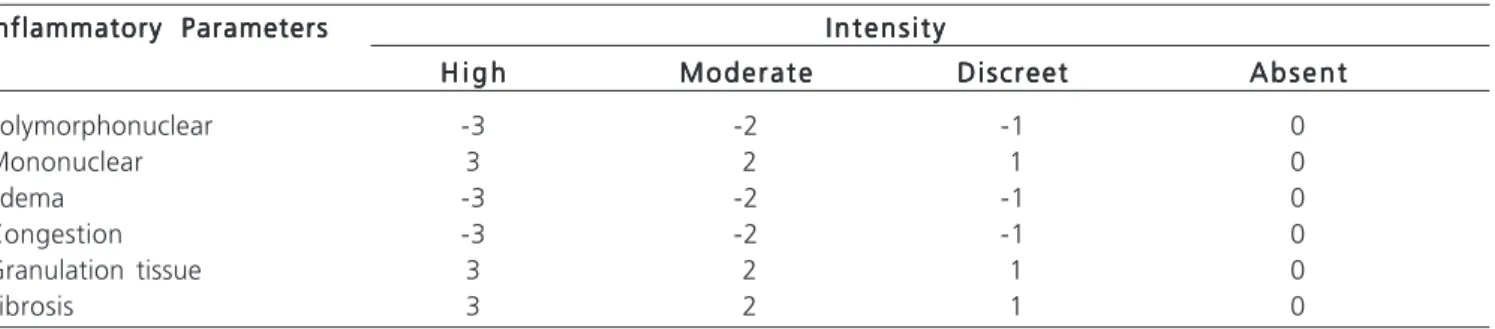
Polymorphonuclear -3 -2 -1 0
Mononuclear 3 2 1 0
Edema -3 -2 -1 0
Congestion -3 -2 -1 0
Granulation tissue 3 2 1 0
Fibrosis 3 2 1 0
Figure 1 Figure 1 Figure 1 Figure 1
Figure 1 – Scheme of the scar fragments used for the study.
Tabela 2 Tabela 2 Tabela 2 Tabela 2
Tabela 2 - Characterization of the phase of the inflammatory process according to the final score.
Final score Final score Final score Final score
Final score Phase of inflammatory processPhase of inflammatory processPhase of inflammatory processPhase of inflammatory processPhase of inflammatory process - 9 a - 3 Acute
- 2,9 a +3 Subacute
+ 3,1 a +9 Chronic
C
A
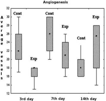
Analysis of angiogenesis demonstrated greater number of vessels in the scars of the Control group at days 3 (p = 0.011) and 7 (p = 0.038). On the 14th day we
observed a tendency to larger number of vessels in the Experiment group (p = 0.181) (Figure 4).
DISCUSSION
DISCUSSION
DISCUSSION
DISCUSSION
DISCUSSION
In spite of the progress of surgical techniques and better postoperative follow-up, literature information shows that the incidence of healing failures has not changed in recent years3,4,9. Works suggest that
post-hepatectomy scars are less resistant than for other surgical procedures4,7,18.
It was demonstrated that there is an increase in collagen synthesis in the remaining liver after partial hepatectomy until the 7th day, this difference no more
existing from the 14th day on. Furthermore, there is decrease
in collagen degradation, suggesting that this process favors the accumulation of this protein during early liver regeneration22.
The analysis of collagen made in the present study showed lower density of collagen in the skin scars of skin/subcutaneous tissue and Aponeurosis. One can also verify that this difference was due to lower density of collagen I, especially in the initial period, which leads to assume that there is delay in this protein synthesis by fibroblasts. One can argue that this lower density can lead to reduced scars’ resistance and be responsible for the larger number of dehiscences and incisional hernias observed in hepatectomized individuals3-9.
The inflammatory response of the Hepatectomized Group has proved to be prolonged, but not so significant. Prolonged inflammatory reaction could be a good rationale to explain the lower density of collagen. However, it was not possible to confirm this information in this study. Perhaps a larger sample can clarify this doubt.
The inflammatory process slows the progression of healing stages, possibly by changes in signaling held by growth factors. Literature data show that the level of
TGF-b2 responsible for the proliferation of fibroblasts and initiation and maintenance of scar response only increases after the proliferation of hepatocytes. Hyperplasia and hepatic mass recovery are prioritized over to healing18.
The hepatectomy and liver regeneration lead to delayed recovery of the abdominal wall incision, possibly by inhibiting the secretion of some cytokines (TGF-b2) over others (HGF), as well as different cell mobilization, demonstrated by increased inflammation12,18.
Figure 4 Figure 4Figure 4
Figure 4Figure 4 – Average number of vessels by field.
Figure 3 Figure 3Figure 3 Figure 3
Figure 3 – Percentage average of the areas examined
represented by total collagen at three, seven and 14 days in the Control and Experiment groups in the aponeurosis.
Figure 2 Figure 2Figure 2 Figure 2
It is not known if a process of healing a tissue interferes with other in the same body, but we know of the importance of cytokines in healing or repair process and that they are expressed in quantities and at different times in each tissue. Some authors suggest that simultaneous procedures lead to increased risk of failure and may hinder the recovery of the abdominal wall1,3,5.
Considering the high incidence of complications of abdominal wound healing and changes of concentrations of growth factors, we note the importance of studying the healing of abdominal wall after hepatectomies. Further studies with dosages of the main factors involved are necessary to better understanding
and possibly reversing the healing delay caused by liver regeneration.
It was observed on the 3rd and 7th days that both
type I collagen and angiogenesis were increased in the Control group. This fact may be explained by the increased concentration of growth factors and oxygen in the site, stimulating fibroblasts.
It would be interesting to test the strength of scars, but for technical reasons this was not held at this time, being the object of subsequent studies.
The data analyzed in this study suggest that hepatectomy leads to delayed healing process, interfering with the synthesis of collagen and angiogenesis.
R E S U M O R E S U M O R E S U M O R E S U M O R E S U M O
Objetivo: Objetivo: Objetivo: Objetivo:
Objetivo: Avaliar a cicatrização da ferida incisional da parede abdominal de ratos hepatectomizados quanto à concentração de colágeno, reação inflamatória e angiogênese. Métodos:Métodos:Métodos:Métodos: Utilizaram-se 48 ratos distribuídos aleatoriamente para laparotomia comMétodos: e sem hepatectomia. As cicatrizes foram estudadas no 3°, 7° e 14° dia de pós-operatório. Analisou-se a densidade do colágeno por método histoquímico e a angiogênese por método imunohistoquímico. Resultados:Resultados:Resultados:Resultados:Resultados: A análise do colágeno total mostrou menor concentração no plano da pele e da tela subcutânea, nas cicatrizes abdominais do grupo experimento (p3=0,011; p7=0,004 e p14=0,008). A densidade de colágeno I foi inferior no grupo hepatectomizado, principalmente no 3° dia, tanto na pele e tela subcutânea (p=0,038) quanto no plano aponeurótico (p=0,026). Houve menor concentração de colágeno III nos dois planos estuda-dos, embora não significante. A resposta inflamatória foi semelhante em todos os tempos, nos dois grupos. Verificou-se que a angiogênese desenvolveu-se mais precocemente no grupo controle (p3=0,005 e p7=0,012) e mais tardiamente no grupo experimen-to (p14=0,048). Conclusão:Conclusão:Conclusão:Conclusão:Conclusão: A hepatectomia leva ao atraso do processo cicatricial, interferindo na síntese do colágeno e na angiogênese.
Descritores: Descritores: Descritores: Descritores:
Descritores: Fígado. Regeneração. Hepatectomia. Cicatrização de feridas.
REFERENCES
REFERENCES
REFERENCES
REFERENCES
REFERENCES
1. Luijendijk RW, Hop WC, van den Tol MP, de Lange DC, Braaksma MM, Ijzermans JN, Boelhouwer RU, de Vries BC, Salu MK, Wereldsma JC, Bruijninckx CM, Jeekel J. A comparision of suture repair with mesh repair for incisional hernia. N Engl J Med 2000; 343(6):392-8.
2. Carlson MA. Acute wound failure. Surg Clin North Am 1997; 77(3):607-36.
3. Gómez R, Hidalgo M, Marques E, Marin L, Loinaz C, Gonzalez I, Garcia I, Moreno E. Incidence and predisposing factors for incisional hernia in patients with liver transplantation. Hernia 2001; 5(4):172-6.
4. Müller V, Lehner M, Klein P, Hohenberger W, Ott R. Incisional hernia repair after orthotopic liver transplantation: a technique employing an inlay/onlay polypropylene mesh. Langenbecks Arch Surg 2003; 388(3):167-73.
5. Piazzese E, Montalti R, Beltempo P, Bertelli R, Puviani L, Pacilè V, Nardo B, Cavallari A. Incidence, predisposing factors, and results of surgical treatment of incisional hernia after orthotopic liver transplantation. Transplant Proc 2004; 36(10):3097-8.
6. Vardanian AJ, Farmer DG, Ghobrial RM, Busuttil RW, Hiatt JR. Incisional hernia after liver transplantation. J Am Coll Surg 2006; 203(4):421-5.
7. Rudow DL, Brown RS Jr, Emond JC, Marratta D, Bellemare S, Kinkhabwala M. One-year morbidity after donor right hepatectomy. Liver Transpl 2004; 10(11):1428-31.
8. D’Angelica M, Maddineni S, Fong Y, Martin RC, Cohen MS, Ben-Porat L, Gonen M, DeMatteo RP, Blumgart LH, Jarnagin WR. Optimal abdominal incision for partial hepatectomy: increased late
complications with Mercedes-type incisions compared to extended right subcostal incisions. World J Surg 2006; 30(3):410-8. 9. van’t RM, De Vos Van Steenwijk PJ, Bonjer HJ, Steyerberg EW,
Jeekel J. Incisional hernia after repair of wound dehiscense: incidence and risk factors. Am Surg 2004; 70(4):281-6.
10. Ramalho FS, Ramalho LNZ, Zucoloto S, Silva Jr OC. Regeneração hepática: algumas definições num universo de incertezas. Acta Cir Bras 1993; 8(4):177-89.
11. Michalopoulos GK, DeFrances MC. Liver regeneration. Science 1997; 276(5309):60-6.
12. Liu ML, Mars WM, Zarnegar R, Michalopoulos GK. Uptake and distribution of hepatocyte growth factor in normal and regenerating adult rat liver. Am J Pathol 1994; 144(1):129-40. 13. Goupil D, Ethier C, Zarnegar R, Gascon-Barré M. Hepatic expression
of regeneration marker genes following partial hepatectomy in the rat. Influence of 1,25-dihydroxyvitamin D3 in hypocalcemia. J Hepatol 1997; 26(3):659-68.
14. Wright T, Hill D, Polo M, Soler P, Pratt B, Nichols E. The modulation of acute incisional wound healing with rTGF-â2 and fibrin sealant [abstract]. Wound Repair Regen 1997; 5:A128.
15. Polo M, Smith PD, Kim YJ, Wang X, Ko F, Robson MC. Effect of TGF-beta2 on proliferative scar fibroblast cell kinetics. Ann Plast Surg 1999; 43(2):185-90.
16. Bennett NT, Schultz GS. Growth factors and wound healing: biochemical properties of growth factors and their receptors. Am J Surg 1993; 165(6):728-37.
17. Steer CJ. Liver regeneration. FASEB J 1995; 9(14):1396-400. 18. Kuhn MA, Smith PD, Wachtel TL, Wright TE, Rogazewski A, Nguyen
19. Higgins GM, Anderson RM. Experimental pathology of the liver: Restoration of the liver of the white rats following partial surgical removal. Arch Pathol 1931; 12:186-202.
20. Vizzotto Jr AO, Noronha L, Scheffel DLH, Campos ACI. Influência da cisplatina administrada no pré e no pós-operatório sobre a cicatrização de anastomoses colônicas em ratos. J Bras Patol Med Lab 2003; 39(2):143-9.
21. Junqueira LC, Cossermelli W, Brentani R. Differential staining of collagen type I, II and III by Sirius Red and polarization microscopy. Arch Histol Jpn 1978; 41(3):267-74.
22. Yamamoto H, Murawaki Y, Kawasaki H. Hepatic collagen synthesis and degradation during liver regeneration after partial hepatectomy. Hepatology 1995; 21(1):155-61.
Received on: 19/03/2010
Accepted for publication: 14/05/2010 Conflict of interest: none
Funding source: none
How to cite this article: How to cite this article:How to cite this article: How to cite this article:How to cite this article:
Biondo-Simões MLP, Bonato FT, Menacho AM, Drechmer M, Cavalcanti TCS, Felizola SJA. Cicatrização da parede abdominal após hepatectomia parcial. Rev Col Bras Cir. [periódico na Internet] 2011; 38(2). Disponível em URL: http://www.scielo.br/rcbc
Mailing address: Mailing address:Mailing address: Mailing address:Mailing address: