Major Article
INTRODUCTION
Corresponding author: Prof. Luiz Carlos Rodrigues Junior. Laboratório de Imunologia/UFCSPA. Av. Sarmento Leite 245, 90050-170 Porto Alegre, Rio Grande do Sul, Brasil.
Phone: 55 51 9153-6749; Fax: 55 51 3303-9000 e-mail: [email protected]
Received 27 May 2015 Accepted 21 July 2015
Comparative study of lymphocytes from individuals
that were vaccinated and unvaccinated against the pandemic
2009-2011 H1N1 infl uenza virus in Southern Brazil
Deise Nascimento de Freitas
[1], Henrique Ataíde Isaía
[1], Andréia Henzel
[2], Eder Simão
[1],
Rodrigo Benedetti Gassen
[1]and Luiz Carlos Rodrigues Junior
[1],[3][1]. Laboratório de Biologia Molecular e Cultivo Celular, Centro Universitário Franciscano, Santa Maria, Rio Grande do Sul, Brazil. [2]. Laboratório de Microbiologia Molecular, Instituto de Ciências da Saúde, Universidade Feevale, Novo Hamburgo, Rio Grande do Sul, Brazil. [3]. Laboratório de Imunologia, Universidade Federal de Ciências da Saúde de Porto Alegre, Porto Alegre, Rio Grande do Sul, Brazil.
ABSTRACT
Introduction: While no single factor is suffi cient to guarantee the success of infl uenza vaccine programs, knowledge of the levels
of immunity in local populations is critical. Here, we analyzed infl uenza immunity in a population from Southern Brazil, a region with weather conditions that are distinct from those in the rest of country, where infl uenza infections are endemic, and where greater than 50% of the population is vaccinated annually. Methods: Peripheral blood mononuclear cells were isolated from 40
individuals. Of these, 20 had received the H1N1 vaccine, while the remaining 20 were unvaccinated against the disease. Cells were stimulated in vitro with the trivalent post-pandemic infl uenza vaccine or with conserved major histocompatibility complex I (MHC I) peptides derived from hemagglutinin and neuraminidase. Cell viability was then analyzed by [3-(4,5-dimethylthiazol-2-yl)-2,5- diphenyltetrazolium bromide)]-based colorimetric assay (MTT), and culture supernatants were assayed for helper T type 1 (Th1) and Th2-specifi c cytokine levels. Results: Peripheral blood lymphocytes from vaccinated, but not unvaccinated,
individuals exhibited signifi cant proliferation in vitro in the presence of a cognate infl uenza antigen. After culturing with vaccine antigens, cells from vaccinated individuals produced similar levels of interleukin (IL)-10 and interferon (IFN)-γ, while those from unvaccinated individuals produced higher levels of IFN-γ than of IL-10. Conclusions: Our data indicate that peripheral blood
lymphocytes from vaccinated individuals are stimulated upon encountering a cognate antigen, but did not support the hypothesis that cross-reactive responses related to previous infections can ameliorate the immune response. Moreover, monitoring IL-10 production in vaccinated individuals could comprise a valuable tool for predicting disease evolution.
Keywords: Infl uenza virus. Vaccine. Peripheral blood lymphocytes. Tropical area.
While the success of a vaccination program for infl uenza is determined, at least in part, by the immunogenicity of the circulating strain, it also depends on the level of local immunity, particularly in areas where respiratory infections are historically prevalent. During epidemics of swine origin infl uenza A (swH1N1), states in Southern Brazil exhibited outbreak characteristics that did not correlate with those of the rest of the country. Throughout the winter, the weather in these states is cold and humid, and there is a high incidence of infl uenza infections. As such, many people are vaccinated
each year(1). However, despite the administration of a high
number of vaccine doses for infl uenza H1N1, cases of severe swH1N1 infections were more prevalent in these states than in the northern and Northeastern states(2).
Enumeration of the serum titers of antibodies specifi c to the viral glycoproteins hemagglutinin (HA) and neuraminidase (NA) is a parameter frequently used to evaluate the immune response to infl uenza(3) (4) (5). Notably, antibodies generated
against these glycoproteins in response to a particular strain of infl uenza do not always guarantee protection to other closely related strains(6) (7). Meanwhile, seroepidemiological studies
indicate that there is a lack of widespread pre-existing antibodies against swH1N1 in the general population(8) (9) (10). In addition to
antibodies, T cells are also involved in the response to infl uenza viruses(3) (11). Several studies have demonstrated that infections
by, or vaccination to, specifi c infl uenza strains can induce the production of cross-reactive T cells that can potentially recognize conserved epitopes in other strains(12). Recent studies,
however, claim that this cross-reactivity does not guarantee an effi cient cross-response(11). Furthermore, T cells that were
METHODS
compared to those of naïve cells, upon antigen stimulation(13).
Indeed, in a study of Brazilian school children, frequent exposure to seasonal infl uenza virus strains was not a relevant factor for preventing infection by swH1N1 in 2010(14). These
fi ndings demonstrate that the relationship between previous exposures to closely related strains of the infl uenza virus and protection during an epidemic is not applicable to all situations. To examine the effects of previous exposures to infl uenza antigens on the immune response to H1N1, we analyzed the response of lymphocytes harvested from the peripheral blood of individuals in Southern Brazil that were either vaccinated or unvaccinated against H1N1 to the post-pandemic infl uenza trivalent vaccine (Fluvac). Additionally, we determined the specifi city of the stimulation using infl uenza H1N1 major histocompatibility class I (MHC class I) conserved epitopes. These analyses were complemented by quantifi cation of the levels of interleukin 10 (IL-10) and interferon (IFN)-γ produced by cells after culturing in the presence of infl uenza antigens. The results of this study enhance our knowledge of the lymphocyte response to infl uenza in people from an endemic region, and may facilitate the development of improved protocols for infl uenza immunization and therapies.
Study population
In total, 40 healthcare professionals between the ages of 20 and 36 years were recruited for this cross-sectional study. Of these, 20 received the infl uenza vaccine during or after the 2009 H1N1 infl uenza pandemic, while the remaining 20 had never received the vaccine. Each subject completed a written inquiry regarding their health and vaccination history, and all subjects had a prior history of respiratory infection. Patients suffering from the fl u or a cold, and those exhibiting a cough or clinical signs of allergic disorders were excluded from the study.
Antigen stimulation
Fluvac vaccine formulated using the A/California/7/2009, A/Perth/16/2009, and B/Brisbane/60/2008 infl uenza strains was obtained from the Multivacin Immunization Clinic (Santa Maria, RS, Brazil). The dose used for stimulation was determined via a dose-response experiment using concentrations of 0.125, 0.25, 0.03, and 0.06µg/well. Moreover, cells were stimulated with HA and NA peptides corresponding to the amino acid sequences present in infl uenza H1N1/California/4/2009, as characterized using immunoinformatics tools(15), which bind multiples alleles
of the human leukocyte antigen (HLA) protein of humans
(Table 1). Synthetic peptides (Genemed Synthesis, Inc., San
Antonio, TX, USA) were diluted in water and then in Roswell Park Memorial Institute(RPMI) 1640 medium (Gibco, Grand Island, NY, USA). The peptide dose used for stimulation was optimized via a dose-response curve experiment using 2.5, 5.0, 10, 20, and 25µM concentrations of each of the following epitopes: HA1, LSSVSSFER; HA2, GMVDGWYGY; HA3, MESVKNGTY; NA1, VSFNQNLEY; and NA2, RPWVSFNQN.
Isolation of peripheral blood mononuclear cells
Eight milliliters of peripheral blood was collected from each volunteer by venipuncture, and samples were stored in ethylenediaminetetraacetic acid (EDTA) tubes (Vacuette®, Campinas, SP, Brazil). Peripheral blood mononuclear cells (PBMCs) were then isolated by centrifugation over a 7mL Ficoll-Hypaque (Sigma-Aldrich, St. Louis, MO, USA) gradient (900 × g, 30 min). After washing with RPMI, cells were viewed microscopically (40×), and viability was determined by trypan blue (Sigma, St Louis, MO, USA) exclusion assay analyses. PBMCs were then resuspended in complete culture medium [RPMI-1640, supplemented with 0.55g/L gentamicin, 1% glutamine, and 10% fetal bovine serum (FBS)] and adjusted to a concentration of 2 × 10⁵ cells/mL with RPMI.
In vitro stimulation of PBMCs
Peripheral blood mononuclear cells were stimulated in vitro with Fluvac or with HA and NA epitopes. Briefl y, 2 × 10⁵ cells
were cultured in flat-bottomed 96-well microplates [Techno Plastic Products (TPP) AG, Trasadingen, Switzerland] with RPMI 1640 supplemented with 10% FBS for 96 h at 37ºC in an atmosphere containing 5% CO2. A 1.5% concentration of the selective T cell mitogen phytohemagglutinin (PHA; Gibco) was used as a positive control, while medium alone was used as a negative control. To assess in vitro stimulation, the vaccine was diluted in RPMI and added to the cultures at a fi nal concentration of 0.125µg/well. Meanwhile, the HA and NA peptides were each added at fi nal concentrations of 5μM, as previously optimized, and the cultures were incubated at 37ºC with 5% CO2 for 120h.
For certain experiments, the cultures were also supplemented with 10 international units (IU)/well of human recombinant interleukin (IL)-2 (BD Biosciences®, Franklin Lakes, NJ, USA). After antigen
stimulation, lymphocyte viability was estimated using an MTT [3-(4,5-dimethylthiazol-2-yl)-2,5- diphenyltetrazolium bromide)]-based colorimetric assay(16) (Invitrogen, Carlsbad, CA, USA). The
optical density at 560nm (OD560) of the resulting reactions was measured using a plate reader (Thermoplate®, RS, Brazil).
Analysis of cytokine production
The concentrations of interferon (IFN)-γ, IL-10, IL-6, IL-4, and IL-2 within the culture supernatants harvested from stimulated cells were measured using a Cytometric Bead Array (CBA) Th1/ Th2 kit (BD Biosciences®). Because the cells from each of the 40
individuals were subjected to 10 different stimuli, we chose to pool the supernatants from those exposed to the same stimulus.
Statistical analyses
RESULTS
TABLE 1 - Conserved hemagglutinin and neuraminidase peptides that bind to multiple human leukocyte antigen class I alleles.
HLA class I Allele A*0101 A*0301 A*2402 B*0702 B*4403
HA LSSVSSFER LSSVSSFER --- --- GMVDGWYGY
conserved GMVDGWYGY GMVDGWYGY MESVKNGTY
epitopes MESVKNGTY
NA VSFNQNLEY VSFNQNLEY RPWVFNQN RPWVFNQN
---conserved
epitopes
HLA: human leukocyte antigen; HA: hemagglutinin; NA: neuraminidase.
Ethical considerations
The study protocol was approved by the Ethics Committee from Universidade Federal de Santa Maria, Rio Grande do Sul/ Brazil, and experiments were performed in accordance with the ethical standards of the responsible committee on human experimentation (institutional, regional, or national), and in keeping with the Helsinki Declaration of 1964, as revised in 1975, 1983, 1989, 1996, and 2000. All subjects agreed to participate, and each participant provided informed written consent. The protocol approval number is 23081.011864/2010-8.
Analysis of the response of circulating lymphocytes from vaccinated and unvaccinated individuals to Fluvac antigens
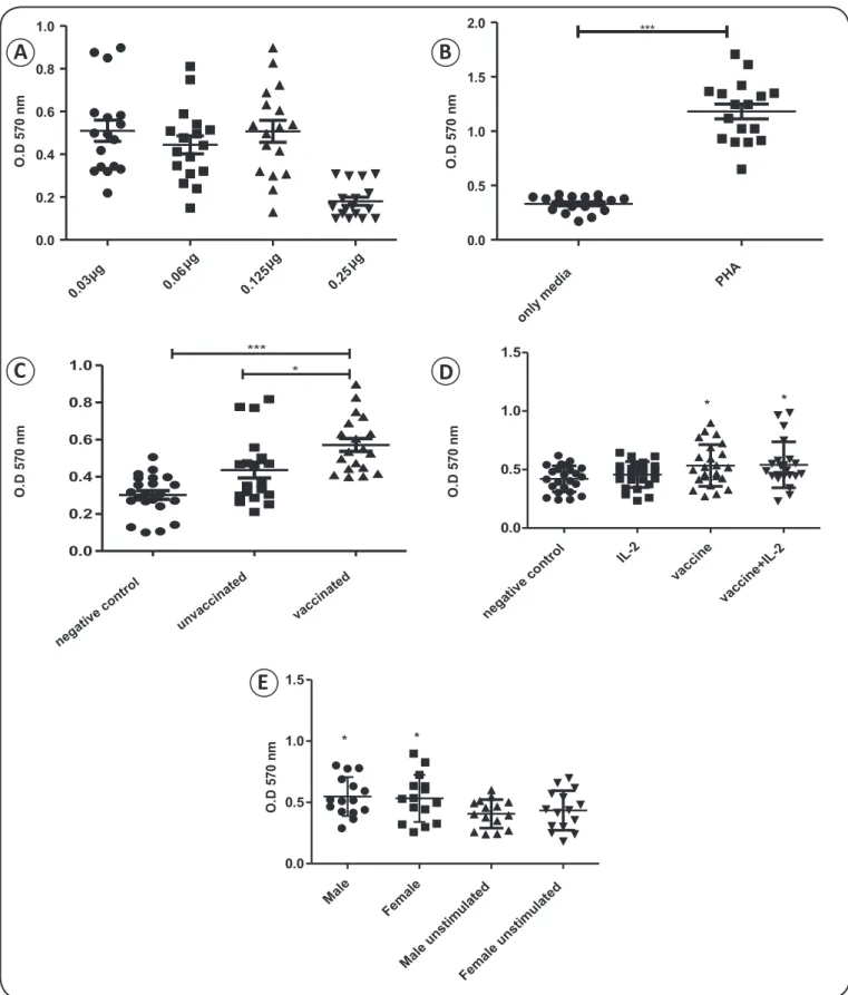
Peripheral blood mononuclear cells from fi fteen vaccinated individuals were isolated and stimulated, as described in the Material and Methods. Because a dose-dependent increase in cell viability was not observed within a range of 0.06µg to 0.125µg (Figure 1A), a dosage of 0.125µg was used for all
subsequent experiments. Meanwhile, the absorbance value obtained for the PHA-treated cells in the MTT assay was three-fold higher than that for the unstimulated cells, indicating that this compound comprised a valid positive control (Figure 1B).
Next, we examined the in vitro response of PBMCs from vaccinated and unvaccinated populations to stimulation with the Fluvac vaccine. As expected, cells from the vaccinated group exhibited signifi cantly higher viability than unstimulated cells (negative control) upon treatment with Fluvac (0.562± 0.032 vs. 0.363 ± 0.017, respectively; p = 0.0019; Figure 1C). While the viability of the cells from the unvaccinated group (0.471 ± 0.029) was also higher than that of the negative control cells (0.363 ± 0.017), this difference was not statistically signifi cant. Antigen-experienced lymphocytes produced during previous infections typically increase the expression of IL-2 receptor upon re-stimulation. To examine whether cells produced in response to vaccination contribute to in vitro stimulation, we evaluated the response of cells from the vaccinated group to stimulation with Fluvac and IL-2. No signifi cant change in viability was observed in cells from the vaccinated group after treatment with IL-2, compared with that observed in cells stimulated with Fluvac alone (Figure 1D). Similarly, while PBMCs from
the unvaccinated group treated with IL-2 exhibited higher levels of stimulation than the negative control cells, this difference was not signifi cant (data not shown). Lastly, there was no difference in the level of lymphocyte stimulation obtained by exposure to Fluvac between the males and females of the vaccinated population (Figure 1E).
Together, these results indicate that, in the population studied, only those individuals that were vaccinated with the monovalent infl uenza post-pandemic vaccine contained blood circulating lymphocytes that could be promptly stimulated with antigens from the post-pandemic fl u vaccine.
Quantifi cation of IFN-γ and IL-10 production
by lymphocytes from the vaccinated and unvaccinated populations
Evaluation of lymphocyte viability alone is not a suitable method for predicting the lymphocyte response. Therefore, to complement the MTT assay results, we evaluated the cytokine profi le induced by in vitro stimulation of PBMCs from vaccinated and unvaccinated individuals. Specifi cally, we measured the levels of helper T type 1 (Th1) [interferon gamma (IFN)-γ; interleukin-2 (IL-2); interleukin-6 (IL-6) and tumor necrosis factor (TNF)-α] and helper T type 2 (Th2) (IL-4, IL-5 and IL-10) specifi c cytokines in the supernatants from stimulated cells. There were no differences in the levels of IL-2, IL-6, TNF-α, IL-4, or IL-5 between the stimulated and negative control cells from either the vaccinated or the unvaccinated group (data not shown). Conversely, there was a dose-dependent increase in the level of IFN-γ secretion from the Fluvac-stimulated cells of the vaccinated group, compared to that observed in cells treated with media alone (negative control; Figure 2A). Similar results were obtained using the
cells of the unvaccinated group. Surprisingly, however, there were higher levels of IFN-γ in the supernatants harvested from the unvaccinated group than in those harvested from the vaccinated group after stimulation with 0.03 and 0.06µg Fluvac.
As observed for IFN-γ, cells from both the vaccinated and unvaccinated groups produced higher amounts of IL-10 in vitro than did the negative control cells after stimulation with Fluvac
(Figure 2B). Notably, while similar levels of IFN-γ and IL-10
were produced by the cells from the vaccinated group at each dose tested (Figure 2C), the cells in the unvaccinated group
0.03μg
μg 0.0
6 μg
0.12
5 μg
0.2 5
0.0 0.2 0.4 0.6 0.8 1.0
O.
D
5
7
0
n
m
O.
D
5
7
0
n
m
O.
D
5
7
0
n
m
only
media P
HA 0.0
0.5 1.0 1.5
2.0 ***
O.
D
5
7
0
n
m
0.0 0.2 0.4 0.6 0.8
1.0 *** *
nega tive c
ontr
ol IL-2
vacc ine
vaccin e+IL-2 0.0
0.5 1.0 1.5
* *
Ma le
Femal e
Ma le u
ns tim
ula ted
Female unsti mulated 0.0
0.5 1.0 1.5
* *
O.
D
5
7
0
n
m
FIGURE 1 - Stimulation of PBMCs from unvaccinated or vaccinated individuals with Fluvac antigens in vitro. A: Dose response analysis (0.03g to 0.25µg). B: Comparison of the viability of cells treated with the positive control (PHA) with that of unstimulated cells (media only). C: Viability of PBMCs stimulated with Fluvac (0.125µg). D: Viability of PBMCs from the vaccinated group after stimulation with IL-2 and/or Fluvac. E: Viability of unstimulated PBMCs and of Fluvac-stimulated PBMCs harvested from vaccinated males or females. Results were analyzed by one-way ANOVA with Dunnett’s and Tukey’s post hoc multiple comparison tests. OD: optic density; PBMCs: peripheral blood mononuclear cells; PHA: phytohemagglutinin; IL-2: interleukin-2; ANOVA: analysis of variance. *p < 0.01. **p < 0.05. ***p < 0.001.
A
B
C
D
E
negative contr
ol
unva ccin
ated
vacc inate
IL-10
0 500 1,000 1,500 2,000 2,500
unvaccinated vaccinated
p
g
/m
L
Vaccinated
nega
tive
control g
0.03
g
0.06
g
0.125
g
0.25 0
1,000 2,000 3,000
IL-10
IFN-p
g
/m
L
unvaccinated
neg ative
cont
rol g
0.03 g
0.06 g
0.125 g
0.25 0
500 1,000 1,500 2,000 2,500
IL-10
IFN-p
g
/m
L
0 1,000 2,000 3,000
pg/
mL
unvaccinated vaccinated
IF
N-A
B
C
D
FIGURE 2 - Quantifi cation of the levels of IFN-γ and IL-10 in supernatants from PBMCs stimulated with Fluvac or with HA or NA peptides. A: Quantifi cation of IFN-γ in the supernatants harvested from the vaccinated and unvaccinated populations after stimulation with whole vaccine (samples were pooled). B: Quantifi cation of IL-10 in the supernatants harvested from the vaccinated and unvaccinated groups after stimulation with Fluvac (samples were pooled). C: and D: Quantifi cation of IFN-γ and IL-10 in the supernatants harvested from the (C) unvaccinated and (D) vaccinated groups after stimulation with Fluvac (samples were pooled). OD: optic density, IFN-γ: interferon gamma; IL-10: interleukin; PBMCs: peripheral blood mononuclear cells; HA: hemagglutinin; NA: neuraminidase.
(Figure 2D). These data indicate that stimulation of PBMCs
from vaccinated individuals with Fluvac antigens results in a balance in the production of IL-10 and IFN-γ, which is not achieved by unprimed (unvaccinated) cells.
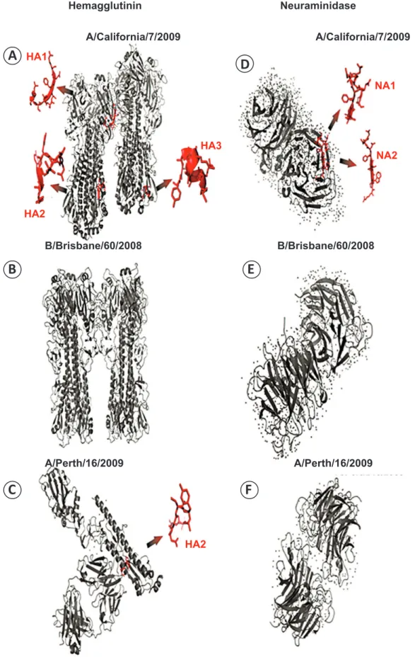
Determination of the specifi city of the immune response of
lymphocytes from the vaccinated group
The trivalent vaccine used for the stimulation studies was composed of antigens from two strains of infl uenza A (A/California/7/2009 and A/Perth/16/2009) and one strain of infl uenza B (B/Brisbane/60/2008); however, the individuals in the vaccination group were vaccinated with the monovalent vaccine, which contains only antigens from swH1N1 (A/California). To determine whether the A/California/4/2009
peptides, which were previously characterized as ligands for multiple HLA alleles, were present in the Fluvac vaccine, we analyzed the crystal structures of both HA and NA. The HA1, HA2, HA3, NA1, and NA2 peptides from A/California/4/2009 were present in A/California/7/2009 (trivalent vaccine). Furthermore, while the HA2 peptide was conserved in A/ Perth/16/2009 and A/California/7/2009, this region was
structurally different from that in A/California/4/2009.
Meanwhile, there was no peptide similarity among other infl uenza strains (Figure 3).
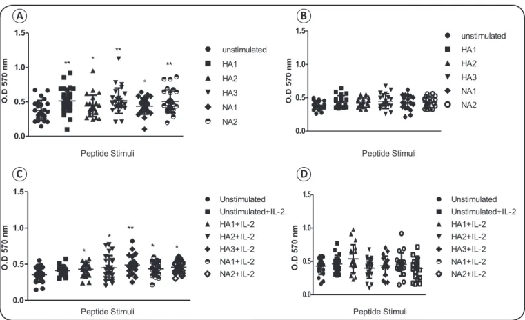
To evaluate whether cells from the vaccinated and unvaccinated groups were capable of responding to peptides from swH1N1, cells were stimulated with the HA1, HA2, HA3, NA1, and NA2 peptides in vitro. The concentration
nega tive
contr ol
0.03
μg 0.06
μg 0.12
5μg 0.25
μg
nega tive
contr ol
0.03
μg 0.06
μg 0.12
5μg 0.25
FIGURE 3 - Crystal structure images of HA and NA indicating the location of the peptides used in this study. A: HA1, HA2, and HA3 peptides from infl uenza A/California/7/2008, and B: B/Brisbane/60/2008. C: HA2 peptide from infl uenza A/Perth/16/2009. D: NA1 and NA2 peptides from infl uenza A/California/7/2008, E: B/Brisbane/60/2008, and F: A/Perth/16/2009. The structures were built using sequences extracted from the PDB database and the PyMOL molecular graphics system. HA: hemagglutinin; NA: neuraminidase; PDB: protein data bank.
Hemagglutinin Neuraminidase
A/California/7/2009 A/California/7/2009
B/Brisbane/60/2008 B/Brisbane/60/2008
A/Perth/16/2009 A/Perth/16/2009
A
B
C
D
E
F
HA1HA2
HA3
NA1
NA2
of HA and NA peptides used for stimulation was optimized
by fi rst examining the effects of a range of concentrations (2.5μM to 40μM) on cell viability. As no signifi cant differences were observed among the doses tested (data not shown), a dose of 2.5μM was used for each peptide in all subsequent experiments. Cells from the vaccinated group exhibited higher levels of viability after treatment with each of the HA or NA peptides than with media alone (Figure 4A). In contrast, neither the HA nor the NA peptides enhanced the viability of cells in the unvaccinated group compared to unstimulated cells (Figure 4B).
The infl uence of IL-2 on the stimulation of lymphocytes was also tested for each epitope. While cells from the vaccinated group were significantly stimulated by both the HA and NA peptides in the presence of IL-2 (Figure 4C), there was
no improvement in the stimulation index of cells from the unvaccinated group after stimulation with HA and NA in the presence of IL-2 (Figure 4D). These results indicate that the
stimulatory response observed in PBMCs from the vaccinated group was due to the exposure of these individuals to swH1N1 cognate antigens via vaccination, and not to exposure to antigens from other infl uenza viruses.
FIGURE 4 - Stimulation of PBMCs from unvaccinated or vaccinated individuals with HA and NA epitopes. A and B: PBMCs
from the vaccinated (A) and unvaccinated (B) groups were stimulated with a 5μM concentration of the HA1, HA2, HA3, NA1, or NA2 infl uenza A conserved peptides. C and D: PBMCs from the (C) unvaccinated and (D) vaccinated groups were stimulated with a 5μM concentration of HA1, HA2, HA3, NA1, or NA2 infl uenza A conserved peptides, as well as IL-2. Results were analyzed by one-way ANOVA with Dunnett’s post hoc multiple comparison tests. OD: optic density; PBMCs: peripheral blood
mononuclear cells; HA: hemagglutinin; NA: neuraminidase; ANOVA: analysis of variance. *p < 0.01, **p < 0.05. ***p < 0.001.
A
B
C
D
0.0 0.5 1.0 1.5
*
*
**
**
**
unstimulated
HA1
HA2
HA3
NA1
NA2
Peptide Stimuli
Peptide Stimuli Peptide Stimuli
Peptide Stimuli
0.0 0.5 1.0 1.5
unstimulated
HA1
HA2
HA3
NA1
NA2
O
.D
5
7
0
n
m
O.
D
5
7
0
n
m
0.0 0.5 1.0 1.5
*
**
* *
*
Unstimulated Unstimulated
Unstimulated+IL-2 Unstimulated+IL-2
HA1+IL-2 HA1+IL-2
HA2+IL-2 HA2+IL-2
HA3+IL-2 HA3+IL-2
NA1+IL-2 NA1+IL-2
NA2+IL-2 NA2+IL-2
O.
D
5
7
0
n
m
O.
D
5
7
0
n
m
0.0 0.5 1.0 1.5
DISCUSSION
Despite numerous efforts to develop an effective vaccine for combating influenza infections, the disease remains a serious health problem, causing frequent epidemics. During the 2009-2011 swH1N1 pandemic, the near total absence of neutralizing antibodies within the general population should have led to a catastrophic infl uenza pandemic; however, the outcome was less devastating than originally predicted(17).
CD8+ T lymphocytes persist for prolonged periods within the
peripheral blood of adult subjects(18) (19) (20). These observations
corroborate our results, as we examined lymphocytes from subjects that were vaccinated against infl uenza after the swH1N1 epidemic and the production of an swH1N1-specifi c vaccine. Furthermore, as in these recent studies, we detected an IFN-γ-mediated recall response in lymphocytes after stimulation with a cognate antigen. Other studies of distinct populations have also demonstrated that vaccination to infl uenza provides long-term immunity; however, these studies typically evaluated cell frequencies or the levels of cytokine production by certain cell types such as IFN-γ+ IL-2+ cells, TNF-α CD4+ T cells, CD107+
CD8+ T cells, and IFN-γ+ CD49+ CD8+ T cells stimulated in the
presence of infl uenza peptide antigens(21).
Numerous studies have demonstrated that previous infl uenza vaccinations, as well as infection by seasonal infl uenza or other infl uenza virus strains, can generate antibodies and lymphocytes that are cross-reactive to another strain(22) (23) (24). However, the
results of our MTT-based cell viability analyses do not support the hypothesis that previous exposures to infl uenza antigens result in improved responses to other closely related infl uenza antigens. The infl uence of previous exposures on the response to infl uenza is likely more evident, however, when examining cell markers and cytokine levels. In a previous study, Iorio et al. detected blood circulating cells that were reactive against infl uenza A (H1N1) 2009 antigens in unvaccinated individuals. This group also demonstrated that in vitro re-stimulation of lymphocytes from subjects immunized with a vaccine composed of antigens from strains A/Wisconsin/67/05, A/ Solomon Island/3/06, and B/Malaysia/2506/04 with noncognate antigens resulted in nearly a three-fold increase in the numbers of CD69+ CD3+ and CD69+ CD8+ T cells present within the
blood. Moreover, stimulation of peripheral CD4+ and CD8+ T
cells from people who received the monovalent vaccine with pandemic A/H1N1 peptides resulted in a signifi cant proliferative response in CD8+ cells.
Because the immune response is influenced by sexual hormones(25), we also compared the response of lymphocytes
from males and females after stimulation with influenza antigens; however, nearly identical stimulation patterns were observed in the cells from both populations, indicating that gender does not infl uence the immune response to infl uenza.
SwH1N1 infection and vaccination induce different patterns of cytokine production, depending on the disease severity or the type of antigen used(26). We observed an increase in IL-10
production in the cells from the vaccinated group, compared to those from unvaccinated individuals, indicating that circulating cells that are reactive against infl uenza antigens are programed to produce IL-10 upon the second exposure to a specific antigen. By comparing IL-10 and IFN-γ production within each group, we observed a balance between the two cytokines in the vaccinated group, while the cells in the unvaccinated group produced higher levels of IFN-γ than IL-10. Meanwhile, cells from the unvaccinated group produced higher levels of IFN-γ in response to Fluvac than did the cells from the vaccinated group. CD4+ T cells are an important source of IL-10, which
is essential for regulatory T cell (Treg) biology, particularly natural CD4+ CD25+ Foxp3+ Treg(27). In a previous study, there
was an increase in the frequency of CD4+ CD25+ and CD4+
Foxp3+ cells in the peripheral blood, followed by an increase
in the plasma levels of IL-10, of individuals vaccinated against different strains of infl uenza(28). In addition, Yu et al detected
elevated levels of IL-6 and IL-10 in the sera of patients with severe H1N1 infection during the 2009 epidemic. This increase in IL-10 production was also observed in the blood of subjects that were recently immunized with the H1N1 vaccine(29), as well
as in the pulmonary tissue of mice experimentally infected with infl uenza A/California/04/09(30) (31).
After examining the effects of Fluvac antigens, we used infl uenza CD8+-conserved peptides to investigate whether
at least part of the observed response was generated against influenza A-specific antigens. Significant stimulation was observed in cells from the vaccinated group to the HA1, HA2, HA3, NA1, and NA2 peptides; however, there were no signifi cant differences in the levels of viability between the cells stimulated with distinct peptides. A recent study demonstrated that the variability observed in the response to infl uenza is not due to differences between antigens, but due to differences between individuals or groups of individuals. This fi nding indicates that previous exposures to particular antigens do not necessarily determine the response to another antigen(32).
As such, two conclusions can be drawn from our results obtained using infl uenza peptides: fi rst, the clonal response of lymphocytes generated in response to the monovalent vaccine can be rescued from the peripheral blood, even after a long period, using the trivalent vaccine; second, this clonal response is not specifi c to a particular epitope, as exposure to all HA and NA peptides tested induced a response. Other studies have recently shown that CD4+ and CD8+ T cells induced
by exposure to seasonal infl uenza virus can cross-recognize internal peptides from H1N1. Such cross-reactive T cells might mediate a heterosubtypic immunity that confers protection against symptomatic illness in individuals lacking pre-existing humoral immunity to the pandemic virus(33) (34). Indeed, Sridhar
and collaborators (2012) detected high frequencies of cross-reactive CD8+ IFN-γ+ IL-2+ T cells against live swH1N1 virus
in individuals that were seronegative for swH1N1(35).
therefore diffi cult to fi nd unvaccinated subjects. In addition, we were unable to examine a group of infected subjects, as there were no cases of H1N1 reported in the area studied, likely due to the high proportion of vaccinated individuals. However, the fi ndings of this study can be used to predict and design more effi cient strategies for infl uenza immunization and therapy by identifying by considering at-risk groups and endemic areas.
ACKNOWLEDGMENTS
The authors declare that there is no confl ict of interest. CONFLICT OF INTEREST
FINANCIAL SUPPORT
REFERENCES
Fundação de Amparo a Pesquisa no Rio Grande do Sul (FAPERGS) for financial support trough the Program for Research in Respiratory Infection (PPIR). Conselho Nacional
de Desenvolvimento Científi co e Tecnológico (CNPq).
Fundação de Amparo a Pesquisa no Rio Grande do Sul.
1. Cerbino Neto J, Penna GO, Werneck GL. Regional differences in mortality associated with pandemic infl uenza A H1N1 in Brazil. Cad Saude Publica 2013; 29:189-194.
2. Domingues CM, de Oliveira WK. Uptake of pandemic infl uenza (H1N1)-2009 vaccines in Brazil, 2010. Vaccine 2012; 30:4744-4751.
3. Hancock K, Veguilla V, Lu X, Zhong W, Butler EN, Sun H, et al. Cross-reactive antibody responses to the 2009 pandemic H1N1 infl uenza virus. N Engl J Med 2009; 361:1945-1952.
4. Miller E, Hoschler K, Hardelid P, Stanford E, Andrews N, Zambon M. Incidence of 2009 pandemic infl uenza A H1N1 infection in England: a cross-sectional serological study. Lancet 2010; 375:1100-1108.
5. Gilbert GL, Cretikos MA, Hueston L, Doukas G, O'Toole B, Dwyer DE. Infl uenza A (H1N1) 2009 antibodies in residents of New South Wales, Australia, after the fi rst pandemic wave in the 2009 southern hemisphere winter. PLoS One 2010; 5:e12562. 6. Tandale BV, Pawar SD, Gurav YK, Parkhi SS, Mishra AC.
Antibody persistence after pandemic H1N1 2009 infl uenza vaccination among healthcare workers in Pune, India. Hum Vaccin Immunother 2013; 9:125-127.
7. Pauksens K. Long-term follow-up in patients with HIV vaccinated with pandemic infl uenza A (H1N1)/09 AS03-adjuvanted split virion vaccine and seasonal trivalent infl uenza split virion vaccine. Infect Ecol Epidemiol 2013; 3:10.3402/iee.v3i0.20766.
8. Vergara-Alert J, Argilaguet JM, Busquets N, Ballester M, Martin-Valls GE, Rivas R, et al. Conserved synthetic peptides from the hemagglutinin of infl uenza viruses induce broad humoral and T-cell responses in a pig model. PLoS One 2012; 7:e40524. 9. Buricchi F, Bardelli M, Malzone C, Capecchi B, Nicolay U,
Fragapane E, et al. Impact of preexisting memory to seasonal A/H1N1 infl uenza virus on the immune response following
vaccination against avian A/H5N1 virus. Eur J Immunol 2013; 43:641-648.
10. Hoschler K, Thompson C, Andrews N, Galiano M, Pebody R, Ellis J, et al. Seroprevalence of infl uenza A(H1N1)pdm09 virus antibody, England, 2010 and 2011. Emerg Infect Dis 2012; 18:1894-1897.
11. Tu W, Mao H, Zheng J, Liu Y, Chiu SS, Qin G, et al. Cytotoxic T lymphocytes established by seasonal human infl uenza cross-react against 2009 pandemic H1N1 infl uenza virus. J Virol 2010; 84:6527-6535.
12. Altenburg AF, Rimmelzwaan G, de Vries RD. Virus-specifi c T cells as correlate of (cross-) protective immunity against Infl uenza. Vaccine 2015; 33:500-506.
13. Yang J, James E, Gates TJ, DeLong JH, LaFond RE, Malhotra U, et al. CD4+ T cells recognize unique and conserved 2009 H1N1 infl uenza hemagglutinin epitopes after natural infection and vaccination. Int Immunol 2013; 25:447-457.
14. Guatura SB, Watanabe AS, Camargo CN, Passos AM, Parmezan SN, Tomazella TK, et al. Surveillance of infl uenza A H1N1 2009 among school children during 2009 and 2010 in São Paulo, Brazil. Rev Soc Bras Med Trop 2012; 45:563-566.
15. De Groot AS, Ardito M, McClaine EM, Moise L, Martin WD. Immunoinformatic comparison of T-cell epitopes contained in novel swine-origin infl uenza A (H1N1) virus with epitopes in 2008-2009 conventional infl uenza vaccine. Vaccine 2008-2009; 27:5740-5747. 16. Mosmann T. Rapid colorimetric assay for cellular growth and
survival: application to proliferation and cytotoxicity assays. J Immunol Methods 1983; 65:55-63.
17. Girard MP, Tam JS, Assossou OM, Kieny MP. The 2009 A (H1N1) infl uenza virus pandemic: A review. Vaccine 2010; 28:4895-4902. 18. McMaster SR, Gabbard JD, Koutsonanos DG, Compans RW, Tripp RA, Tompkins SM, et al. Memory T cells generated by prior exposure to infl uenza cross react with the novel H7N9 infl uenza virus and confer protective heterosubtypic immunity. PLoS One 2015; 10:1-11. 19. Li J, Arévalo MT, Chen Y, Chen S, Zeng M. T-cell-mediated
cross-strain protective immunity elicited by prime-boost vaccination with a live attenuated infl uenza vaccine. Int J Infe Dis 2014; 27:37-43.
20. Landry N, Pillet S, Favre D, Poulin JF, Trépanier S, Yassine-Diab B, et al. Infl uenza virus-like particle vaccines made in Nicotiana
benthamiana elicit durable, poly-functional and cross-reactive
T cell responses to infl uenza HA antigens. Clin Immunol 2014; 154:164-177.
21. Bonduelle O, Carrat F, Luyt C-E, Leport C, Mosnier A, Benhabiles N, et al. Characterization of pandemic infl uenza immune memory signature after vaccination or infection. J Clin Invest 2014; 124:3129-3136.
22. Zens KD, Farber DL. Memory CD4 T cells in infl uenza. Curr Top Microbiol Immunol 2015; 386:399-421.
23. Liu J, Wu B, Zhang S, Tan S, Sun Y, Chen Z, et al. Conserved epitopes dominate cross-CD8+ T-cell responses against infl uenza A H1N1 virus among Asian populations. Eur J Immunol 2013; 43:2055-2069.
24. Chiu C, Wrammert J, Li GM, McCausland M, Wilson PC, Ahmed R. Cross-reactive humoral responses to infl uenza and their implications for a universal vaccine. Ann N Y Acad Sci 2013; 1283:13-21.
25. Oertelt-Prigione S. The infl uence of sex and gender on the immune response. Autoimmun Rev 2012; 11:A479-A485.
27. Pontoux C, Banz A, Papiernik M. Natural CD4 CD25(+) regulatory T cells control the burst of superantigen-induced cytokine production: the role of IL-10. Int Immunol 2002; 14:233-239. 28. Wang SM, Tsai MH, Lei HY, Wang JR, Liu CC. The regulatory
T cells in anti-infl uenza antibody response post infl uenza vaccination. Hum Vaccin Immunother 2012; 8:1243-1249. 29. Iorio AM, Bistoni O, Galdiero M, Lepri E, Camilloni B, Russano
AM, et al. Infl uenza viruses and cross-reactivity in healthy adults: humoral and cellular immunity induced by seasonal 2007/2008 infl uenza vaccination against vaccine antigens and 2009 A(H1N1) pandemic infl uenza virus. Vaccine 2012; 30:1617-1623.
30. Yu X, Zhang X, Zhao B, Wang J, Zhu Z, Teng Z, et al. Intensive cytokine induction in pandemic H1N1 infl uenza virus infection accompanied by robust production of IL-10 and IL-6. PLoS One 2011; 6:e28680.
31. Itoh Y, Shinya K, Kiso M, Watanabe T, Sakoda Y, Hatta M, et al.
In vitro and in vivo characterization of new swine-origin H1N1
infl uenza viruses. Nature 2009; 460:1021-1025.
32. Richards KA, Topham D, Chaves FA, Sant AJ. Cutting edge: CD4 T cells generated from encounter with seasonal infl uenza viruses and vaccines have broad protein specifi city and can directly recognize naturally generated epitopes derived from the live pandemic H1N1 virus. J Immunol 2010; 185:4998-5002.
33. Sridhar S, Begom S, Bermingham A, Ziegler T, Roberts KL, Barclay WS, et al. Predominance of heterosubtypic IFN-gamma-only-secreting effector memory T cells in pandemic H1N1 naive adults. Eur J Immunol 2012; 42:2913-2924.