Vancomycin-Resistant Enterococci, Colonizing the Intestinal Tract
of Patients in a University Hospital in Greece
Metallidis Simeon1, Chatzidimitriou Maria2, 1First Internal Medicine Department, Infectious Diseases Division, AHEPA Tsona Afroditi1, Bisiklis Alexandros2, University Hospital, Thessaloniki; 2Microbiology Department, AHEPA Lazaraki Georgia1, Koumentaki Eleni1, University Hospital, Thessaloniki; 3Department of Clinical Bacteriology, Gikas Ahilleas3, Alexiou-Daniel Stela2 Parasitology, Zoonoses, and Geographical Medicine (Collaborating and Nikolaidis Pavlos1 Center of WHO), University of Heraklion; Greece
Objectives. Determine the prevalence of Vancomycin-resistant enterococci (VRE) colonizing the intestinal tract of hospitalized patients and define risk factors. Material and Methods. A point prevalence survey of VRE fecal carriage was carried out among patients who stayed at a 600-bed teaching hospital for at least two days. Resistance to vancomycin was detected by the E-test method. Epidemiological data was recorded for all patients included in the study and was used for the risk factor analysis. Results. A total of 128 patients hospitalized for at least two days were enrolled in this investigation. Thirty-nine patients (30.5%) were colonized with vancomycin-resistant enterococci. Twenty-three of the 39 strains were identified as Enterococcus faecium, 13 were identified as
Enterococcus gallinarum and three strains as Enterococcus casseliflavus. The risk factors that were significantly associated with VRE colonization included length of hospital stay (13.2 days vs. 8.6 days), age (60.7 years vs. 47.7 years) and the presence of underlying malignancies (28.2% vs. 11.2%). An association was found between VRE colonization and the use of antimicrobials with anaerobic activity, such as metronidazole, piperacillin/tazobactam and imipenem. The use of vancomycin was associated with VRE colonization in the intensive care unit. Conclusions. VRE colonization must be monitored, and risk factors should be determined, because they are useful for screening hospitalized patients for VRE colonization in order to establish prevention and control measures.
Key Words: Vancomycin-resistant enterococci, inestinal tract, hospitalized patients.
Received on 22 November 2005; revised 28 April 2006.
Address for correspondence: Dr. Professor Nikolaidis Pavlos. First Department of Internal Medicine, Infectious Diseases Division, AHEPA University Hospital, 1 Stilponos Kyriakidi Str, PC 54006, Thessaloniki, Greece. Phone. 00302310994619, Fax. 00302310994615. E-mail: [email protected]
The Brazilian Journal of Infectious Diseases 2006;10(3):179-184. © 2006 by The Brazilian Journal of Infectious Diseases and Contexto Publishing. All rights reserved.
Enterococcus species have recently emerged as important nosocomial pathogens [1]. According to data from the National Nosocomial Infections Surveillance System (NNIS), enterococci are the fourth-leading cause of nosocomial infections (third among bacteremias and second among urinary tract infections) in the United States [2-4]. Their resistance to several antimicrobial agents, whether intrinsic (low-level resistance to penicillin, cephalosporins, and aminoglycocides), or acquired (glycopeptides, high concentrations of aminglycosides), is of great concern. Vancomycin-resistant enterococci(VRE) were first isolated in 1986 in Europe [5] and in1987 in the United States[6]; since then, their presence has increasingly been detectedthroughout the world. In the United States, the Centers for Disease Control and Prevention recorded a 20-fold increase in the incidence of vancomycin-resistant enterococci (VRE) associated with nosocomial infections between 1989 and 1993 [7]. Since the VanA and VanB vancomycin resistance determinants are transferable, glycopeptideresistance could be passed on to other
pathogens, such as methicillin-resistantStaphylococcus aureus, thus creating a highly dangerous pathogendifficult to treat with currently-availableantibiotics.
There had been only one report of VRE infection in our hospital [8], until recently, when three patients in ICU were identified as having a bloodstream infection with VRE.We conducted a point prevalence culture survey to investigate intestinal colonization with VRE among hospitalized patients. We also recorded epidemiological data in order to determine which patients are at high risk for VRE colonization.
Material and Methods
Epidemiological investigation. Epidemiological data was recorded for each patient, including age, sex, prior hospitalization during the last month, prior hospitalization in other departments of our hospital, antibiotic treatment, length of hospital stay, primary diagnosis, surgery, invasive procedures, endoscopies, antibiotic treatment during the last 30 days, use of steroids and medical history.
Selective culture and biochemical identification of vancomycin-resistant enterococci. All samples were plated onto selective D-Enterococcosel agar plates (bioMe´rieux, Marcy l’Etoile, France). They were supplemented with 8 mg of vancomycin per mL agar. All plates were incubated aerobically at 35.8oC for 48 h. From each plate, one or more colonies morphologically resembling enterococci (i.e., dark brown halo) were initially identified by Gram staining, growth in 6.5% NaCl broth, and bile esculin hydrolysis. All presumed enterococci were further identified, as described by Facklam and Collins [9], by u s i n g t h e f o l l o w i n g p h y s i o l o g i c a l t e s t s : l a c k o f pigmentation and motility, pyrrolidonyl arylamidase; leucine aminopeptidase; acid formation from mannitol, sorbitol, sorbose, arabinose, raffinose, and sucrose; arginine hydrolysis; and tolerance to tellurite. All isolates were also identified in parallel with the API-20 S Streptococcus system (bioMe´rieux).
Susceptibility testing. Resistance to vancomycin was detected by the E-test (AB Biodisk, Solna, Sweden). An inoculum with turbidity equivalent to that of a 0.5 McFarland standard and Mueller-Hinton agar were used. Plates were read after incubation at 37°C for 24 h, and the minimum inhibitory concentrations (MICs) obtained by the E-test were rounded to the nearest higher doubling dilution. All enterococci with decreased susceptibility to vancomycin (MICs>4 mg/liter) were subjected to further susceptibility tests by standard agar dilution and broth dilution methods, according to the guidelines of the National Committee for Clinical Laboratory Standards (NCCLS) [10]. Enterococcus faecalis ATCC 29212 and Staphylococcus aureus ATCC 29213 were used as reference strains. The glycopeptide agents, vancomycin and teicoplanin, were tested. Results were interpreted according to the standards of the NCCLS: for vancomycin, resistance was defined as an MIC of >32 μg/ mL and susceptibility was an MIC of <4 μg/mL; for teicoplanin, resistance was an MIC of >32 μg/mL and susceptibility was an MIC of <8 μg/mL [11].
Statistical analysis. Statistical analysis was preformed using the SPSS software. Categorical variables were compared using either Fisher’s exact or the χ2 test. Continuous variables were compared using the Kruskal-Wallis nonparametric test. The standard significance level, p<0.05, was used, and all tests of statistical significance were two-tailed.
Results
One-hundred-twenty-eight patients hospitalized for at least two days were enrolled in this investigation. The mean age of the study participants was 51.6 (SD=24.2) years; 72 of them (56.3%) were male. Thirty-nine patients (30.5%) were colonized with VRE strains: 10 (7.8%) in the ICU, 14 (10.9%) in a surgical ward and 15 (11.7%) in an internal medicine ward. No VRE strain was isolated in the pediatric ward.
Twenty-three of the 39 strains were identified as Enterococcus faecium, 13 were identified as Enterococcus gallinarum and three strains as Enterococcus casseliflavus. Table 1 provides the results of the in vitro susceptibility tests. Four E. faecium strains were resistant to vancomycin and teicoplanin (MICs > 32 μg/mL, VanA phenotype). Nineteen E. faecium strains had intermediate resistance to vancomycin (MICs from 6-12 μg/mL) and were sensitive to teicoplanin (MICs from 0.25 to 1 μg/mL, VanB phenotype). Thirteen E. gallinarum and three E. casseliflavus strains had intermediate resistance to vancomycin (MICs from 6-12 μg/mL) and were sensitive to teicoplanin (MICs from 0.25 to 1 μg/mL, VanC phenotype).
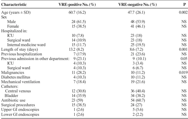
The risk factors that were significantly associated with VRE colonization included length of hospital stay (13.2 days vs. 8.6 days, p=0.001), age (60.7 years vs. 47.7 years, p=0.002) and the presence of underlying malignancies (28.2% vs. 11.2%). Table 2 summarizes the characteristics and risk factors of patients with and without VRE. Isolation of VRE was not significantly associated with stay in the intensive care unit (ICU), surgical ward or internal medicine ward. Prior admission to other medical departments (23.1% vs. 10.1%) was associated with VRE colonization, but no specific correlation was identified with a specific department.
The use of antimicrobial agents is summarized in Table 3. The only association found was between VRE colonization and use of antimicrobials with anaerobic activity, such as metronidazole, piperacillin/tazobactam and imipenem. No single antimicrobial agent was associated with VRE acquisition, although prior carbapenem and ureidopenicillin use approached significance (p=0.06). Thus, we explored the possibility of relationships within the different departments. In ICU’s, vancomycin use was associated with VRE colonization (40% of patients with VRE in ICU had been treated with vancomycin vs. 8.7% of patients not colonized, p=0.03). Surgical procedures, use of central venous catheters, corticosteroid use, diabetes mellitus, mechanical ventilation and gastrointestinal endoscopies were not associated with an increased risk of VRE colonization.
Discussion
National Nosocomial Infections Surveillance System (NNIS), the percentage of enterococcal isolates exhibiting vancomycin resistance in ICUs in the USA reached 25.2% of 1,579 isolates tested by the year 1999, a 43% increase in the mean rate of resistance compared with the years 1994-1998 [13]. By the year 2000, the percentage reached 26.3% out of 2,575 isolates tested [14]. The prevalence of VRE in Europe is still very low. VRE can cause important nosocomial epidemics and can increase morbidity, mortality and costs [15,16].
Intermediate resistance to vancomycin in Staphylococcus aureus (VISA) was first described in 1996 [17]. It is possible that VanA resistance genes could be transferred from enterococci to S. aureus via plasmid-mediated conjugation. In June 2002, the first documented case of infection caused by vancomycin-resistant S. aureus (VRSA) was reported in a patient from Michigan, USA [18]. Cultures also identified concomitant infection due to VRE. This VRSA isolate contained the VanA gene, which suggests that the resistance determinant Table 1. Antimicrobial susceptibilities of 39 vancomycin-resistant entercoccus strains
Vancomycin Teicoplanin
MIC(μμμμμg/mL) S/I/R MIC (μμμμμg/mL) S/I/R
E. faecium ICU 7 27 256 R 256 R VanA
E. faecium Int. medicine 12 72 256 R 32 R VanA
E. faecium Surgical 11 70 256 R 32 R VanA
E. faecium Int. medicine 20 55 64 R 256 R VanA
E. faecium ICU 2 70 8 I 0.75 S VanB
E. faecium Surgical 27 72 8 I 0.19 S VanB
E. faecium ICU 7 41 6 I 0.5 S VanB
E. faecium ICU 3 76 6 I 0.19 S VanB
E. faecium ICU 19 31 8 I 0.25 S VanB
E. faecium Int. medicine 14 41 12 I 1 S VanB
E. faecium Int. medicine 23 19 8 I 0.75 S VanB
E. faecium Int. medicine 13 67 8 I 1 S VanB
E. faecium Int. medicine 11 75 8 I 0.75 S VanB
E. faecium Int. medicine 20 54 8 I 0.75 S VanB
E. faecium Surgical 25 35 8 I 0.25 S VanB
E. faecium Surgical 29 65 8 I 0.5 S VanB
E. faecium Surgical 14 51 8 I 0.75 S VanB
E. faecium Surgical 26 68 8 I 1 S VanB
E. faecium Surgical 15 52 8 I 0.5 S VanB
E. faecium Int. medicine 11 72 8 I 0.5 S VanB
E. faecium Int. medicine 17 76 8 I 0.75 S VanB
E. faecium Int. medicine 6 57 6 I 0.5 S VanB
E. faecium Surgical 14 72 8 I 0.38 S VanB
E. gallinarum Surgical 10 67 12 I 0.5 S VanC
E. gallinarum ICU 32 70 8 I 0.5 S VanC
E. gallinarum ICU 16 70 8 I 0.38 S VanC
E. gallinarum ICU 30 55 8 I 0.5 S VanC
E. gallinarum Int. medicine 4 92 8 I 0.5 S VanC
E. gallinarum Surgical 4 75 6 I 0.19 S VanC
E. gallinarum Int. medicine 8 46 8 I 0.38 S VanC
E. gallinarum Int. medicine 8 71 8 I 0.38 S VanC
E. gallinarum Surgical 8 58 8 I 0.75 S VanC
E. gallinarum Surgical 6 66 6 I 1 S VanC
E. gallinarum Int. medicine 11 73 8 I 0.75 S VanC
E. gallinarum Int. medicine 11 70 8 I 1 S VanC
E. gallinarum ICU 6 70 8 I 0.5 S VanC
E. casseliflavus ICU 6 68 8 I 0.19 S VanC
E. casseliflavus Surgical 8 70 8 I 1 S VanC
E. casseliflavus Int. medicine 2 31 8 I 0.75 S VanC
LOS = Length of Stay; ICU = intensive care unit; S = susceptible, intermediate, resistant.
Phenotype Age
Years LOS
Days
Enterococcuss
Table 2. Epidemiological data of patients with and without vancomycin-resistant entercoccus (VRE) strain colonization
Table 3. Antimicrobial agents administrated to patients within the previous 30 days
might have been acquired from VRE. The potential for VRE to pass genes conferring vanvomycin-resistance to S. aureus provoked a concern for understanding VRE epidemiology.
VRE infections are very low in Greece, as has been sporadically reported [19], in spite of the fact that it is one of the countries with the highest prevalence of high-level gentamycin-resistant enterococci, as reported recently in a European prevalence study [20]. Little is known about VRE colonization in Greece and no study has ever been contacted. In our study, 39 patients (30.5%) were found to have VRE: 10 (7.8%) in the ICU, 14 (10.9%) in surgical wards and 15 (11.7%) in internal medicine wards. The prevalence of fecal colonization by VRE reported in other European studies was: 2% in the Netherlands [21], 4.9% in ICUs of French general hospitals [22] and 3.5% in Belgium [23].
Most clinical isolates were Enterococcus faecium (58.9%), while E. gallinarum accounted for 33.3% and E. casseliflavus accounted for 7.6% of the isolates. No E. faecalis strains were detected. In the USA, the number of E. gallinarum and E. casseliflavus strains among VRE strains is very low, from 0.5-1% [24]. These species are not always taken into account, because their resistance to glycopeptides is intrinsic and their pathogenicities are very low. Four E. faecium strains had a VanA phenotype and the rest had a VanB phenotype.
The mean length of stay in our hospital is seven days. Colonized patients in our study had been hospitalized longer (13.2 days) than the average patient. Patients with VRE were older, 60.7 years vs. 47.7 years for VRE-negative patients. Prior antimicrobial therapy has been known to be a risk factor for VRE acquisition. The only association found in our study
Characteristic VRE-positive No. (%) VRE-negative No. (%) P
Age (years ± SD) 60.7 (16.2) 47.7 (26.1) 0.002
Sex
Male 24 (61.5) 48 (53.9) NS
Female 15 (38.5) 41 (46.1) NS
Hospitalized in:
ICU 10 (7.8) 23 (18) NS
Surgical ward 14 (10.9) 23 (18) NS
Internal medicine ward 15 (11.7) 25 (19.5) NS
Length of stay (days) 13.2 (8.2) 8.6 (7.2) 0.001
Previous hospitalization 7 (17.9) 21 (23.6) NS
Previous admission in other department: 9 (23.1) 9 (10.1) 0.05
ICU 4 (10.3) 3 (3.4) NS
Surgical ward 4 (10.3) 6 (6.7) NS
Malignancies 11 (28.2) 10 (11.2) 0.019
Diabetes mellitus 4 (10.3) 10 (11.2) NS
Mechanical ventilation 7 (18.4) 19 (21.6) NS
Catheters:
Central venous 12 (30.8) 36 (40.4) NS
Bladder 14 (35.9) 34 (38.2) NS
Antibiotic use 23 (59) 54 (60.7) NS
Surgical procedures 15 (38.5) 24 (27) NS
Upper GI endoscopies 1 (2.6) 5 (5.6) NS
Lower GI endoscopies 1 (2.6) 2 (2.2) NS
GI = gastrointestinal tract.
Antimicrobials VRE-positive VRE-negative P
Antianaerobic agents* 10 (25.6%) 10 (11.2%) 0.03
Aminoglycosides 6 (15.4%) 14 (15.7) NS
Quinolones 4 (10.3%) 5 (5.6%) NS
Carbapenems 3 (7.7%) 1 (1.1%) 0.06
Ureidopenicillins 8 (20.5%) 8 (9%) 0.06
Vancomycin 4 (10.3%) 6 (6.7%) NS
Cephalosporins 6 (16.4%) 15 (16.9%) NS
was between antimicrobials with anaerobic activity (such as metronidazole, piperacillin/tazobactam, imipenem) and VRE colonization. The use of carbapenems and ureidopenicillins as risk factors approached significance. In the subpopulation of ICU patients, vancomycin use was related with VRE colonization. In studies during VRE epidemics, a clear relationship between antibiotic use, including especially vancomycin, with VRE colonization has been reported [25,26]. Patients with malignancies were more often colonized with VRE (28.2% vs. 11.2%). Malignancies are a risk factor for VRE colonization and infection, as reported by other authors, especially because of the prolonged hospitalization and the extensive use of antibiotics by these patients [27,28]. Many authors have reported that prior ICU admission is a risk factor for VRE colonization, but we were unable to identify such a relationship in our study.
Most VRE isolates were Van B (there where only four van A and 16 van C isolates), thus stratification for risk factors was not possible.
Whereas VRE in the USA seems to be a hospital problem, probably caused by the extensive use of vancomycin and other antimicrobial agents, such as cephalosporins [29,30], the occurrence of VRE in Europe is possibly boosted by the use of glycopeptide analogs as growth factors in bioindustry and the consequent transmission of VRE via the food chain [31-33]. Torres et al. have suggested that VRE can be part of the intestinal microflora of patients inside and outside the hospital [34].
All patients in our survey were hospitalized for at least two days. Our study was designed as a one-day prevalence study, so we were not able to acquire any data about the previous status of the patients. We assumed that the patients were colonized during their hospital stay, but the fact that two patients with VRE were hospitalized for only two days and one patient with VRE for only three days (none of these patients had any previous hospitalization) means that these particular patients might have been colonized in the community. VRE colonization is an important factor leading to nosocomial dissemination of the organism. Also, VRE colonization independently increases a patient’s risk of developing infections, such as bloodstream infections [35,36].
The Centers for Disease Control and Prevention recommends that hospitals develop a comprehensive plan to prevent and control infection and colonization of patients with VRE [37]. This plan should include prompt identification of VRE-colonized/infected patients, initiation of isolation precautions to prevent patient-to-patient transmission of VRE and prudent use of antimicrobials, especially vancomycin. Isolation of colonized patients is very difficult, because of the large number of patients, as seen in our study; thus great emphasis must be given to hygiene measures. The Infectious Diseases Control Committee of our hospital is taking into account the risk factors found in our study in order to identify patients who may be colonized with VRE. Patients who are
hospitalized for a long time, patients with malignancies, and those who have extensively used vancomycin or antimicrobials with anaerobic activity are now routinely tested for VRE colonization.
References
1. Spera, R.V., Farber B.F. Multiply resistant Enterococcus
faecium: the nosocomial pathogen of the 1990s. JAMA
1992;26:2563-4.
2. Emori T.G., Gaynes R. P. An overview of nosocomial infections, including the role of the microbiology laboratory. Clinical Microbio Reviews 1992;6:428-42.
3. Chenoweth C., Schaberg D.R. The epidemiology of enterococci. Eur J Clin Microbiol Infect Dis 1990;9:80-90.
4. Moellering R.C. Emergence of enterococcus as a significant pathogen. Clin Inf Dis 1992;14:1173-8.
5. Leclercq R., Derlot E., Duval J., Courvalin P. Plasmid-mediated resistance to vancomycin and teicoplanin in E. faecium. New England. J Medicine 1988;319:157-61.
6. Kaplan R.R., Gilligan P.H., Facklam R.R. Recovery of resistant enterococci during vancomycin prophylaxis. J Clin Microbiol 1988;26:1216-8.
7. Centers for Disease Control and Prevention. Nosocomial enterococci resistant to vancomycin—United States 1989–1993. Morbidity and Mortality Weekly Report 1992;42:597-600. 8. Tsakris A., Pournaras S., Douboyas J. Changes in antimicrobial
resistance of enterococci isolated in Greece. J Antimicrob Chemother 1997;40:735-7.
9. Facklam R R., Collins M.D. Identification of Enterococcus
species isolated from human infections by a conventional test scheme. J Clin Microbiol 1989;27:731-4.
10. National Committee for Clinical Laboratory Standards. Methods for dilution antimicrobial susceptibility tests for bacteria that grow aerobically, 3rd ed. Approved standard M7-A3. National Committee for Clinical Laboratory Standards, Villanova, Pa, 1993.
11. National Committee for Clinical Laboratory Standards. Performance standards for antimicrobial susceptibility tests, 4th ed. Approved standards. NCCLS document M7-A2. National Committee for Clinical Laboratory Standards, Villanova, Pa, 1990.
12. Chavers L.S., Moser S.A., Benjamin W.H., et al. Vancomycin resistant enterococci: 15 years and counting. J Hosp Inf 2003;53:159-71.
13. National Nosocomial Infections Surveillance (NNIS) System report, data summary from January 1990-May 1999. Amer J Inf Control 1999;27:520-32.
14. National Nosocomial Infections Surveillance (NNIS) System Report, Data Summary from January 1992-June 2001. Amer J Inf Control 2001;29:404-2l.
15. Nosocomial enterococci resistant to vancomycin United States, 1989–1993. Morbidity and Mortality Weekly Report 1993;42:597-9.
16. Edmond M.B., Ober J.F., Dawson J.D., et al. Vancomycin-resistant enterococcal bacteremia: natural history and attributable mortality. Clin Infect Dis 1996;23:1234-9. 17. Hiramatsu K. The emergence of Staphylococcus aureus with
18. Staphylococcus aureus resistant to vancomycin–United States, 2002. Morbidity and Mortality Weekly Report 2002;51: 565-567.
19. Antonios N. Maniatis, Spyros Pournaras, Maria Kanellopoulou, et al. Dissemination of Clonally Unrelated Erythromycin-and Glycopeptide-Resistant Enterococcus faecium Isolates in a Tertiary Greek Hospital. J Clin Microbiol 2001; 39(12):4571-4.
20. Schouten M.A., Hoogkamp-Korstanje J.A.A., Meis F.G., A. Voss and the European VRE Study Group. (2000). Prevalence of Vancomycin-Resistant Enterococci in Europe. Eur J Clin Microbiol Inf Dis 2000;19:816-22.
21. Endtz H.P., N.Van den Braak, A. Van Belkum, et al. Buitting, A. Van Duin, and H. A. Verbrugh. Fecal carriage of vancomycin-resistant enterococci in hospitalized patients and those living in the community in The Netherlands. J Clin Microbiol 1997; 35:3026-31.
22. Boisivon A., Thibault M., Leclercq R. Colonization by vancomycin-resistant enterococci of the intestinal tract of patients in intensive care units from French general hospitals. Clin Microbiol Inf 1997;3:175-9.
23. Gordts B., Van Landuyt H., Ieven M., et al. Vancomycin-resistant enterococci colonizing the intestinal tracts of hospitalized patients. J Clin Microbiol 1995;33:2842-6. 24. Wells C.L., Juni B.A., Cameron S.B., et al.Stool carriage, clinical
isolation, and mortality during an outbreak of vancomycin-resistant enterococci in hospitalized medical and/or surgical patients. Clin Infect Dis 1995;21:45-50.
25. Boyle J.F., Soumakis S.A., Rendo A., et al. Epidemiologic analysis and genotypic characterization of a nosocomial outbreak of vancomycin-resistant enterococci. J Clin Microbiol 1993;31:1280-5.
26. Handwerger S., Raucher B., Altarac D., et al. Nosocomial outbreak due to Enterococcus faecium highly resistant to vancomycin, penicillin and gentamicin. Clin Inf Dis 1993;16:750-5.
27. Edmond M.B., Ober J.F., Weinbaum D.L., et al. Vancomycin-resistant Enterococcus faecium bacteremia: risk factors for infection. Clin Infect Dis 1995;20:1126-33.
28. Montecalvo M.A., Horowitz H., Gedris C., et al. Outbreak of vancomycin-, ampicillin-, and aminoglycoside-resistant
Enterococcus faecium bacteremia in an adult oncology unit.
Antimicrob Agents Chemother 1994;38:1363-7.
29. McDonald L.C., Kuehnert M.J., Tenover F.C., Jarvis W.R. Vancomycin-resistant enterococci outside the health-care setting: prevalence, sources, and public health implications. Emerg Infect Dis 1997;3:311-7.
30. Woodford N., Johnson A.P., Morrison D., et al. Current perspectives on glycopeptide resistance. Clin Microbiol Rev 1993;8:585-615.
31. Bates J., Jordens Z., Selkon J.B. Evidence for an animal origin of vancomycin-resistant enterococci. Lancet 1993;342:490-1.
32. Donnelly J.P., Voss A., Witte W., Murray B.E. Does the use of antimicrobial agents, including glycopeptide antibiotics, influence the efficacy of antimicrobial therapy in humans? J Antimicrob Chemother 1996;37:389-90.
33. Chadwick P.R., Woodford N., Kaczmarski E.B., et al. Glycopeptide-resistant enterococci isolated from uncooked meat. J Antimicrob Chemother 1996;38:908-9.
34. Torres C.J.A., Reguera M.J., Sanmartin J.C., et al. Van-A mediated vancomycin resistant Enterococcus spp. in sewage. J Antimicrob Chemother 1994;33:553-61.
35. M o n t e c a l v o M . A . , H o r o w i t z H . , G e d r i s C . , e t a l . O u t b r e a k o f v a n c o m y c i n - , a m p i c i l l i n - , a n d a m i n o g l y c o s i d e - r e s i s t a n t E n t e r o c o c c u s f a e c i u m
bacteremia in an adult oncology unit. Antimicrob Agents Chemother 1994;38:1363-7.
36. Roghmann M.C., McCarter R.J., Brewrink J., et al.
Clostridium difficile infection is a risk factor for bacteremia due to vancomycin- resistant enterococci (VRE) in VRE-colonized patients with acute leukemia. Clin Infect Dis 1997;25:1056- 9.