Address to: Dra. Yara de Miranda Gomes. CPqAM/FIOCRUZ. Caixa Postal 7472. Av. Moraes Rego s/n, 50670-420 Recife, PE, Brasil.
Phone: 55 81 2101-2500; Fax: 55 81 3453-1911
e-mail: yara@cpqam.fi ocruz.br Received 23 August 2011 Accepted 06 February 2012
Evaluation of CD4
+CD25
+T lymphocyte response
time kinetics in patients with chronic Chagas disease
after
in vitro
stimulation with recombinant
Trypanosoma cruzi
antigens
Suellen Carvalho de Moura Braz
[1], Virginia Maria Barros de Lorena
[1],[2],
Adriene Siqueira Melo
[1], Maria da Glória Aureliano Melo Cavalcanti
[3]and Yara de Miranda Gomes
[1],[2][1]. Centro de Pesquisas Aggeu Magalhães, Fundação Oswaldo Cruz, Recife, PE. [2]. Programa Integrado de Doença de Chagas, Fundação Oswaldo Cruz, Rio de Janeiro, RJ. [3]. Pronto-socorro Cardiológico de Pernambuco, Universidade de Pernambuco, Recife, PE.
ABSTRACT
Introduction: CD4+CD25+ T lymphocytes have been implicated in the regulation of host infl ammatory response against
Trypanosoma cruzi, and may be involved in theclinical courseof the disease. Methods:Peripheral blood mononuclear cells from patients with chronic Chagas disease were culturedin the presence of T. cruzirecombinant antigensand assayed for lymphocytes
at distinct time points. Results: It was possible to differentiate clinical forms of chronicChagas disease at days 3 and 5 according to presence of CD4+CD25+ T cells in cell cultures. Conclusions: Longer periods of cell culture proved to be potentially valuable for prospective evaluations of CD4+CD25+ T lymphocytes in patients with chronic Chagas disease.
Keywords: Chronic Chagas disease. Recombinant antigens. CD4+CD25+ T lymphocytes.
The clinical course of chronic Chagas disease (ChD) varies from absence of symptoms (indeterminate form - IND) to severe manifestations with cardiovascular (cardiac form - CARD) and/ or digestive (digestive form) impairment1. It has been widely accepted in the scientifi c community that symptomatic forms of chronic ChD are associated with the establishment of an exacerbated cytotoxic cellular immune response that leads to infl ammatory tissue damage and development of symptoms2,3. Nevertheless, there is evidence of a specifi c CD4+ T lymphocyte lineage, characterized by expression of CD25 surface markers a nd the FoxP3 transcription factor, that is involved in the regulation of the cytotoxic response in chronic ChD4-6. Furthermore, developmental or functional disturbances in these cells have been linked to autoimmune and infl ammatory diseases in humans and animals7. Higher percentages of CD4+CD25+ T lymphocytes are observed in peripheral blood from patients with IND forms of ChD4-6, which indicates that these cells have a role in controlling development of the morbidities common to human ChD. These lymphocytes produce cellular factors such as interleukin 10 (IL10) and transforming growth factor beta (TGF-β), whichare among the host immunosuppression
mechanisms employed during the host immune response to
Trypanosoma cruzi4. While the role of these cytokines in immune modulation guided by CD4+CD25+FoxP3+ T cells has been reported4,8,9, there is still no consensus about their mechanisms of action.
The development of specifi c recombinant antigens of T. cruzi
represents an important step in better understanding the role of the cellular responses in ChD10. Our group has been evaluating the use of the recombinant antigens CRA (cytoplasmic repetitive antigen) and FRA (fl agellar repetitive antigen) as immunological markers of the clinical evolution of ChD11-13, demonstrating signifi cant results for the differentiation of distinct clinical manifestations of the chronic phase of ChD.
for 30 min with intracellular antibodies (FoxP3-PE/BD Pharmingen; IL10-PE/Caltag). After another round of wash by centrifugation (300 × g/5 min), cells were fi xed (BD Cytofi x™) and 30,000 events/sample were acquired in a FACScalibur® fl ow cytometer (Becton Dickinson) and analyzed using the CellQuest Pro Software. Detection of TGF-β in supernatants from days 1 and 5 was performed using the Human TGF-β1 Immunoassay
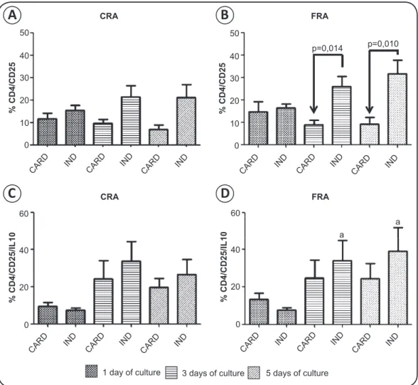
difference among cell culture time points (1, 3, and 5 culture days) after stimulation with either recombinant antigen (Figures 1A
and 1B). However, an increase in the mean number of
CD4+CD25+ T cells in IND patients was observed over the course of the cultures, mainly after FRA stimulation, while in the CARD group, there was a decrease in the mean number of CD4+CD25+ T cells from the fi rst to the fi fth day of culture.
CRA
CRA
FRA
FRA 50
% CD4/CD2
5
40
30
20
10
0
CARD IND CARD IND CARD IND
50
p=0,014
a
a p=0,010
% CD4/CD2
5
% CD4/CD25/IL10 % CD4/CD25/IL10
40
30
20
10
60 60
40 40
20 20
0 0
0
CARD
CARD CARD
IND
IND IND
IND
IND IND
IND
IND IND
CARD
CARD CARD
CARD
CARD CARD
1 day of culture 3 days of culture 5 days of culture
FIGURE 1 - Detection of CD4+CD25+ (A and B) and CD4+CD25+IL10+ (C and D) T lymphocytes in peripheral blood mononuclear cells of patients with chronic Chagas disease afterin vitrostimulation with CRA and FRA. Signifi cant differences are indicated in the fi gures with the respective p-values.An “a” indicates signifi cant differences in comparison with day 1. CRA: cytoplasmic repetitive antigen; FRA:fl agellar repetitive antigen; CARD: patients with cardiac clinical form of chronic Chagas disease; IND: patients with indeterminate clinical form of chronic Chagas disease; CD:cluster of differentiation; IL10: interleukin 10.
A
B
The means of CD4+CD25+ T cell counts between the 2 groups (CARD versus IND) for each culture time point (culture day 1, 3, or 5) were also compared. Differences between CARD and IND were statistically signifi cant at days 3 (p= 0.014) and 5 (p= 0.010) after in vitro stimulation with FRA (Figure 1B). However, no
differences were observed after stimulation with CRA (Figure 1A). In regard to IL10-producing CD4+CD25+ T lymphocytes, the mean values of CD4+CD25+IL10+ T cells in the IND group, in the presence of FRA, were statistically different between days 1 and 3 (p= 0.031) and between days 1 and 5 (p= 0.031). Comparison of the CARD and IND forms showed that the mean number of CD4+CD25+IL10+ T cells was higher in the IND group than in the CARD patients after stimulation with CRA and FRA, however, this difference was non-signifi cant (Figures 1C
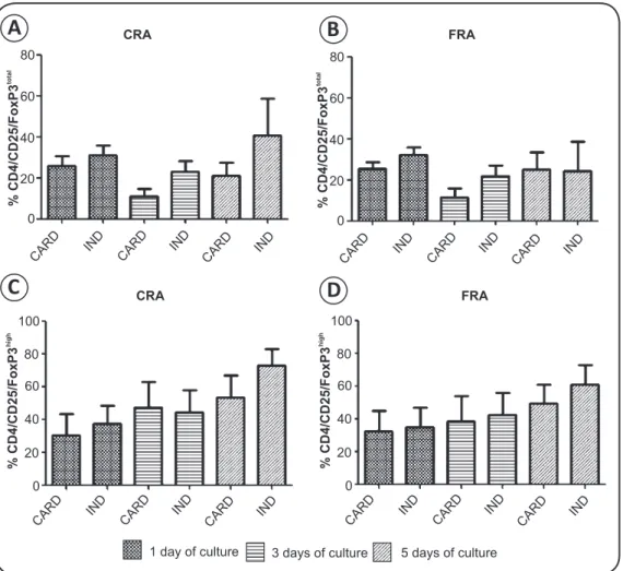
and 1D). With regard to the presence of CD4+CD25+FoxP3total and CD4+CD25+FoxP3high T cells, there was no statistical
difference observed between the groups or among the cell culture time points within each group (Figure 2).
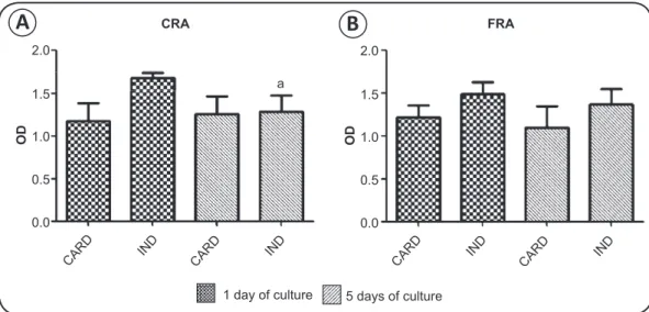
The presence of TGF-β in supernatants was not signifi cantly different between patients with CARD and IND forms of chronic
ChD. Nevertheless, a reduction in TGF-β production after 5 days was observed upon stimulation with CRA (p = 0.043) (Figure 3).
Thus, in the present study we found that in vitro culture of
PBMC for 3 and 5 days allowed differentiation between CARD or IND forms of ChD through evaluation of CD4+CD25+ T lymphocyte counts after antigenic stimulation. Previous studies using whole blood (ex vivo)5,6 and after 22 hours of culture in the presence of epimastigote antigens7 were also able to differentiate CARD and IND forms by analyzing the percentage of these cells through fl ow cytometry. Because CD4+CD25+ T cells represent a small percentage of total circulating CD4+ T lymphocytes7, we believe that longer periods of cell culture may improve studies regarding the possible roles of these cells in the clinical course of ChD. In regard to IL10 production by CD4+CD25+ T lymphocytes in chronic ChD, our data corroborated with those presented by Araújo et al.6, in that no difference was found between patients with CARD and IND clinical forms. However, the increased numbers of CD4+CD25+IL10+ T lymphocytes throughout the cell culture period reinforced the importance of considering longer periods of cultivation.
CRA
CRA
FRA
FRA
% CD4/CD25/FoxP3
total
% CD4/CD25/FoxP3
high
% CD4/CD25/FoxP3
high
% CD4/CD25/FoxP3
total
80
80 80
100 100
80
60
60 60
60
40
40 40
40
20
20 20
20
0
0 0
0
CARD
CARD CARD
CARD IND
IND IND
IND IND
IND IND
IND IND
IND IND
IND CARD
CARD CARD
CARD CARD
CARD CARD
CARD
1 day of culture 3 days of culture 5 days of culture
FIGURE 2 - Detection of CD4+CD25+FoxP3total (A and B) and CD4+CD25+ FoxP3high (C and D) T lymphocytes in peripheral blood mononuclear cells of patients
with chronic Chagas disease afterin vitrostimulation with CRA and FRA. Signifi cant differences are indicated in the fi gures with the respective p-values.
CRA: cytoplasmic repetitive antigen; FRA: fl agellar repetitive antigen; CARD: patients with cardiac clinical form of chronic Chagas disease; IND: patients with indeterminate clinical form of chronic Chagas disease; CD:cluster of differentiation.
A
B
1 day of culture 5 days of culture
FIGURE 3 - Detection of transforming growth factor-β in supernatant of cultures from patients with chronic Chagas disease afterin vitrostimulation with CRA
(A) and FRA (B). Signifi cant differences are indicated in the fi gures with the respective p-values.The “a” represents a signifi cant difference in comparison with day 1. CRA: cytoplasmic repetitive antigen; FRA: fl agellar repetitive antigen; CARD: patients with cardiac clinical form of chronic Chagas disease; IND: patients with indeterminate clinical form of chronic Chagas disease; OD: optical density.
The observation that there were no differences in TGF-β production among patients with CARD and IND forms of ChD may have been due to the lower sample size evaluated. Moreover, with the chosen method (capture ELISA), the source of cytokine production is not identifi ed. With regard to the reports citing TGF-β as a pathway of immunoregulation by CD4+CD25+FoxP3+ T lymphocytes7,8, the technique reported here did not clarify whether CD4+CD25+FoxP3+ T cells were responsible for TGF-β production. Thus, the evolution of these growth factors under the regulation of CD4+CD25+FoxP3+ T cells in individuals with chronic ChD will only be confi rmed upon further study.
We conclude that CD4+CD25+ T lymphocyte cultures derived from serum of patients chronic ChD that are maintained for longer periods have the potential to be used in prospective studies aimed at understanding the clinical evolution of the disease. Moreover, this approach may also aid in the identifi cation of cellular patterns that can serve as biological markers to monitor patients. Further, it is important to mention that a greater number of individuals should be evaluated to prove the importance of this cell population in immunoregulation in individuals with the IND form of ChD.
REFERENCES
The authors declare that there is no confl ict of interest.
CONFLICT OF INTEREST
ACKNOWLEDGMENTS
FINANCIAL SUPPORT
We are thankful to the staff of Ambulatório de Doença de Chagas e Insuficiência Cardíaca (PROCAPE/UPE) for assistance in selecting the patients included in this study, to the Programa de Desenvolvimento Tecnológico em Insumos para Saúde (PDTIS/FIOCRUZ) for use of its facilities, to Antônio Ferreira and Edimilson Silva at Biomanguinhos/FIOCRUZ for providing recombinant antigens, to George Diniz at CPqAM⁄ FIOCRUZ for assistance with statistics, and to Mineo Nakazawa at CPqAM⁄FIOCRUZ for technical assistance.
This research received fi nancial support by the Conselho
Nacional de Desenvolvimento Científi co e Tecnológico (CNPq)
(Universal Edital n° 478572/2009-3) and Coordenação de Aperfeiçoamento de pessoal de Nível Superior (Capes). Y.M.
Gomes is a CNPq fellow (number 306427/2006-0). V.M.B. Lorena is a postdoctoral CNPq fellow. S.C.M. Braz and A.S Melo were candidates for the Master’s degree in Public Health (CPqAM-FIOCRUZ) and were CNPq (number133106/2009-8) and Capes fellows, respectively.
1. Rassi Jr A, Rassi A, Marin-Neto JA. Chagas disease. Lancet 2010; 375:1388-1402.
2. Dutra OD, Gollob KJ. Current concepts in imunoregulation and pathology of human Chagas disease. Curr Opin Infect Dis 2008; 21:287-292. 3. Lescure FX, Le Loup G, Freilij H, Develoux M, Paris L, Brutus L, et al. Chagas
disease: changes in knowledge and management. Lancet Infect Dis 2010; 10: 556-570.
4. Vitelli-Avelar DM, Sathler-Avelar R, Dias JCP, Pascoaly VPM, Teixeira-Carvalho A, Lage PS, et al. Chagasic patients with indeterminate clinical form of the disease have high frequencies of circulating CD3+CD16-CD56+ natural killer T cells and CD4+CD25High regulatory T lymphocytes. Scand
J Immunol 2005; 62: 297-308.
5. Vitelli-Avelar DM, Sathler-Avelar R, Massar RL, Borges JD, Lage PS, Lana M, et al. Are increased frequency of macrophage-like and natural killer (NK) cells, together with high levels of NKT and CD4+CD25high T cells balancing
6. Araujo FF, Gomes JAS, Rocha MOC, Williams-Blangero S, Pinheiro VM, Morato MJS, et al. Potencial role of CD4+CD25high regulatory T cells in morbidity in Chagas disease. Front Biosci 2007; 12: 2797-806.
7. Sakaguchi S, Tomoyuki Y, Nomura T, Ono M. Regulatory T cells and Immune Tolerance. The Cell 2008; 133:775-787.
8. Nakamura K, Kitani A, Strober W. Cell contact-dependent immunosuppression by cell surface-bound transforming growth factor β. J Exp Med 2001; 194: 629-644.
9. Piccirillo CA, Letterio JJ, Thornton AM, McHugh RS, Mamura M, Mizuhara H et al. CD4+CD25+ regulatory T cells can mediate suppressor function in the absence of transforming growth factor β1 production and responsiveness. J Exp Med 2002; 196:237-245.
10. Lorca M, Gonzalez A, Veloso C, Reyes V, Vergara U. Immunodetection of antibodies in sera from symptomatic and asymptomatic Chilean Chagas’
disease patients with Trypanosoma cruzi recombinant antigens. Am J Trop
Med Hyg 1992; 46:44-49.
11. Lorena VMB, Lorena IMB, Braz SCM, Melo AS, Melo MFAD, Melo MGAC, et al. Cytokine levels in serious cardiopathy of Chagas disease after in vitro stimulation with recombinant antigens from Trypanosoma cruzi. Scand J Immunol 2010; 72:529-539.
12. Vasconcelos RHT, Amaral FN, Cavalcanti MGAM, Silva ED, Ferreira AGP, Morais CNL, et al. Increased levels of IgA antibodies against CRA and FRA recombinant antigens of Trypanosoma cruzi differentiate digestive forms of Chagas disease. Hum Immunol 2010; 71: 964-967.
13. Verçosa AFA, Lorena VMB, Carvalho CL, Melo MFAD, Cavalcanti MGA, Silva ED, et al. Chagas' disease: IgG isotypes against cytoplasmic (CRA) and fl agellar (FRA) recombinant repetitive antigens of Trypanosoma cruzi