INTRODUCTION
1. Instituto de Patologia Tropical e Saúde Pública, Universidade Federal de Goiás, Goiânia, GO. 2.Instituto de Química, Universidade Federal de Goiás, Goiânia, GO.
Address to: Dra. Maria do Rosário Rodrigues Silva. IPTSP/UFG. Rua 235 s/n, Setor Universitário, 74605-050 Goiânia, GO, Brasil.
Phone: 55 62 3209-6127; Fax 55 62 3209-6363. e-mail: [email protected]
Received in 12/05/2011 Accepted in 16/06/2011
Molecular analysis and dimorphism of azole-susceptible and resistant
Candida albicans
isolates
Análise molecular e dimorismo de isolados de
Candida albicans suscetíveis e resistentes aos azólicos
Carolina Rodrigues Costa
1, Lúcia Kioko Hasimoto e Souza
1, Fábio Silvestre Ataídes
1, Pedro Henrique Ferri
2,
Maysa Paula da Costa
1, Orionalda de Fátima Lisboa Fernanades
1and Maria do Rosário Rodrigues Silva
1ABSTACT
Introduction: Candida albicans is responsible for supericial or systemic infections known as candidiasis, which may be found in infected tissue as unicellular budding yeasts, hyphae, or pseudohyphae. In this study, the efects of both luconazole and itraconazole antifungal agents on the hyphal formation and genotypic characterization of C. albicans isolates classiied as either susceptible or resistant were investigated. Methods: he hyphal production of ive C. albicans
isolates under the action of antifungal agents was investigated by culturing yeast on growth medium and on hyphal induction medium. he genotypic characterization was carried out for 13 isolates of C. albicans using the random ampliied polymorphic DNA-polymerase chain reaction (APD-PCR) method. Results: he dimorphism analysis showed that the hyphal formation was higher in resistant than in the susceptible isolates to both azoles. he APD-PCR method identiied the formation of two diferent groups. In group A, four resistant and two susceptible isolates were clustered, and in group B, one resistant and six susceptible isolates were clustered. Conclusions:Considering that hyphal formation was higher in resistant isolates in the presence of azole drugs, we conirmed that the hyphal production is closely related to susceptibility to azoles. hese drugs may afect the morphogenesis of C. albicans depending on their susceptibility to these drugs. In relation to APD-PCR, most resistant isolates classiied in group A and susceptible isolates in group B demonstrated that this method presented a similar standard between the two groups, suggesting that by this technique, a strong correlation between genotypes and luconazole-resistant samples may be found.
Keywords:Candida albicans. Azole antifungals. Hyphal formation. In vitro susceptibility. APD-PCR.
RESUMO
Introdução:Candida albicans é responsável por infecções supericiais ou sistêmicas conhecidas como candidíase, encontrada em tecidos infectados na forma de leveduras brotantes unicelulares, hifas ou pseudohifas. Neste estudo, os efeitos de agentes antifúngicos como o luconazol e o itraconazol sobre a formação de hifas e caracterização genotípica de isolados de C. albicans
suscetíveis ou resistentes foram investigados. Métodos: A produção de hifas de cinco isolados de C. albicans, sob a ação de antifúngicos foi investigada pelo cultivo da levedura em meios de crescimento e de indução de hifas. A caracterização genotípica foi realizada para 13 isolados de
C. albicans pelo método de APD-PCR. Resultados: A análise do dimorismo mostrou que a formação de hifas foi maior nos isolados resistentes do que nos suscetíveis aos antifúngicos. O método de APD-PCR identiicou a formação de dois diferentes grupos. No grupo A, foram agrupados quatro isolados resistentes e dois suscetíveis e no grupo B um resistente e seis suscetíveis. Conclusões: Considerando que a formação hifal foi maior em isolados resistentes na presença de azólicos, concluimos que a produção hifal está muito relacionada a suscetibilidade a estes fámacos. Estes antifúngicos podem alterar a morfologia de C. albicans em dependência da sua suscetibilidade.No método de APD-PCR, o encontro da maioria dos isolados resistentes classiicados como pertencentes ao grupo A e suscetíveis ao grupo B demonstrou que este método apresentou um padrão semelhante entre os dois grupos, sugerindo que por este método pode ser detectado uma estreita correlação entre genótipos e amostras resistentes ao luconazol.
Palavras-chaves: Candida albicans. Antifúngicos azólicos. Formação hifal. Suscetibilidade
in vitro. APD-PCR.
Candida albicans is responsible for supericial or systemic infections known as candidiasis, which is frequently associated with immunocompromised patients1. It may be found in infected tissue as
unicellular budding yeasts, hyphae, or pseudohyphae. he dimorphism facilitates tissue invasion, increases tissue viability, and allows it to escape from macrophage and neutrophil engulfment2, 3.
Some antifungal agents afect the morphology of
C. albicans cells. According to Gil et al.4, hyphal forms
are predominant in azole-resistant isolates. Hyphal formation in susceptible strains is inhibited at clinically signiicant concentrations of azole drugs.5
Genetic analysis of Candida isolates reveals that virulence atributes may be intimately related to genotypic proiles. Molecular methods have been used for such analysis because of their sensitivity and speciicity, allowing intraspeciic identiication of isolates as well as detection of small diferences in nucleic acid content among species and ancestries of the same species6. he analysis of the random
ampliied polymorphic DNA (APD) is used for characterization of fungi, especially C. albicans7.
his technique presents speed, simplicity, and power discriminatory among the isolated ones6, 8.
Evidence shows that azole-resistant isolates, especially luconazole, result from genomic alterations9, 10.
The emergence of fluconazole resistance may be associated with continued exposure of C. albicans
population to this drug, leading to a gradual elevation of minimal inhibitory concentration (MIC) of some isolates9-11. However, there is little information
about the genomic diversity of resistant isolates to antifungals compared with those that are susceptible to the drugs.
hus, this study investigates C. albicans hyphal formation of susceptible and resistant isolates in the presence of azole drugs and genotypically characterizes luconazole-susceptible and -resistant isolates.
METHODS
Costa CR et al - APD and dimorphism of Candida albicans
Candida albicans identiication
Candida albicans isolates from oral cavities of human immunodeiciency virus (HIV)-infected patients used in this study were previously identiied using germ tube test, chlamidoconidia formation, carbon and nitrogen assimilation, and sugar fermentation12. Furthermore, all isolates were
streaked onto CHROMagar®Candida medium plates (CHROMagar,
Paris, France) for presumptive identiication of C. albicans. API 20
Candida identiication kit (BioMerieux, France) was used when the yeasts could not be identiied by any of the methods previously mentioned. Growth at 45oC on Sabouraud dextrose broth and assimilation tests with
xylose and α-methyl-D-glucoside were used for discrimination between
C. albicans and Candida dubliniensis13, 14. All isolates were stored at -70oC
in yeast extract peptone dextrose agar (YEPD-Difco) with 10% glycerol and subcultured twice on YEPD agar before testing to ensure purity of cultures.
In vitro antifungal susceptibility testing
Minimal inhibitory concentrations were determined using broth microdilution method in RPMI 1640 medium, standardized by Clinical and Laboratory Standards Institute (CLSI)15.
Antifungal agents
Itraconazole and luconazole were tested against 13 C. albicans
isolates. The final concentration of drugs ranged from 0.06 to 64µg/ml for luconazole and from 0.015 to 16µg/ml for itraconazole.
In vitro susceptibility tests were performed to yield eight times the inal concentration when the MICs were >64µg/ml for luconazole and >16µg/ml for itraconazole. Candida parapsilosis ATCC 22019 was included as a quality control strain to check the accuracy of drug dilutions and reproducibility of results.
Determination of resistance
Candida albicans isolates were considered resistant when MIC values were ≥64µg/ml for luconazole and ≥1µg/ml for itraconazole, according to breakpoints established by CLSI15.
Dimorphism of azole-resistant and -susceptible strains
To determine the efects of azole antifungal agents on hyphal production, five isolates of C. albicans classified as susceptible (numbers 2, 8, and 15) and resistant (numbers 13 and 23) to both azoles were used.
Yeast growth and hyphal induction in the presence of itraconazole or luconazole
Hyphal induction under the action of antifungal agents was performed according to Ha and White5 with slight modiications.
Briefly, the isolates stored at -70oC in YEPD medium were
subcultured in an agar plate of this medium for 24h. Single yeast colonies were inoculated in 5ml of YAD medium (1.7g of yeast nitrogen base without amino acids and ammonium sulfate, 5g of ammonium sulfate, and 5.4g of dextrose per liter) for 48h in the presence of diferent concentrations of luconazole or itraconazole (1/2, 1/4, and 1×MIC) as well as in the absence of these drugs.
Ater incubation with shaking (180 rpm) at 30oC, cell cultures
were centrifuged for 5 min at 10oC, washed twice with 5ml of
phosphate-bufered saline, and then transferred to 5ml of hyphal induction medium (M199) containing bovine calf serum (10%) to a inal concentration of 3×10⁶ cells/ml. he same concentrations of
antifungal agents (0, 1/2, 1/4, and 1×MIC) were added to the hyphal induction medium. Cells were incubated at 37oC with shaking at
240rpm. Ater 3h, 250 cells (yeast and hyphae) were counted using the Neubauer chamber. hese experiments were repeated in two diferent occasions, and the mean was calculated.
Genotypic characterization
A total of 13 C. albicans isolates characterized as susceptible or resistant to luconazole were submited to genotypic identiication using the APD-PCR technique.
Preparation of DNA
Genomic DNA extraction was based on the method described by Del Poeta et al16. and modiied by Casali et al17. Briely, a heavy
inoculum of C. albicans strains grown in YEPD agar at 37oC for 24
to 48h was suspended in 0.5ml of TENTS (10mM of Tris, pH 7.5; 1mM of EDTA, pH 8.0; 200mM of NaCl; 2% Triton; 1% SDS), containing 0.2ml of 0.45-mm glass beads and 0.5ml of phenol-chloroform, which were vortexed for 2min. Ater centrifugation for 10 min at 13,000rpm, the aqueous phase was transferred to a new tube, and the same volume of ethanol (100%) was added. It was then incubated at -20oC for 1h for DNA precipitation. he precipitated
DNA was resuspended in 0.5ml TE (10mM of Tris HCl, pH 8.0; 1mM of EDTA, pH 8.0), containing 50µg/ml of RNAse, and was incubated at 37oC for 30 min. he yeast DNA was extracted once
with phenol and chloroform, reprecipitated with 20µl of NaCl (5 mol/l) and 1 volume of 100% ethanol, washed with 70% ethanol, resuspended in 100µl of TE bufer, and stored at -20oC.
Random ampliied polymorphic DNAanalysis Ampliication reactions using primer RSD6 (5’-GCGATCCCCA-3’)
were performed as described by Dassanayake et al18. The
reactions were performed in volume of 25µl, including about 50ng of DNA template, 10mM of Tris-HCl, pH 8.3 (2.5µl), 50mM of KCl (1µl), 1.5mM of MgCl2, 0.2mM of each dNTP (2µl), and 2.5 U (0.5µl) of Taq DNA polymerase (Invitrogen), added with 1.5µM of RSD6 (1.25µl). Ampliication was performed in a PCR MJ Research hermal Cycler model PTC-100TM programmed
as follows: he irst ive cycles included denaturation at 94oC for
30s, annealing at 27oC for 2 min, and primer extension at 72oC for 2
min, followed by 45 denaturation cycles at 94oC for 30s, annealing
at 32oC for 2 min, and a inal extension at 72oC for 2 min, held at this
temperature for 15 min. Ampliication products were separated using electrophoresis in 1.2% agarose gel containing 1X Tris-borate-EDTA bufer, stained with blue green (LGC Biotechnology) and visualized under UV light.
Statistical analysis
The results obtained on dimorphism were analyzed by a proportion test using the Epi-info version 6.0. p values <0.05 were considered statistically signiicant. he dendrogram of the genetic similarity of fluconazole-susceptible and resistant isolates was performed by PAST version 1.9119 via the unweighed pair-group
average method (UPGMA), using the Raup-Crick similarity index20.
Cophenetic correlation was used to check the representativeness of the clustering procedure21.
Ethical considerations
RESULTS
Susceptibility testing
According to the interpretive criteria used for antifungal drug resistance described in the Materials and Methods section, ive resistant and eight susceptible C. albicans isolates for each one of the drugs were identiied. Fluconazole MIC values were 64µg/ml in one isolate (34), 128µg/ml in four (1, 13, 23, and 24), and ≤4µg/ ml for the other eight. Itraconazole MIC values were 64µg/ml in two isolates (13 and 23), 16µg/ml in three (5, 36, and 115), and ≤0.5µg/ml for the other eight. Antifungal susceptibility of
C. parapsilosis ATCC 22019 was within the ranges previously reported.
Dimorphism
Among the ive C. albicans isolates analyzed, diferences at the hyphal formation because ofconcentration of antifungal drugs and azole susceptibility were observed. In all isolates, the reduction of hyphal formation was directly related to drug concentration. here was a higher reduction of hyphae in 1×MIC values than in 1/2 MIC and 1/4 MIC values. For susceptible isolates, a statistically signiicant diference was observed between 1×MIC and 1/2 MIC, and between 1×MIC and 1/4 MIC (p<0.001), but not between 1/2 MIC and 1/4 MIC.
For the resistant isolates, statistical diference in relation to the reduction of hyphae between the MIC values tested was not observed. In the absence of antifungal drugs (control), more than 90% of hyphal formation was observed in all susceptible or resistant isolates.
he hyphal formation also varied according to the drug; it was more susceptible to inhibition by itraconazole than luconazole. For instance, in one isolate (13), hyphae formation reached 28.8% under the efect of itraconazole 1×MIC, whereas it reached 50% with luconazole under similar conditions. Nevertheless, there was no statistically signiicant diference between the two antifungal agents.
Hyphal formation was higher in resistant isolates in the presence of azole drugs (p<0.001). Figures 1 and 2 show the hyphal production by susceptible and resistant isolates induced by incubation with both luconazole and itraconazole MICs and subMICs.
Genotypic characterization of luconazole-resistant and -susceptible C. albicans isolates using APD-PCR
Candida albicans isolates showed a number of bands ranging from seven to 11 using primer RSD6 by the APD-PCR method
(Figure 3). Data interpretation criteria were related to diferences in position of the bands. he generated proile of the bands allowed the identiication of eight diferent genotypes among the 13 C. albicans
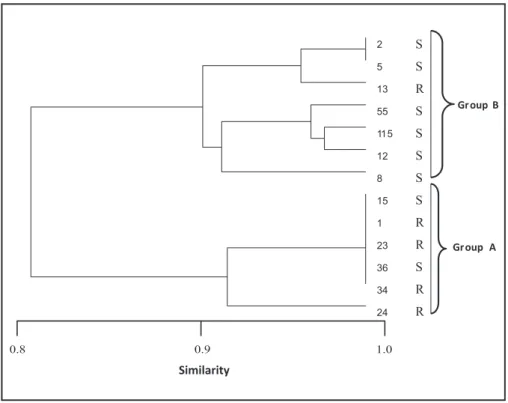
isolates - luconazole-resistant or -susceptible. hey were classiied as groups A and B in agreement with the dendrogram of genetic similarity using the Raup-Crick coeicient index, a probabilistic measure based on presence-absence data. he UPGMA had a high cophenetic correlation of 0.86, suggesting that the dendrogram preserves the pairwise distances between the original unmodeled data points. Group A included susceptible isolates 15 and 36 and resistant isolates 1, 23, 24, and 34. Group B included susceptible isolates 2, 5, 12, 55, and 115 and resistant isolate 13, as it is shown in Figure 4. hree (1, 23, and 34) of the ive luconazole-resistant isolates produced the same genotypic proile (A1). However, only two (2 and 5) produced the same genotypic proile (B1) among luconazole-susceptible isolates. Fluconazole-susceptible isolates, 15 and 36, presented the same genotypic proile as the resistant isolates, classiied as A1. he other isolates produced independent genotypes. he molecular proiles of isolates are shown in Table 1.
FIGURE 1 - Efects of diferent concentrations of itraconazole (1xMIC, 1/2 and 1/4 MIC, and control = absence of drug) on hyphal formation (M199 medium added with 10% bovine calf serum) in both susceptible (2, 8, and 15) and resistant (13 and 23) Candida albicans isolates.
FIGURE 2 - Efects of diferent concentrations of luconazole (1xMIC, 1/2 and 1/4 MIC, and control = absence of drug) on hyphal formation (M199 medium added with 10% bovine calf serum) in both susceptible (2, 8, and 15) and resistant (13 and 23) Candida albicans isolates.
FIGURE 3 - Random ampliied polymorphic DNA ingerprinting of luconazole-susceptible Candida albicans isolates (2, 5, 8, 12, 15, 36, 55, and 115) and
FIGURE 4 - Dendrogram of 13 luconazole-resistant Candida albicans isolates (1, 13, 23, 24, and 34) and luconazole-susceptible isolates (2, 5, 8, 12, 15, 36, 55, and 115) obtained by ampliication using primerRSD6.
TABLE 1 - Molecular proile by APD method of resistant and susceptible
Candida albicans isolates to luconazole.
Isolates Molecular proile
1 A1
13 B2
Resistant 23 A1
24 A2
34 A1
2 B1
5 B1
8 B6
Susceptible 12 B5
15 A1
36 A1
55 B3
115 B4
APD: random ampliied polymorphic DNA.
24 34 36 23 1 15 8 12 115 55 13 5
2 S
S
R
S
S
S
S
S
R
R
S
R
R
Gr oup B
Gr oup A
0.8 0.9 1.0
Similarity
DISCUSSION
Exoenzyme production, adherence ability of host cells, the ability to produce hyphae, and antifungal drug resistance are considered important virulence factors of Candida species, which facilitate the establishment of infection22, 23.
The transition from spherical cells to hyphae expressed by
C. albicans is strictly related to yeast pathogenicity. Filamentous fungi have a structure that allows penetration into the host tissue more easily than spherical cell forms23, 24. Enzymes are mainly secreted
from hyphae tips and are able to degrade proteins, lipids, and other cellular components that facilitate tissue iniltration25. Mutant yeasts
are unable to form filaments, thus presenting reduced invasion capacity and virulence in mouse models23, 26. Spherical cellsshow less
in vitro adhesion — initial phase for penetration of a microorganism — to buccal epithelial cells than ilamentous forms1.
he inluence of azole antifungals related to the ability of fungi to form hyphae by susceptible or resistant isolates to these antifungal has been litle investigated. here are indications that luconazole-susceptible yeasts present reduced hyphae production, whereas resistant isolates have been litle affected by this antifungal agent5. In this
study, inhibition of hyphal growth of azole-susceptible strains of C. albicans was observed at higher levels than resistant isolates in the presence of both itraconazole and/ or luconazole. Such inhibition depended on antifungal concentration. Interestingly, growth inhibition in susceptible isolates was accompanied by reduction of hyphal formation. his result allowed suggesting that no inhibition of growth of the fungi under the efects of the antifungal in resistant isolates presents a strong correlation with the filamentous production. It is known that azoles act by inhibiting production of ergosterol, which is the main constituent of a fungal cell membrane. hese antifungal agents probably reduce the production of hyphae because of the increased surface area of hyphal cells in comparison to those of spherical forms. he hyphal structure requires more plasma membrane to the cell. Previous works show that alterations in the ERG11 gene, responsible for ergosterol production, limit hyphal formation presumably because of the lack of ergosterol27. Although
luconazole is the most used antifungal agent for the treatment of infections caused by C. albicans, an increased number of resistant isolates have been observed18. herefore, detection of resistance
among isolates is considered of great importance.
Yeast genotypes may vary according to drug susceptibility. he genetic diversity among luconazole-resistant C. albicans isolates has revealed controversial results. Genotypic characterization in groups has been deined by Xu et al.28 for resistant isolates to luconazole,
who observed that resistant strains to this drug were genetically more similar to each other than they were to susceptible strains. Dassanayake et al.18 showed that, of the 20 isolates studied, only one
resistant isolate presented the same proile as the susceptible one. he molecular proile found in this work using the primer RSD6 by APD-PCR, with eight diferent genotypes being four of the ive resistant isolates to the luconazole classiied in the same group (A), showing the same original clonal and one isolate in another group (B) shared with susceptible isolates, proved that the behaviors of the resistant strains were similar but may also be another clonal source. In a previous experiment with primer RSD6, 15 diferent genotypes were identiied in 20 isolates, showing independent genotypes for each one of the 10 luconazole-susceptible isolates but only six genotypes for the 10 luconazole-resistant isolates18.
In conclusion, considering that hyphal formation was higher in resistant isolates in the presence of azole drugs, we conirmed that the hyphal production is closely related to susceptibility to azoles. According to Ha and White5, the azoles may afect the morphogenesis
he authors declare that there is no conlict of interest.
CONFLICT OF INTEREST
FINANCIAL SUPPORT
REFERENCES
of C. albicans depending on their susceptibility to these drugs. Most resistant isolates classiied in group A and susceptible isolates in group B demonstrated that APD-PCR typing method presented a similar standard between the two groups, suggesting that, by this technique, a strong correlation between genotypes and luconazole-resistant samples may be found.
Conselho Nacional de Desenvolvimento Cientíico e Tecnológico
(CNPq). Process number 474837/2006-8.
1. Kriznik A, Bouillot M, Coulon J, Gaboriaud F. Morphological speciicity of yeast and ilamentous Candida albicans forms on surface properties. Biologies 2005; 328:928-935.
2. Braga PC, Alieri M, Culici M, Dal Dasso M. Inhibitory activity of thymol against the formation and viability of Candida albicans hyphae. Mycoses 2007; 50:502-506.
3. Watamoto T, Samaranayake LP, Jayatilake JAMS, Egusa H, Yatani H, Seneviratne CJ. Efect of ilamentation and mode of growth on antifungal susceptibility of Candida albicans. Int J Antimicrob Agents 2009; 34:333-339.
4. Gil C, Perez-Diaz R, Nombela C. Inhibitory and morphological efects of several antifungal agents on three types of C. albicans morphological mutants. J Med Vet Mycol1994; 32:151-162.
5. Ha KC, White TC. Efects of azole antifungal drugs on the transition from yeast cells to hyphae in susceptible and resistant isolates of the pathogenic yeast Candida albicans. Antimicrob Agents Chemother 1999; 43:763-768.
6. Enache-Soare S, Pelinescu D, Ionescu R, Avram I, Stoica I, Vassu-Dimov T. Molecular identiication of some yeast strains involved in oral candidosis. Rom Biotechnol Let2009; 14:4180-4186.
7. Boriollo MFG, Holing JF, Mendes A, Rosa EAR. Ferramentas moleculares para caracterização de Candida albicans (Robin) Berkhout (1923) em estudos epidemiológicos. Est Biol 2005; 27:21-47.
8. Soll DR. The ins and outs of DNA fingerprinting the infectious fungi. Clin Microbiol Rev 2000; 13:332-370.
9. Franz R, Kelly SL, Lamb DC, Kelly DE, Ruhnke M, Morschhauser J. Multiple molecular mechanisms contribute to a stepwise development of luconazole resistance in clinical Candida albicans strains. Antimicrob Agents Chemother 1998; 42:3065-3072.
10. Ribeiro MA, Paula CR, Perfect JR, Cox GM. Phenotypic and genotypic evaluation of luconazole resistance in vaginal Candida strains isolated from HIV-infected women from Brazil. Med Mycol2005; 43:647-650.
11. Barchiesi F, Hollis RJ, McGough DA, Scalise G, Rinaldi MG, Pfaller MA. DNA subtypes and luconazole susceptibilities of Candida albicans isolates from the oral cavities of patients with AIDS. Clin Inf Dis 1995; 20:634-640. 12. Kurtzman CP, Fell JW. he Yeasts, A Taxonomic Study. 4th edition. Amsterdam,
he Netherlands: Elsevier Science BV; 1998.
13. Dolapacci I, Tekeli A, Gocmen JS, Aysev D, Guriz H. Investigation of Candida dubliniensis in Candida spp.-positive hemocultures. Acta Path Microbial Scand 2002; 110:391-395.
14. Faggi E, Pini G, Campisi E, Martinelli C, Difonzo E. Detection of Candida dubliniensis in oropharyngeal samples from human immunodeiciency virus infected and non-infected patients and in a yeast culture collection. Mycoses 2005; 48:211-215.
15. Clinical and Laboratory Standards Institute. Reference Method for Broth Dilution Antifungal Susceptibility Testing of Yeasts, Approved Standard. CLSI document M27-A3. hird edition. Wayne, PA. CLSI 2008; 28:1-25.
16. Del Poeta M, Tofaletim DL, Rude TH, Dykstra CC, Heitman J, Perfect JR. Topoisomerase I is essential in Cryptococcus neoformans: role in pathology and as an antifungal target. Genetics 1999; 152:176-178.
17. Casali AK, Goulart L, Silva LKR, Ribeiro AM, Amaral AA, Alves SH, et al. Molecular typing of clinical and environmental Cryptococcus neoformans isolates in the Brazilian state Rio Grande do Sul. FEMS Yeast Res 2003; 33:405-415. 18. Dassanayake RS, Ellepola ANB, Samaranayake YH, Samaranayake LP. Molecular
heterogeneity of luconazole-resistant and susceptible oral Candida albicans isolates within a single geographic locale. APMIS 2002; 110:315-321. 19. Hammer O, Harper DAT, Ryan PD. PAST: Paleontological Statistics Sotware
Package for Education and Data Analysis. Palaeontologia Electronica [Internet]. Version 2.10; 2001. Available from: htp://folk.uio.no/ohammer/past 2009. 20. Harper DAT. Numerical Palaeobiology. Computer-Based Modelling and
Analysis of Fossils and their Distributions. Media CD, version: 1.11. John Wiley & Sons; 1999.
21. Sokal RR, Rohlf FJ. he comparison of dendrograms by objective methods. Taxon 1962; 11:33-40.
22. Calderone A, Fonzi WA. Virulence factors of Candida albicans. Trend Microb 2001; 9:327-335.
23. Yang YL. Virulence factors of Candida species. J Microbiol Immunol Infect 2003; 36:223-228.
24. Gow NAR, Brown AJP, Odds FC. Fungal morphogenesis and host invasion. Curr Opin Microbiol 2002; 5:366-371.
25. Hube B, Naglik J. Candida albicans proteinases: resolving the mystery of a gene family. Microbiology 2001; 147:1997-2005.
26. Felk A, Kretschmar M, Albrecht A, Schaller M, Beinhauer S, Nichterlein T, et al. Candida albicans hyphal formation and the expression of the Efg1-Regulated proteinases Sap4 to Sap6 are required for the invasion of parenchymal organs. Infect Immun2002; 70:3689-3700.
27. Lees ND, Broughton MC, Sanglard D, Bard M. Azole susceptibility and hyphal formation in cytochrome P-450-deicient mutant of Candida albicans. Antimicrob Agents Chemother1990; 34:831-836.