○ ○ ○ ○ ○ ○ ○ ○ ○ ○ ○ ○ABSTRACT○ ○ ○ ○ ○ ○ ○ ○ ○ ○ ○ ○ ○ ○ ○ ○ ○ ○ ○ ○ ○ ○ ○ ○ ○ ○
INTRODUCTION
Aspergillosis of the central nervous sys-tem is an uncommon infection, mainly oc-curring in immunocompromised patients. There are various clinical presentation forms of this infection: aseptic1,2 and persistent men-ingitis, mycotic aneurysms,2 ischemic and hemorrhagic infarcts and the tumor-like form or aspergilloma. In this article we present a case of a patient with cerebral aspergilloma.
○ ○ ○ ○ ○ ○ ○ ○ ○ ○ ○ ○ ○ ○ ○ ○ ○ ○ ○ ○
CASE REPORT
A forty-two-year-old woman was brought to the emergency department. She had diabetes that was under poor control and she had been having progressive head-aches over a three-week period. Her mental state was impaired, and she had right-sided body weakness and loss of vision. The neu-rological examination showed right hemi-paresis, decreased visual acuity in the right eye and blindness in the left eye. There was no eye deviation or anisocoria. Further ex-amination revealed comprehension aphasia, thus accounting for the impairment of her mental state.
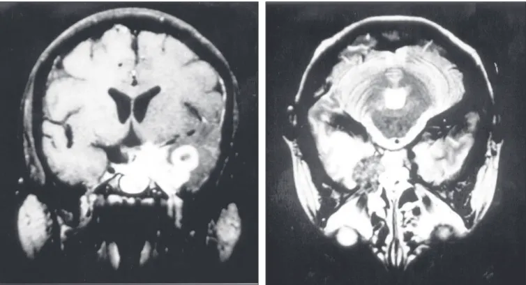
A computed tomographic scan revealed a tumor-like lesion with high density and mod-erate mass effect on the anterior left temporal lobe with edema in the surrounding area. It appeared to have moderate heterogeneous enhancement with iodide contrast solution. Magnetic resonance imaging showed also a mass over the left temporal lobe in its mesial aspects and infiltrating the left cavernous si-nus, with a high signal in the T1-weighted
• Eberval Gadelha Figueiredo • Erich Fonoff
• Marcos Gomes • Emílio Macedo • Raul Marino Júnior
Tumoral form of aspergillosis
in central nervous system
(cerebral aspergilloma):
case report
Division of Neurosurgical Clinic, Hospital das Clínicas, Faculdade de
Medicina, Universidade de São Paulo, São Paulo, Brazil
Aspergillosis of the central nervous system is an un-common infection, mainly occurring in immuno-compromised patients. It may be presented in several forms: meningitis, mycotic aneurysms, infarcts and the tumoral form (aspergilloma). The authors report a case of a diabetic patient with cerebral aspergilloma.
KEY WORDS: Aspergillosis. Diabetes mellitus. As-pergillus. Central nervous system neoplasms.
series (Figure 1). The surrounding edema was best observed in the T2 series (Figure 2).
After compensating for her diabetes, she underwent surgery by means of temporopari-etal craniotomy. The lesion encountered had a light yellowish color, hard consistency and very significant bleeding when manipulated. The intraoperative pathological examination revealed fungal hyphae. The operation became protracted and resection was only partially performed.
During the postoperative period, she was given intravenous amphotericin B. She re-quired long treatment due to postoperative fungal meningitis that was probably elicited by the surgical manipulation.
○ ○ ○ ○ ○ ○ ○ ○ ○ ○ ○ ○ ○ ○ ○ ○ ○ ○ ○ ○
DISCUSSION
Cerebral aspergillosis is an infrequent medical condition, but it is still a potentially fatal disease. However, it has become more frequently diagnosed because of high rates of suspicion among immunocompromised pa-tients such as those with transplants and HIV-positive or aids patients, who show higher in-cidence of the condition. In a series of 1,730 transplanted patients, 60 were neurologically compromised and 18.3% of these had aspergilloma.2 A few more cases with the tumoral form in immunocompromised pa-tients have been reported by Shuper.3
The lungs are probably the entry por-tal. However, isolated central nervous sys-tem aspergillosis (no detectable pulmonary disease) has already been reported.4 The prognosis in such patients is very poor even with surgical and anti-fungal treatment, es-pecially when there is an association with
C
ase Repor
t
São Paulo Medical Journal — Revista Paulista de Medicina
252
C
ase Repor
t
other bacteria or infections.1,4
Aspergilloma is even rarer in patients who are apparently immunocompetent, although all cases described have had associated diseases such as diabetes mellitus or unhealthy habits like drug abuse.1,4
Diagnosis of aspergilloma in immuno-competent patients remains difficult because medical staff rarely suspects this condition.
Figure 1. Coronal magnetic resonance imaging of the brain in the T1-weighted series after
gadolinium injection, showing a lesion over the left temporal lobe in its mesial aspect and
infiltrating the left cavernous sinus, with high signal.
Figure 2. Magnetic resonance imaging of the brain in the T2 series, showing left temporal
lobe edema.
Although such patients have much better prognosis than for immunocompromised patients, the diagnosis is frequently missed or delayed.3
Neuroimaging gives some backing for considering such a diagnosis, although it does not bring any specific findings to light. The radiological findings in aspergilloma cases are the same as for brain abscesses and may have
an appearance similar to that of many tumors. There is frequently a ring-like en-hancement when contrast medium is in-jected. With magnetic resonance imaging, it appears as a tumoral lesion with a low signal in the T1-weighted series, with variable edema on the lesion periphery. Routine analysis of cerebrospinal fluid does not help in elucidating the etiology.
São Paulo Medical Journal — Revista Paulista de Medicina
253
Aspergiloma cerebral: Relato de caso
Aspergilose do sistema nervoso central é uma infecção rara, acometendo principalmente pa-cientes imunossuprimidos. Clinicamente pode se apresentar sob várias formas: menin-gite, aneurisma micótico, infartos cerebrais e
○ ○ ○ ○ ○ ○ ○ ○ ○ ○ ○ ○ ○ ○ ○ ○ ○ ○ ○ ○ ○ ○ ○ ○ ○ ○ ○ ○ ○ ○ ○ ○ ○ ○ ○ ○ ○ ○ ○ ○ ○ ○
RESUMO
Eberval Gadelha Figueiredo, MD. Assistant neurosur-geon, Clinical Neurosurgery Division, Hospital das Clínicas, Faculdade de Medicina, Universidade de São Paulo, São Paulo, Brazil.
Erich Fonoff, MD. Resident doctor, Clinical Neurosurgery Division, Hospital das Clínicas, Faculdade de Medicina, Universidade de São Paulo, São Paulo, Brazil.
Marcos Gomes, MD. Assistant neurosurgeon, Clinical Neu-rosurgery Division, Hospital das Clínicas, Faculdade de Medicina, Universidade de São Paulo, São Paulo, Brazil.
Emílio Macedo, MD. Resident doctor, Clinical Neurosur-gery Division, Hospital das Clínicas, Faculdade de Medicina, Universidade de São Paulo, São Paulo, Brazil.
Raul Marino Júnior, MD. Professor, Clinical Neurosur-gery Division, Hospital das Clínicas, Faculdade de Medicina, Universidade de São Paulo, São Paulo, Brazil.
Sources of funding: None
Conflict of interest: None
Date of first submission: April 22, 2003
Last received: May 26, 2003
Accepted: May 30, 2003
Address for correspondence:
Eberval Gadelha Figueiredo 3848, N 3rd Avenue Apt. 3069
Phoenix - Arizona 85013 USA
E-mail:[email protected]
COPYRIGHT © 2003, Associação Paulista de Medicina
○ ○ ○ ○ ○ ○ ○ ○ ○ ○ ○ ○ ○ ○ ○ ○ ○ ○ ○ ○
Publishing information
como lesão tumoral (aspergiloma). Os auto-res apauto-resentam o caso de um paciente diabé-tico com aspergiloma cerebral.
PALAVRAS-CHAVE: Aspergilose. Diabetes
mellitus. Aspergillus. Tumores cerebrais. Sis-tema nervoso central.
1. Boes B, Bashir R, Boes C, Hahn F, McConnell JR, McComb R. Central nervous system aspergillosis. Analysis of 26 patients. J Neuroimaging 1994;4(3):123-9.
2. Breneman E, Colford JM. Aspergillosis of the CNS
pre-○ ○ ○ ○ ○ ○ ○ ○ ○ ○ ○ ○ ○ ○ ○ ○ ○ ○ ○ ○ ○ ○ ○ ○ ○ ○ ○ ○ ○ ○ ○ ○ ○ ○ ○ ○ ○ ○ ○ ○ ○ ○ ○ ○ ○ ○ ○ ○ ○ ○ ○ ○ ○ ○ ○ ○ ○ ○ ○ ○ ○ ○ ○ ○
REFERENCES
senting as aseptic meningitis. Clin Infect Dis 1992; 15(4):737-8.
3. Shuper A, Levitsky HI, Cornblath DR. Early invasive CNS as-pergillosis. An easily missed diagnosis. Neuroradiology
1991;33(2):183-5.
4. Walsh TJ, Hier DB, Caplan LR. Aspergillosis of the central nervous system: clinicopathological analysis of 17 patients. Ann Neurol, 1985;18(5):574-82.