Ruling out intra-abdominal injuries in blunt trauma patients using
clinical criteria and abdominal ultrasound
Exclusão de lesões intra-abdominais em vítimas de trauma fechado através de
variáveis clínicas e ultrassom abdominal completo
Flávia Helena BarBosa Moura, asCBC-sP1; José Gustavo Parreira, tCBC-sP2,3; tHiara Mattos4; Giovanna ZuCCHini rondini4;
Cristiano Below4; JaCqueline arantes G. PerlinGeiro, tCBC-sP2,3; silvia Cristine soldá, tCBC-sP2,3; José Cesar asseF, tCBC-sP2,3.
INTRODUCTION
I
n Brazil, in 2015, more than 152,000 people lost their lives due to external causes1. However, this is just oneof the aspects of this problem2. During acute phase,
costs of medical care, hospitalization, exams and tre-atment must also be taken into account. It is expected that, in a broad sample of victims with blunt trauma, 2% to 3% have intra-abdominal lesions (IAL)3. Even in
asymptomatic patients with normal physical exam, it is possible to find potentially lethal intra-abdominal le-sions. This is one of the reasons why we consider non-diagnosed intra-abdominal lesions a frequent cause of “preventable” death in trauma patients. Therefore, image exams had been progressively introduced in the evaluation of victims of blunt trauma.
At present, FAST (Focused Assessment Sono-graphy for Trauma) is one of the first methods that can be used. This method uses ultrasound to detect free liquid at the abdominal cavity. However, a nega-tive exam does not exclude IAL. Also, total abdominal ultrasound, aside from free liquid, detects visceral le-sions. It is performed by radiologists, since the lear-ning curve is much longer. Total US presents, as main advantages, its low cost, availability, portability, the chance to reexam several times the same patient, and the lack of use of ionizing radiation or contrast. It may also present false-negative results, since it depends on the examiner and not always detects minor bleeding or intraperitoneal lesions4,5. Its result may be
compro-mised by gas interposition, obesity or empty bladder. Literature data show that US may fail in up to 10%
1 - Irmandade da Santa Casa de Misericórdia de São Paulo, Department of Surgery, São Paulo, SP, Brazil. 2 - Irmandade da Santa Casa de Misericórdia de São Paulo, Emergency Department, São Paulo, SP, Brazil. 3 - Faculty of Medical Sciences of Santa Casa de São Paulo, Department of Surgery, São Paulo, SP, Brazil. 4 - Faculty of Medical Sciences of Santa Casa de São Paulo, Medical School, São Paulo, SP, Brazil.
A B S T R A C T
Objective: to identify victims of blunt abdominal trauma in which intra-abdominal injuries can be excluded by clinical criteria and by com-plete abdominal ultrasonography. Methods: retrospective analysis of victims of blunt trauma in which the following clinical variables were analyzed: hemodynamic stability, normal neurologic exam at admission, normal physical exam of the chest at admission, normal abdomen and pelvis physical exam at admission and absence of distracting lesions (Abbreviated Injury Scale >2 at skull, thorax and/or extremities). The ultrasound results were then studied in the group of patients with all clinical variables evaluated. Results: we studied 5536 victims of blunt trauma. Intra-abdominal lesions with AIS>1 were identified in 144 (2.6%); in patients with hemodynamic stability they were present in 86 (2%); in those with hemodynamic stability and normal neurological exam at admission in 50 (1.8%); in patients with hemodynamic stability and normal neurological and chest physical exam at admission, in 39 (1.5%); in those with hemodynamic stability, normal neurological, chest, abdominal and pelvic physical exam at admission, in 12 (0.5%); in patients with hemodynamic stability, normal neurological, chest, abdominal and pelvic physical exam at admission, and absence of distracting lesions, only two (0.1%) had intra-abdominal lesions. Among those with all clinical variables, 693 had normal total abdominal ultrasound, and, within this group, there were no identified intra-abdom-inal lesions. Conclusion: when all clinical criteria and total abdominal ultrasound are associated, it is possible to identify a group of victims of blunt trauma with low chance of significant intra-abdominal lesions.
patients6,7.
Computer tomography (CT) has a sensitivity of 99% in some studies and is considered a better ac-curate method to diagnose traumatic intra-abdominal lesions, but also with disadvantages and limitations. CT not always detects pancreatic and intestinal le-sions8. Also, when compared to abdominal
ultrasou-nd, is less available, with higher cost, with no portabi-lity and with the risks of ionizing radiation and use of iodinated contrast9-12. Therefore, the attending
physi-cian must know how to use these image methods op-timally, balancing their advantages and disadvantages. In a large trauma center, the optimized use of US saves costs of more expensive exams and non-therapeutic procedures.
We hypothesize that the association of cli-nical variables and total abdominal ultrasound may be used to exclude intra-abdominal lesions in victims of blunt trauma, lowering the need of abdominal Ct. Therefore, the objective of our study is to evaluate the association of several clinical variables and abdominal ultrasound to exclude intra-abdominal lesions in vic-tims of blunt trauma.
METHODS
This study was approved by the Research Ethical Committee of Irmandade da Santa Casa de São Paulo, # 59542816.2.0000.5479. We performed a re-trospective analysis of information at the data bank of the Emergency Department, that were prospectively collected using standardized protocols of quality con-trol, from 2008 to 2010.
In the study, we included victims of blunt trauma with more than 14 years old. We revised iden-tification, trauma mechanism, general condition at ad-mission, exams, lesions, treatment and complications. Severity of trauma and lesions were stratified by the following scores: Coma Glasgow Scale (CGS), Revised Trauma Score (RTS), Abbreviated Injury Scale (AIS), Or-gan Injury Scale (OIS) and Injury Severity Score (ISS)13.
The routine protocol of objective evaluation of abdomen of victims of blunt trauma in our Emer-gency Department includes initial physical exam, ima-ge and laboratory exams. Imaima-ge exams include FAST,
US and CT; the latter is selectively obtained depending on the risk evaluation of abdominal lesion of the at-tending physician. Laboratory tests include white cells count, serum amylase, arterial blood gases, according to severity of trauma. Leukocytosis, hyperamylasemia and metabolic acidosis (base deficit inferior to -6mEq/L) suggest the presence of lesions eventually not identified by the image exams.
In our study, we selected clinical variables evaluated at admission: hemodynamic stability (HS), normal neurological exam at admission (NNEx), nor-mal chest physical exam (NTEx), nornor-mal abdominal and pelvic physical exam (NAPEx) and absence of distracting lesions (ADL). HS was considered when SBP>100mmHg and CR<100bpm. NNEx was consi-dered when patient was conscious, oriented, Coma Glasgow Scale 15. NTEx was considered when phy-sical exam showed no signs of thoracic trauma and patients had no symptoms of thoracic lesions. NAPEx was considered when patient was asymptomatic and abdominal and pelvic physical exams showed no pain or signs of local trauma (bruises, escorting, hemato-mas). Distracting lesions were those AIS>2 at skull, thorax or extremities.
Those criteria were progressively overlapped to select the group with the smallest possibility to pre-sent IAL with AIS>1. Those variables are easily identi-fied during initial evaluation of trauma patients, and are useful tools for decision making. They were de-termined by the most senior surgical resident (R3/R4) along with attending physicians. After that, we analy-zed the US results in the group of patients with all clinical evaluated variables.
We also performed a comparison of frequen-cy of IAL with AIS>1 among patients with or without evaluated variables, using the chi-square test, consi-dering statistically significant when p<0.05. We cal-culated the Odds Ratio and 95% confidence interval for absence of intra-abdominal lesions according to clinical variables.
ac-curacy of the model obtained by logistic regression.
RESULTS
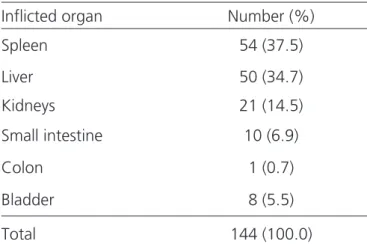
During the studied period, 5536 patients vic-tims of blunt abdominal trauma were consulted. IAL were identified in 172 patients (3.1%). Of patients with abdominal AIS>1, there were 144 (2.6%) patients with IAL, mainly parenchymal organs (Table 1).
Table 1. AIS>1 intra-abdominal lesions in 144 patients victims of blunt trauma.
Inflicted organ Number (%)
Spleen 54 (37.5)
Liver 50 (34.7)
Kidneys 21 (14.5)
Small intestine 10 (6.9)
Colon 1 (0.7)
Bladder 8 (5.5)
Total 144 (100.0)
Table 2. Association of analyzed variables and frequency of intra-abdo-minal lesions.
Variable Number of patients
IAL frequency (%)
HS 4290 86 (2.0)
NNEx 3419 78 (2.3)
NTEx 4998 117 (2,3)
NAPEx 4945 73 (1.5)
ADL 4431 64 (1.4)
HS + NNEx 2834 50 (1.8)
HS + NNEx + NTEx 2577 39 (1.5)
HS + NNEx + NTEx +
NAPEx 2356 12 (0.5)
HS + NNEx + NTEx +
NAPEx + ADL 2031 4 (0.2)
HS: hemodynamic stability; NNEx: normal neurological exam at admission; NTEX: normal thorax exam at ad-mission; NAPEx: normal abdominal and pelvic exams at admission; ADL: absence of distracting lesions.
Table 3. Abdominal ultrasound positivity to intra-abdominal lesions (AIS>1).
Inflicted organ (absolute number)
total US (performed)
Positive US n (%)
Liver (50) 28 27 (96.4)
Spleen (54) 33 32 (96.9)
Kidney (21) 9 8 (88.9)
Small intestine (10) 3 2 (66.7)
Bladder (8) 2 2 (100.0)
Total 75 71 (94.6)
Considering only the 4290 patients with HS, IAB were present in 86 (2%). In 2834 patients with HS and NNEx, IAL were diagnosed in 50 (1.8%). In 2577 patients with HS, NNEX and NTEx , IAL were present in 39 (1.5%). Of 2356 patients with HS, NNEx, NTEx and NAPEx, IAL were found in 12 (0.5%). Of 2031 patients with HS, NNEx, NTEx, NAPEx and ADL, only two had IAL (two splenic lesions: one not treated by surgery and another submitted to splenectomy) (Table 2).
In the group with all clinical variables, 693 had normal US, and, in this group, there were no IAL. In patients with abdominal AIS>1 submitted to US ac-cording to our protocol, US reached 94.6% of positi-vity, identifying 71 of 75 possible IAL (Table 3).
At figure 1, we observed the comparison of frequency of IAL with AIS>1 among patients with and without studied clinical variables. All comparisons were statistically significant, p<0.001. The highest
DISCUSSION
Trauma is a disease where it is observed ex-change of energy between external environment and human body, that may cause lesions in all organism. It is a World epidemics. Trauma disease is the major cause of loss of productive years, since it afflicts mainly young people in most productive years2. Early identification
and treatment of lesions assure better prognosis. Our data confirm low frequency of intra-abdominal lesions in a cohort of victims of blunt trauma (2.6%). However, the analysis of such lesions show that they could add morbidity and mortality if not identified on time.
Many studies tried to stablish guidelines to exclude intra-abdominal lesions using clinical crite-ria3,12,14-18. These clinical markers were capable to
indi-cate the presence of intra-abdominal lesions, but with low performance to exclude them. In clinical practice, “exclusion” of IAL is a very frequent problem. This is the reason why image methods are used to comple-ment correct abdominal evaluation in victims of blunt trauma.
CT is the gold-standard exam to identify pos-sible IAL, with up to 99% of sensitivity. However, it has some disadvantages: higher cost and long hospi-talization time. In centers with high daily number of patients, the use of CT must be efficient, without a significant number of negative exams during evaluation of victims of blunt trauma; also, it exposes patients to ionizing radiation, and present risks inherent to the use of intravenous contrast. At present, the best protocol
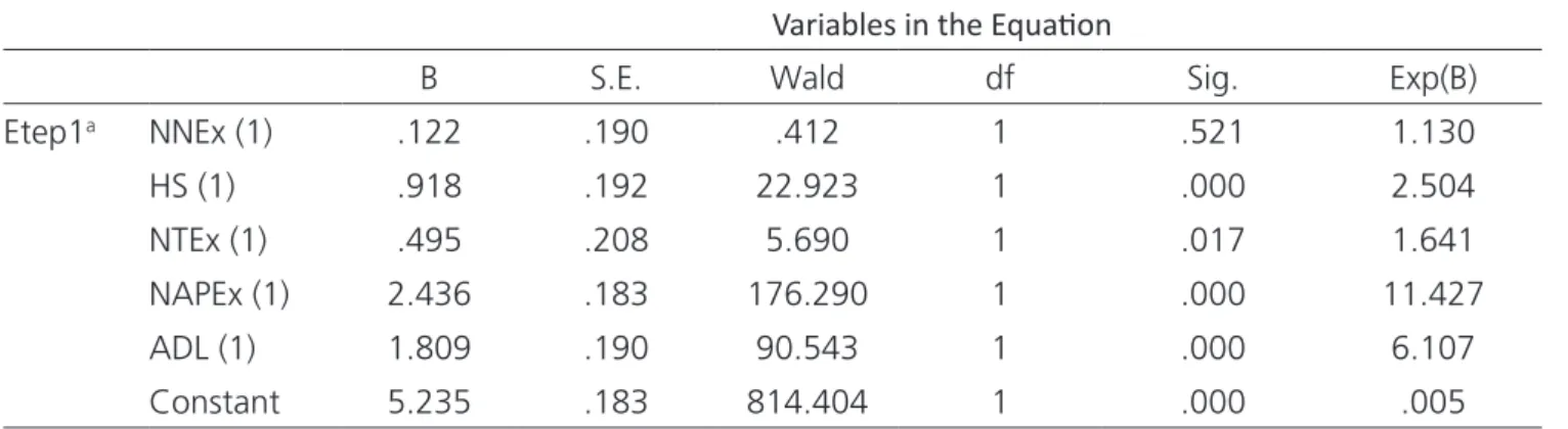
Table 4. Logistic regression by the “Enter” method of analyzed variables.
Variables in the Equaion
B S.E. Wald df Sig. Exp(B)
Etep1a NNEx (1) .122 .190 .412 1 .521 1.130
HS (1) .918 .192 22.923 1 .000 2.504 NTEx (1) .495 .208 5.690 1 .017 1.641
NAPEx (1) 2.436 .183 176.290 1 .000 11.427 ADL (1) 1.809 .190 90.543 1 .000 6.107
Constant 5.235 .183 814.404 1 .000 .005
Variables entered on step 1: NNEx: normal neurological exam at admission; HS: hemodynamic stability; NTEx: nor-mal chest exam at admission; NAPEx: nornor-mal abdominal and pelvic exam at admission; ADL: absence of distractive lesions.
Figure 1. Comparison of frequency of IAL>1 among groups.
O.R.: Odds ratio. 95% Confidence Interval.
Figure 2. ROC curve for the model with clinical variables.
for selective use of CT based on clinical, laboratorial and image methods (such as FAST and US)3,9-12 has not
been determined. Among these, FAST is the method with lower sensitivity. However, it is the only availa-ble resource in the trauma room, important in hemo-dynamically unstable patients. Usually, FAST does not identify 25% of IAL, and its accuracy varies from 60 to 80%3,19. US has a sensitivity of up to 90% if performed
by a talented radiologist. When we associate US to cli-nical variables, sensitivity reach that of CT, as shown by some studies3,5,6,15,20.
Our objective met this necessity. We believe that the use of clinical variables and associated total ab-dominal US may present an adequate accuracy. When we chose those clinical variables, we observed practi-cality. We selected those that could be evaluated at trauma room without the need of complex resources. During initial evaluation, we observe hemodynamics status, and perform thoracic, abdominal, neurological and extremities exams. In front of doubtful evalua-tion of thorax or pelvis exams, X-rays may be ordered. Therefore, during initial consultation, it is possible to identify the following variables: hemodynamic stability (HS), normal neurological exam at admission (NNEx), normal chest exam at admission (NTEx), normal abdo-minal and pelvic physical exam at admission (NAPEx) and absence of distracting lesions (ADL).
The choice of these variables took into ac-count previous studies that associated the presence of intra-abdominal lesions to hemodynamic stability, thoracic lesions, as well as pelvic, extremities and in-tracranial lesions12. It is important to highlight that
ab-dominal physical exam may be normal, even in patients with potentially lethal intra-abdominal lesions. This can be explained by the presence of associated cranial-en-cephalic trauma (CET), distracting lesions or use of sedatives at admission (for example, for oral-tracheal intubation) that can misguide clinical exam18,21.
There-fore, the doubt to perform image exams is observed in patients with normal abdominal exam but with other indicators of abdominal lesion.
Sharples and Brohi22, in 2016, revised
litera-ture and identified seven important studies. Sensitivity of several tools to detect intra-abdominal lesions varied from 86% to 100%. In our study, the sequential
addi-tion of studied variables (HS + NNEx + NTEx + NAPEx + ADL) resulted in a frequency of 0.1% of abdominal le-sions. If associated to US, all lesions with AIS>1 would be identified (100% sensitivity).
Holmes et al.23, in 2009, evaluated a model
that included clinical data (Glasgow Coma Scale <14, pain at costal arches, abdominal pain and femur frac-ture) and laboratory tests such as hematocrit and urine exam. This study, that included 1595 patients during validation phase, presented a tool with negative pre-dictive value of 98.6%. In our analysis, we chose not to include laboratory exams, that would add a significant time to the process. Even without these exams, we had a significant accuracy.
Nishijima et al.24, in 2012, add to clinical and
laboratory exams the results of bedside ultrasound. These authors reported that the presence of intra-ab-dominal free liquid was the best marker of lesion, over-coming clinical data and laboratory exams. However, the absence of liquid did not exclude the presence of intra-abdominal lesions. This study reinforces the idea to associate clinical data and image exams, such as in our study and of other authors15.
Chardoli et al.25, in 2017 used the absence
of clinical markers, ultrasound (FAST) and laboratory alterations in intra-abdominal lesions as a criteria for hospital discharge of patients victims of blunt trauma without CT. These authors interviewed by phone these patients after one week and none of 158 patients had symptoms of undiagnosed abdominal lesions. Our data also favor the identification of a subgroup of victims of blunt trauma with minimal chance of intra-abdomi-nal lesions that could be discharged without CT scan. This would optimize the available resources of crow-ded emergency centers without compromising patient safety. There are data that show that significant IAL are apparent up to nine hours after trauma26. Maybe
this variable can be included in future studies in order to also limit the use of abdominal ultrasound in these patients.
REFERENCES
1. Brasil. Ministério da Saúde. DATASUS. Indicadores de mortalidade: óbitos por causa externa [Internet]. Brasília (DF): Ministério da Saúde; 2015 [citado 2017 Aug 2017]. Disponível em: http://tabnet.datasus.gov. br/cgi/tabcgi.exe?sim/cnv/ext10uf.def
2. Reichenheim ME, de Souza ER, Moraes CL, de Mello Jorge MH, da Silva CM, de Souza Minayo MC. Violence and injuries in Brazil: the effect, progress made, and challenges ahead. Lancet. 2011;377(9781):1962-75. 3. Farrath S, Parreira JG, Olliari CB, Silva MA, Perlingeiro
JA, Soldá SC, et al. Identifying severe abdominal injuries during the initial assessment in blunt trauma patients. Rev Col Bras Cir. 2013;40(4):305-11.
4. Stengel D, Rademacher G, Ekkernkamp A, Güthoff C, Mutze S. Emergency ultrasound-based algorithms for diagnosing blunt abdominal trauma. Cochrane Database Syst Rev. 2015;(9):CD004446.
5. Feyzi A, Rad MP, Ahanchi N, Firoozabadi J. Diagnostic accuracy of ultrasonography in detection of blunt abdominal trauma and comparison of early and late ultrasonography 24 hours after trauma. Pak J Med Sci.
2015;31(4):980-3.
6. Brown MA, Sirlin CB, Hoyt DB, Casola G. Screening ultrasound in blunt abdominal trauma. J Intensive Care Med. 2003;18(5):253-60.
7. Nural MS, Yardan T, Güven H, Baydin A, Bayrak IK, Kati C. Diagnostic value of ultrasonography in the evaluation of blunt abdominal trauma. Diagn Interv Radiol. 2005;11(1):41-4.
8. Melamud K, LeBedis CA, Soto JA. Imaging of Pancreatic and Duodenal Trauma. Radiol Clin North Am. 2015;53(4):757-71, viii.
9. Colling KP, Irwin ED, Byrnes MC, Reicks P, Dellich WA, Reicks K, et al. Computed tomography scans with intravenous contrast: low incidence of contrast-induced nephropathy in blunt trauma patients. J Trauma Acute Care Surg. 2014;77(2):226-30.
10. James MK, Schubl SD, Francois MP, Doughlin GK, Lee SW. Introduction of a pan-scan protocol for blunt trauma activations: what are the consequences? Am J Emerg Med. 2017;35(1):13-19.
11. Radwan MM, Abu-Zidan FM. Focussed Assesment Sonograph Trauma (FAST) and CT scan in blunt abdominal trauma: surgeon’s perspective. Afr Health There may be also possible disagreement of a particular
variable by the attending physicians. One of the major limitations is the lack of a “true” negative result, that is, our protocol may have not identified lesions and we were not informed of new admissions for that reason. Since our team is responsible for daily follow up of all
victims of trauma, probably we would be aware of this fact. Also, other studies are needed, particularly pros-pective, to validate our data.
Our final message is that it is possible to as-sociate clinical variables and US to exclude IAL with AIS>1 in victims of blunt trauma.
Objetivo: identificar vítimas de trauma fechado de abdome nas quais as lesões intra-abdominais possam ser excluídas por critérios clí-nicos e por ultrassonografia abdominal completa. Métodos: análise retrospectiva de vítimas de trauma fechado em que se analisou as seguintes variáveis clínicas: estabilidade hemodinâmica, exame neurológico normal à admissão, exame físico do tórax, do abdome e da pelve normais à admissão e ausência de lesões distrativas (Abbreviated Injury Scale >2 em crânio, tórax e/ou extremidades). Em seguida estudou-se o resultado da ultrassonografia no grupo de pacientes com todas as variáveis clínicas avaliadas. Resultados: estudamos 5536 vítimas de trauma fechado. Lesões intra-abdominais com AIS>1 foram identificadas em 144 (2,6%) casos; em pacientes com estabilidade hemodinâmica, estavam presentes em 86 (2%); naqueles com estabilidade hemodinâmica e exame neurológico normal à admissão em 50 (1,8%); nos casos com estabilidade hemodinâmica, exame neurológico e do tórax normais à admissão em 39 (1,5%); em pacientes com estabilidade hemodinâmica e com exame neurológico, do tórax, do abdome e da pelve normais em 12 (0,5%); naque-les com estabilidade hemodinâmica e com exame neurológico, do tórax, do abdome e da pelve normais e ausência de naque-lesões distrativas, em apenas dois (0,1%) pacientes. Nos pacientes com todas as variáveis clínicas, 693 apresentavam ultrassonografia abdominal completa normal e, neste grupo, não foram identificadas lesões intra-abdominais posteriormente. Conclusão: pela somatória de critérios clínicos e ultrassonografia abdominal completa, é possível identificar um grupo de vítimas de trauma fechado com baixa chance de apresentar lesões intra-abdominais significativas.
Sci. 2006;6(3):187-90.
12. Farrath S, Parreira JG, Perlingeiro JA, Solda SC, Assef JC. Predictors of abdominal injuries in blunt trauma. Rev Col Bras Cir. 2012;39(4):295-300.
13. Pereira Jr GA, Scarpelini S, Basile-Filho A, Andrade JI. Índices de trauma. Medicina, Ribeirão Preto. 1999;32(3):237-50.
14. Deunk J, Brink M, Dekker HM, Kool DR, van Kuijk C, Blickman JG, et al. Routine versus selective computed tomography of the abdomen, pelvis, and lumbar spine in blunt trauma: a prospective evaluation. J Trauma. 2009;66(4):1108-17.
15. Dehqanzada ZA, Meisinger Q, Doucet J, Smith A, Casola G, Coimbra R. Complete ultrasonography of trauma in screening blunt abdominal trauma patients is equivalent to computed tomographic scanning while reducing radiation exposure and cost. J Trauma Acute Care Surg. 2015;79(2):199-205.
16. Mackersie RC, Tiwary AD, Shackford SR, Hoyt DB. Intra-abdominal injury following blunt trauma. Identifying the high-risk patient using objective risk factors. Arch Surg.1989;124(7):809-13.
17. Deunk J, Brink M, Dekker HM, Kool DR, Blickman JG, van Vugt AB, et al. Predictors for the selection of patients for abdominal CT after blunt trauma: a proposal for a diagnostic algorithm. Ann Surg. 2010;251(3):512-20. 18. Karamercan A, Yilmaz TU, Karamercan MA, Aytaç
B. Blunt abdominal trauma: evaluation of diagnostic options and surgical outcomes. Ulus Travma Acil Cerrahi Derg. 2008;14(3):205-10.
19. Richards JR, McGahan JP. Focused Assessment with Sonography in Trauma (FAST) in 2017: what radiologists can learn. Radiology. 2017;283(1):30-48.
20. Brown MA, Casola G, Sirlin CB, Patel NY, Hoyt DB. Blunt
abdominal trauma: screening us in 2,693 patients. Radiology. 2001;218(2):352-8.
21. Parreira JG, Malpaga JMD, Olliari CB, Perlingeiro JAG, Soldá SC, Assef JC. Predictors of “occult” intra-abdominal injuries in blunt trauma patients. Rev Col Bras Cir. 2015;42(5):311-7.
22. Sharples A, Brohi K. Can clinical prediction tools predict the need for computed tomography in blunt abdominal? A systematic review. Injury. 2016;47(8):1811-8.
23. Holmes JF, Wisner DH, McGahan JP, Mower WR, Kuppermann N. Clinical prediction rules for identifying adults at very low risk for intra-abdominal injuries after blunt trauma. Ann Emerg Med. 2009;54(4):575-84. 24. Nishijima DK, Simel DL, Wisner DH, Holmes JF. Does
this adult patient have a blunt intra-abdominal injury? JAMA. 2012;307(14):1517-27.
25. Chardoli M, Rezvani S, Mansouri P, Naderi K, Vafaei A, Khorasanizadeh M, et al. Is it safe to discharge blunt abdominal trauma patients with normal initial findings? Acta Chir Belg. 2017;117(4):211-215.
26. Jones EL, Stovall RT, Jones TS, Bensard DD, Burlew CC, Johnson JL, et al. Intra-abdominal injury following blunt trauma becomes clinically apparent within 9 hours. J Trauma Acute Care Surg. 2014;76(4):1020-3.
Received in: 15/08/2017
Accepted for publication: 28/09/2017 Conflict of interest: none.
Source of funding: none.
Mailing address:
José Gustavo Parreira