65
Pakistan Veterinary Journal
ISSN: 0253-8318 (PRINT), 2074-7764 (ONLINE)
Accessible at: www.pvj.com.pk
Ultrastructure of the Interstitial Tissue in the Testis of the Egyptian Dromedary Camel
(
Camelus dromedarius
)
M. I. Abd-Elaziz, A. M. Kassem, D. M. Zaghloul*, A. E. Derbalah and M. H. Bolefa
Histology and Cytology Department, Faculty of Veterinary Medicine, Alexandria University, Egypt *Corresponding author: [email protected]
A R T I C L E H I S T O R Y A B S T R A C T
Received: Revised: Accepted:
July 06, 2011 August 08, 2011 August 15, 2011 Key words:
Camel
Interstitial tissue Leydig cells Myofibroblasts Seasons Testis
The ultrastructural examination of the testicular interstitial tissue of Egyptian dromedary camel was performed to observe the seasonal changes. The activity of the interstitial tissue increased largely in spring. This was indicated by the large number of mature Leydig cells and two to three layers of myofibroblasts around the basal laminae of the seminiferous tubules with large blood vessels in the interstitial tissue. The testicular activity was moderate in winter as indicated by the lower number of immature Leydig cells. The lowest activity was in summer when Leydig cells became inactive with pyknotic nuclei. The cells of interstitial tissue lost their junctions with each other, leaving large intercellular spaces and myofibroblasts transformed to fibrocytes. The testicular activity began again to increase in autumn. The testicular activity of camel, however, did not stop in any season of the year, because even in non-breeding seasons a part of the interstitial tissue of the testis was active.
©2011 PVJ. All rights reserved To Cite This Article: Abd-Elaziz MI, AM Kassem, DM Zaghloul, AE Derbalah and MH Bolefa, 2012. Ultrastructure of the interstitial tissue in the testis of the Egyptian dromedary camel (Camelus dromedarius). Pak Vet J, 32(1): 65-69.
INTRODUCTION
The camel testis undergoes fluctuations in weight and marked histological changes which are related to age of the animal and season of the year (Abdel-Raouf et al., 1975; Osman et al., 1979; Hafez et al., 2011). Records had shown that testicular weight reached a seasonal peak during the rutting months (Singh and Bharadwaj, 1978ab; Tingari et al., 1979). There was also an increase in the volume and activity of the interstitial cells of Leydig, as revealed by their ultrastructure. Also quantitative and qualitative changes in the morphology of the camel testis were studied by Abdel-Raouf et al. (1975) in relation to age and season. The number of Sertoli cells was almost constant, but the numbers of mature Leydig cells, compared to the numbers of pre-Leydig and immature Leydig cells, increased by the end of winter so that, during the spring, the interstitial cells were mainly of mature type. Degenerative changes with diminished numbers of mature cells were seen in the summer and this trend continued into early and mid-autumn.
The boundary tissue of the semineferous tubules of camel was described by Moniem et al. (1980) that, it consisted of three lamellae; inner fibrous, inner cellular and outer cellular. The inner lamella was subdivided into two homogenous layers enclosing a third one that contained collagenous fibers and fine filaments. The inner cellular
lamella consisted of several layers of myoid cells; each layer was separated from the adjacent layer by homogenous material and varying amounts of collagen. The outer cellular lamella consisted predominantly of fibrocytes together with some fibroblasts and scattered collagen. Tingari et al. (1984) and Zayed et al. (1995) showed that the ratio of semineferous tubules to interstitial tissue was greater in summer than in winter months, and the boundary tissue investing the seminiferous tubules was thicker during the period from June to August as compared to November to January, when the spermatogenic activity was at its peak. The interstitial tissue showed wide variation from month to month in its components. The main components were predominantly Leydig cells with some fibroblasts, collagenous fibers, lymphatic vessels and blood capillaries (Abd-Elmaksoud et al., 2008). Al-Qarawi and El-Belely (2004) reported that there were characteristic interstitial and peritubular groups of mast cells as a component of the testicular interstitium of camel. The aim of the present study was to describe the seasonal effects of the desert environment on the structure of the testicular interstitial tissue of the Egyptian dromedary camel.
MATERIALS AND METHODS
dromedarius). The specimens were collected from Kom-Hamada slaughterhouse of El-Behera province along the four seasons of the year. Specimens were grossly examined for any pathological changes and only the apparently healthy ones were selected. The testes were exposed and dissected free from the surrounding tissues shortly after death. Specimens were taken from the middle portion. Pieces of 1 mm thickness were cut from the testis and quickly fixed in 6% solution of phosphate buffered gluteraldehyde (pH 7.4) for 6 hours at 4ºC (McDowell and Trump, 1976).
After initial fixation, the tissues were washed in several changes of cold (4ºC) 0.1 M phosphate buffer every 15 minutes for 2 hrs. The tissues were post fixed in 1% solution of osmium tetroxide in cold (4ºC) 0.1 M buffer (pH 7.2) for 2 hrs. Then, they were rapidly dehydrated through ascending grades of ethyl alcohol, transferred to propylene oxide and placed in a 1:1 mixture of propylene oxide and epoxy araldite (Hayat, 1986).
Semi-thin sections (1 µm) cut initially were stained with toludine blue and under light microscope to select the suitable areas for the electron microscopic examination. The ultrathin sections (60-100 nm) were cut by a glass knife with LKB microtome, and stained with uranyl acetate followed by lead citrate (Hayat, 1986). These sections were examined with Joel 100 cx transmission electron microscope operating at 100 Kilovolts.
RESULTS
Ultrastructural examination of the testis of camel collected during winter revealed that the interstitial cells consisted mainly of mature Leydig cells and a low number of pre-Leydig or immature Leydig cells (Fig. 1). Leydig cells had a spherical nucleus containing extended chromatin, smooth and rough endoplasmic reticulum and many cytoplasmic vacuoles (Fig. 2). The interstitial tissue also contained fibroblasts and fibrocytes which could be seen just beneath the basal lamina of the seminiferous tubules (Fig. 3). In this season some Leydig cells showed myelin figures indicating a degenerative change. Blood vessels also could be seen in the interstitial tissue (Fig. 4).
Samples of testes collected in spring revealed predominantly mature Leydig cells. These cells were more active with many large vacuoles indicative of the lipids. These vacuoles of different sizes were evenly interspersed throughout the cytoplasm in close association with the smooth endoplasmic reticulum (Fig. 5). Dense cyto- plasmic granules and smooth endoplasmic reticulum were also seen in the cytoplasm of Leydig cells. The nuclei of Leydig cells were large spherical (Fig. 5) and sometimes triangular with a corrugated nuclear envelop and clumps of condensed chromatin, with extended chromatin in between (Fig. 6). About two to three layers of myofibroblasts were present under the basal lamina of the seminiferous tubules. The basal laminae were thick and associated with collagen fibers (Fig. 7). These myofibro- blasts had vacuolated cytoplasm, some dense cytoplasmic granules (Fig. 8) and tonofilaments. Large blood vessels could be seen in the interstitial tissue in this season.
Samples collected during summer showed that Leydig cells decreased in number leaving empty intertubular spaces (Fig. 9). The cells of interstitial tissue were shrunken
with loss of junctions between them and the interstitial tissue became mainly a few connective tissue fibers and fibroblasts. Also some testicular tissue showed pyknotic nuclei of Leydig cells indicating degeneration (Fig. 9) in addition to a few number of pre-Leydig and immature Leydig cells. Myofibroblasts almost disappeared and transformed into fibroblasts and fibrocytes.
In autumn, the interstitial cells began to increase and filled the interstitial spaces. Leydig cells became active with spherical nuclei having extended chromatin and many cytoplasmic vacuoles and dense granules. Blood vessels were clear and large in size. Myofibroblasts were seen with a contracted nuclei and the basal lamina of the seminiferous tubules were thick (Fig. 10).
DISCUSSION
During this study we found that Leydig cells of camel testicular interstitial tissue contained well developed smooth endoplasmic reticulum in winter and spring seasons which reached to maximum number in spring. The most remarkable morphological characteristic found in all interstitial cells of Leydig of the vertebrate testis was the abundance of smooth endoplasmic reticulum, in the interstitial cells as reported in laboratory animals (Goldblatt and Gunning, 1985), mouse (Werner et al., 1991) and camel (Al-Qarawy et al., 2001). The majority of enzymes of steroid biosynthesis were found in smooth endoplasmic reticulum (Baker et al., 2003).
During spring and summer seasons the mature Leydig cell had many vacuoles indicative of extracted lipid, these vacuoles were evenly distributed in cytoplasm with different sizes. The variation of size of vacuoles seemed to be correlated with the phases of the secretory cycle of the individual cells in which cholesterol was utilized as a substrate for steroid biosynthesis (Baker et al., 2003). We found that some vacuoles were in close association with smooth endoplasmic reticulum. Similar results have been reported in camel (Tingari et al., 1979) and mice (Baker et al., 2003).
In the present work we noticed myelin figures in the cytoplasm of Leydig cells in winter season. Friedlander et al. (1994) observed that during the mating season there was a drastic reduction of smooth endoplasmic reticulum and proliferation of myelin figures within Leydig cells which disrupt at the end of their differentiation. Knobil and Neill (2006) stated that poorly digested substances remained within Leydig cells lysosomal system and were recognized as dense membrane bound granules commonly seen as heterogeneous conglomerates of myelin figures.
In summer, Leydig cells decreased in number leaving empty intertubular spaces while in autumn Leydig cells became active again with cytoplasmic vacuoles and dense granules in their cytoplasm. It can, therefore, be suggested that Leydig cells of camel maintained some degree of activity throughout the year being maximal during rutting seasons (spring and winter). Tingari et al. (1979) reported same results in camel and Pasha et al. (2011) proved it by ultrasonography.
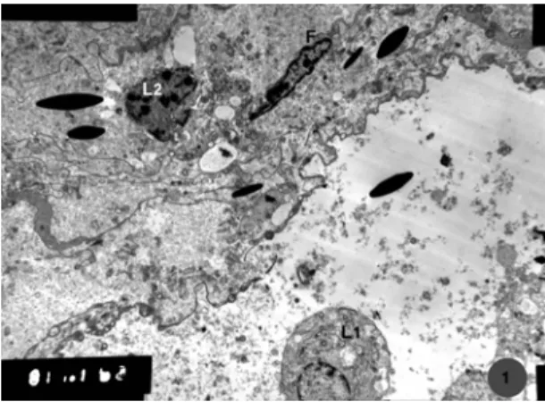
Fig.1: Transmission electron micrograph of the interstitial tissue of camel testis showing, mature (L1) and immature (L2) Leydig cells and fibroblast (F): Winter season (Mic.Mag. X 2500).
Fig. 2: Transmission electron micrograph of a Leydig cell in the testicular
interstitial tissue showing, the spherical nucleus (N), smooth endoplasmic reticulum (sER), rough endoplasmic retriculum (rER) and cytoplasmic vacuoles (v): Winter season (Mic. Mag. X 10000).
Fig. 4: Transmission electron micrograph of the testicular interstitial
tissue showing, Leydig cells (L) with myelin figures (arrows), blood vessel (Bv): Winter season (Mic. Mag. X 4000).
Fig. 5: Transmission electron micrograph of a Leydig cell showing, large
spherical nucleus (N) with a corrugated nuclear envelop (arrows) and clumps of condensed chromatin (asterisk) with extended chromatin in between. Many vacuoles (v) are present with different sizes, large electron dense membranous granules (g) and well developed smooth endoplasmic reticulum (sER): Spring season (Mic. Mag. X 10000).
Fig. 3: Transmission electron micrograph of the testicular interstitial
tissue of camel showing, a fibrocyte (F) under the basal lamina (BL) of the seminiferous tubules. Note the Sertoli cell (S): Winter season (Mic. Mag. X 5000).
Fig. 6: Transmission electron micrograph of another Leydig cell showing,
Fig. 7: Transmission electron micrograph of a myofibroblast (MF) around the basal lamina (BL) of the seminiferous tubules. They form 2-3 layers. Their nuclei (N) are elongated and somewhat contracted: Spring season (Mic. Mag. X 5000).
Fig. 8: Transmission electron micrograph of a myofibroblast in the
testicular interstitial tissue between the seminiferous tubules showing, part of the nucleus (N), with clear nucleoli, smooth (sER) and rough (rER) endoplasmic reticulum, cytoplasmic vesicles (ve) with different sizes and some dense granules (g). Note the Golgi complex (arrow) near the nucleus: Spring season (Mic. Mag. X 10000).
cells) varied from species to species. In rat, hamster and mice only one layer of myoid cells was seen in the testis. They added that myofibroblasts not only provided structural integrity to tubules but also took part in the regulation of spermatogenesis and the testicular function. Verhoeven et al. (2000) added that myofibroblasts contributed to the contractile activity of testicular tubules and maintained mesenchymal epithelial interaction with Sertoli cells both by cooperation in the deposition of extracellular matrix elements and by secretion of paracrine agonist.
Conclusions
The ultrastructure of the testicular interstitial tissue of Egyptian dromedary camel showed activity all over the year but reached its peak at spring, moderate in winter, decreased in autumn and least activity was noted in summer. The basal lamina of the seminiferous tubules were surrounded by two to three layers of myofibroblasts for contractile activity especially in the season of testicular activity and mainly in summer they transformed to thin fibrocytes.
Fig. 9: Transmission electron micrograph of the interstitial tissue of the
seminiferous tubules showing the wide intercellular spaces (asterisks), shrinked cells and loss of junctions between them. The nuclei with condensed chromatin (arrows), in addition to the very low number of cytoplasmic organelles. Some collagen fibers (Co) could be seen: Summer season (Mic. Mag. X 4000).
Fig. 10: Transmission electron micrograph of testicular interstitial
tissue showing, myofibroblast (MF) with a contracted nucleus (N). Note the thick basal lamina (BL) of the seminiferous tubules and collagen fibers (Co) under it: Autumn season (Mic. Mag. X 4000).
REFERENCES
Abd-Elmaksoud A, A Sayed-Ahmed, M Kassab and K Aly, 2008. Histochemical mapping of glycoconjugates in the testis of the one
humped camel (Camelus dromedarius) during rutting and
non-rutting seasons. Acta Histochem, 110: 124-133.
Abdel-Raouf M, MF El-Bab and MM Owaida, 1975. Studies on
reproduction in the camel (Camelus dromedarieus). V. Morphology of
the testis in relation to age and season. J Reprod Fertil, 43: 109-116. Al-Qarawi AA, HA Abdel-Rahman, MS El-Belely and SA El-Mougy, 2001.
Intratesticular morphometric, cellular and endocrine changes around the pubertal period in dromedary camels. Vet J, 162: 241-249.
Al-Qarawi AA and MS El-Belely, 2004. Intratesticular, morphometric, cellular and endocrine changes in dromedary bulls exhibiting azoospermia. Vet J, 167: 194-201.
Baker P, H Johnston, M Abel, HM Charlton and PJ O'Shaughnessy, 2003. Differentiation of adult-type Leydig cells occurs in gonadotrophin-deficient mice. Reprod Biol Endocrinol, 1: 4.
Friedlander M, A Rosenstrauch and E Bedrak, 1994. Leydig cell differentiation during the reproductive cycle of the seasonal
breeder Camelus dromedarius: an ultrastructural analysis. Gen
Comp Endocrinol, 55: 1-11.
Hafez SA, SM El-Shafey and T Caceci, 2011. Histological and lectin histochemical characterization of the efferent ductules in the
dromedary (Camelus dromedarius). J Vet Anat, 4: 51-67.
Hayat MA, 1986. Basic Techniques for Transmission Electron
Microscope. 2nd Ed, Academic Press, Baltimore, USA.
Knobil E and JD Neill, 2006. Knobil and Neill`s Physiology of
Reproduction. Vol 1, 3rd Ed, Gulf Professional Publishing,
Maekawa M, K Kamimura and T Nagano, 1996. Peritubular myoid cells in the testis: their structure and function. Arch Histol Cytol, 59: 1-13. McDowell EM and BF Trump, 1976. Histologic fixatives suitable for
diagnostic light and electron microscopy. Arch Pathol Lab Med, 100: 405-415.
Moniem KA, MD Tingari and E Kunzel, 1980. The fine structure of the
boundary tissue of the seminiferous tubule of the camel (Camelus
dromedarius). Acta Anatomica, 107: 169-176.
Osman DI, KA Moniem and MD Tingari, 1979. Histological observations on the testis of camel, with special emphasis on spermatogenesis. Acta Anatomica, 104: 164-171.
Pasha RH, AS Qureshi, LA Lodhi and H Jamil, 2011. Biometric and ultrasonographic evaluation of the testis of one-humped camel
(Camelus dromedarius). Pak Vet J, 31: 129-133.
Singh UB and MB Bharadwaj, 1978a. Morphological changes in the testis and
epididymis of camel (Camelus dromedarius). Acta Anat, 101: 275-279.
Singh UB and MB Bharadwaj, 1978b. Histological and histochemical
studies on the testis of the camel (Camelus dromedarius) during the
various seasons and ages. Acta Anat, 101: 280-288.
Tingari MD, KA Moniem and E Kunzel, 1979. Observations on the fine
structure of the testicular interstitial cells in the camel (Camelus
dromedarius). Q J Exp Physiol Cong Med Sci, 64: 39-45.
Tingari MD, AS Ramos, ES Gaili, BA Rahma and AH Saad, 1984. Morphology of the testis of the one humped camel in relation to reproductive activity. J Anat, 139: 133-143.
Verhoeven G, E Hoebenand and K De Gendt, 2000. Peritubular cell-Sertoli cell interactions: factors involved in PModS activity. Andrologia, 32: 42-45.
Werner G, B Haben and K Neumann, 1991. The fine structure of testicular interstitial cells in the mouse. A comparison after conventional double fixation versus rapid freezing followed by freeze substitution. J Submicrosc Cytol Pathol, 23: 405-414. Zayed AE, A Hifny, A Abou-Elmagd and KH Wrobel, 1995. The
seasonal changes in the intertubular tissue of the camel testis