Investigation of the Genes Involved in Antigenic
Switching at the
vlsE
Locus in
Borrelia burgdorferi
: An
Essential Role for the RuvAB Branch Migrase
Ashley R. Dresser1, Pierre-Olivier Hardy2, George Chaconas1,2*
1Department of Biochemistry & Molecular Biology, The University of Calgary, Calgary, Alberta, Canada,2Department of Microbiology & Infectious Diseases, The
University of Calgary, Calgary, Alberta, Canada
Abstract
Persistent infection by pathogenic organisms requires effective strategies for the defense of these organisms against the host immune response. A common strategy employed by many pathogens to escape immune recognition and clearance is
to continually vary surface epitopes through recombinational shuffling of genetic information. Borrelia burgdorferi, a
causative agent of Lyme borreliosis, encodes a surface-bound lipoprotein, VlsE. This protein is encoded by thevlsElocus
carried at the right end of the linear plasmid lp28-1. Adjacent to the expression locus are 15 silent cassettes carrying
information that is moved into thevlsElocus through segmental gene conversion events. The protein players and molecular
mechanism of recombinational switching atvlsEhave not been characterized. In this study, we analyzed the effect of the
independent disruption of 17 genes that encode factors involved in DNA recombination, repair or replication on
recombinational switching at thevlsElocus during murine infection. InNeisseria gonorrhoeae, 10 such genes have been
implicated in recombinational switching at thepilElocus. Eight of these genes, includingrecA, are either absent fromB. burgdorferi, or do not show an obvious requirement for switching at vlsE. The only genes that are required in both
organisms areruvA and ruvB, which encode subunits of a Holliday junction branch migrase. Disruption of these genes
results in a dramatic decrease invlsErecombination with a phenotype similar to that observed for lp28-1 orvls-minus
spirochetes: productive infection at week 1 with clearance by day 21. In SCID mice, the persistence defect observed with
ruvAandruvBmutants was fully rescued as previously observed forvlsE-deficientB. burgdorferi. We report the requirement of the RuvAB branch migrase in recombinational switching atvlsE, the first essential factor to be identified in this process. These findings are supported by the independent work of Lin et al. in the accompanying article, who also found a
requirement for the RuvAB branch migrase. Our results also indicate that the mechanism of switching at vlsE in B.
burgdorferiis distinct from switching atpilEinN. gonorrhoeae, which is the only other organism analyzed genetically in
detail. Finally, our findings suggest a unique mechanism for switching at vlsE and a role for currently unidentified B.
burgdorferiproteins in this process.
Citation:Dresser AR, Hardy P-O, Chaconas G (2009) Investigation of the Genes Involved in Antigenic Switching at thevlsELocus inBorrelia burgdorferi: An Essential Role for the RuvAB Branch Migrase. PLoS Pathog 5(12): e1000680. doi:10.1371/journal.ppat.1000680
Editor:Jenifer Coburn, Medical College of Wisconsin, United States of America
ReceivedJune 3, 2009;AcceptedNovember 4, 2009;PublishedDecember 4, 2009
Copyright:ß2009 Dresser et al. This is an open-access article distributed under the terms of the Creative Commons Attribution License, which permits unrestricted use, distribution, and reproduction in any medium, provided the original author and source are credited.
Funding:This work was supported by a grant from the Canadian Institutes of Health Research (MOP-53086). G.C. is Scientist of the Alberta Heritage Foundation for Medical Research and holds a Canada Research Chair in the Molecular Biology of Lyme Borreliosis. The funders had no role in study design, data collection and analysis, decision to publish, or preparation of the manuscript.
Competing Interests:The authors have declared that no competing interests exist.
* E-mail: chaconas@ucalgary.ca
Introduction
Antigenic variation through targeted genome rearrangements is a common strategy for immune evasion and has been identified in many important pathogens including protozoa [1,2,3,4], bacteria [5,6,7,8,9,10,11] and fungi [12]. In spite of the common occurrence of this strategy for immune evasion amongst pathogens, few molecular details of the recombinational switching processes that generate diversity in antigen-expressing genes have been reported for any organism.
Lyme borreliosis is a world wide health problem. It is a
multisystemic illness caused by the spirochete Borrelia burgdorferi,
and related species. Disease progression occurs through three stages: early, disseminated and persistent and can result in various arthritic, cardiac and neurological concerns if left untreated
[13,14,15]. Persistent infection byB. burgdorferirequires continual
segmental gene conversion at the vlsE locus, which encodes a
35 kDa membrane lipoprotein [9,16,17,18,19]. ThevlsEgene, or
expression locus is carried at the right end of the linear plasmid
lp28-1. In the absence of lp28-1 or when thevlslocus is deleted a
productive murine infection ensues, but the spirochetes are cleared between days 8 and 21 post-infection [16,17,20,21]. Adjacent to
vlsE(also referred to asvls1), is a contiguous upstream array of 15
silent cassettes separated from each other by 17 bp direct repeats,
which also flank the vlsE variable region (see Fig. 1C in [18]).
During murine infection (and probably in other mammals) information is transferred unidirectionally from the silent cassettes into the expression site to generate diversity at six regions (VR1–
VR6) within the central region of thevlsEgene [17,18,19]. These
regions correspond to highly exposed regions of the VlsE protein and are believed to be prominently displayed antigenic areas [22]. Generation of antigen diversity occurs through segmental gene
conversion such that information from several silent cassettes can
be transferred into the singlevlsElocus to generate a mosaic gene
with possibilities for the production of myriad unique VlsE proteins. All silent cassettes are utilized as sequence donors in the
gene conversion events atvlsEand the majority of recombination
events are short, ranging from 1–22 codon changes [17]. Similarly, the requirement for flanking sequence homology is also short, in the neighborhood of approximately 10 nucleotides.
An interesting feature of switching atvlsE is that it does not
occur when spirochetes are grown in culture or when they reside in the tick midgut. [18,23]. Moreover, the acquired immune response is not required, as switching occurs in SCID mice, which lack the ability to mount an acquired immune response to antigenic challenge [9,16,17,20]. The mammalian signal that triggers recombinational switching remains unknown at this time.
These features make the study of antigenic variation in B.
burgdorferi difficult and limit these studies to animal infection models. In the mouse, antigenic switching can be detected four
days after infection and by 28 days no parental vlsE sequences
remain in the population of spirochetes recovered from some tissues in infected animals [9,17,20].
Even though B. burgdorferi has a small genome [24,25],
genetic manipulation is time consuming, inefficient and sometimes difficult [26]. The protein machinery that promotes
recombinational switching atvlsEis, therefore, unknown at this
time. A single study towards this end has reported that theB.
burgdorferi recAgene is not required for antigenic switching [27]. In this study we generated 17 mutants carrying disruptions in known DNA recombination, repair and replication genes in the hopes of identifying proteins involved in recombinational
switching atvlsE. A single recombination function, the RuvAB
Holliday junction branch migrase encoded by theruvAandruvB
genes, was unambiguously identified as a requirement for
switching at vlsE, a result also reported in the accompanying
paper by Lin et al [28]. In contrast, 10 known recombination, repair, or replication genes are required in recombinational
events underlying antigenic switching at pilE in N. gonorrhoeae
[29,30,31,32,33,34,35,36,37,38,39]. Eight of those genes are
either missing or not required for switching at vlsE in B.
burgdorferi. Our results point towards a unique mechanism for
switching atvlsEin and suggest that it may involve specialized
proteins that help to mediate the process.
Results
Construction of DNA repair and replication gene disruptions inB. burgdorferi
A systematic approach was undertaken to disrupt 21 different
genes in order to investigate their role invlsErecombination inB.
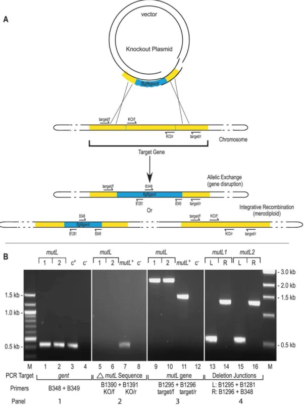
burgdorferi. Knockout plasmids were constructed (Figure S1) and
used to transform the infectiousB. burgdorferiB31 clone 5A4 [40].
Following transformation, allelic exchange results in successful
gene disruption (Fig. 1A). However two other transformation
outcomes can arise: integretative recombination, which results in merodiploid formation, and cases where no recombinants are
recovered [26]. To investigate the structure of the B. burgdorferi
transformants, they were screened using PCR with various primer
combinations (Fig. 1B). The presence of the gentamicin resistance
cassette (Panel 1) and the absence of the expected deleted
sequences (,500 bp) from the disrupted target gene (Panel 2)
were first confirmed using the indicated primer sets. The target
gene was also amplified (Panel 3) to confirm the approximate
0.7kb size increase relative to wild-type DNA due to the insertion of the gentamicin resistance cassette. Finally, the correct insertion site was verified using combinations of the target and knockout
primers to amplify the insertion boundaries (Panel 4). In addition
to the PCR analyses, gene disruptions were independently confirmed by Southern hybridizations using probes specific to the gentamicin resistance cassette and the deleted portion of the
target gene (seeFig. S2andTable S2).
Of the 21 DNA replication, repair and recombination gene
knockouts attempted, 17 were successful (Table 1). When a
disruption attempt was unsuccessful, the knockout plasmid was re-constructed in an effort to minimize possible effects on adjacent gene expression from read-through of transcription
from thegentcassette. This was accomplished by either changing
the polarity of the gentamicin resistance cassette relative to the gene target, or by adding (or removing) a T7 transcriptional terminator. Three gene targets required reconstruction of the
knockout plasmid in order to successfully obtainB. burgdorferigene
disruptions.recJwas first attempted without the T7 terminator in
the reverse orientation and resulted only in merodiploids. The gentamicin resistance cassette in the forward orientation with the T7 terminator did result in knockouts and further attempts were
halted. ThesbcDknockout was first attempted with a construct
containing the T7 terminator and the gentamicin resistance cassette in the reverse orientation. This attempt resulted only in merodiploids; however, when the polarity was changed to the
forward orientation, allelic exchange was successful. The recA
disruption was also difficult to obtain. Unsuccessful attempts were first made with the gentamicin resistance cassette in the forward orientation with and without the T7 terminator. When the T7
terminator was removed and the gent gene was in the reverse
polarity, true knockouts of therecAgene were obtained. Difficulty
in obtaining a recA gene disruption has also been previously
reported [41], however, a single recA null mutant has been
previously constructed with the insertion of a kanamycin
resistance cassette in the forward orientation [27]. Finally,dnaB,
hbb,recBandrecCknockouts were not obtained despite changing
the polarity of the gentamicin resistance cassette and adding or removing a T7 transcriptional terminator.
Effect ofB. burgdorferi genedisruptions on C3H/HeN mouse infections: an essential role forruvABin recombinational switching
For each gene disruption two clones were chosen which contained the full plasmid complement required for infectivity, Author Summary
A common strategy for evasion of the host immune system is the continuous variation of a major surface protein that elicits a dominant immune response (anti-genic variation). Many pathogens accomplish this goal by unidirectional movement of DNA sequence information from silent or archival gene copies into an expression site. The molecular details of how this gene shuffling is accomplished are not understood for any organism. In the flat-wave shaped bacterium causing Lyme disease, information is moved from 15 silent cassettes into thevlsE
gene to promote antigenic variation. In this work we have investigated the effect of independent mutation of 17 DNA replication, recombination and repair genes on the
movement of genetic information intovlsE. We found that
Figure 1. Gene disruption and confirmation. A)Gene disruption strategy. The infectiousB. burgdorferistrain B31, clone 5A4 (B31-5A4) was transformed with a knockout plasmid carrying a one kb gentamicin cassette (blue) that replaced the central portion of the target gene (yellow) as described in Materials and Methods. The two possible outcomes of recombination events with the target gene are shown: allelic exchange would result in gene disruption while integrative recombination of the knockout plasmid would result in merodiploid formation. The position of PCR primers used for construct verification are shown by arrows on the schematic.B)Construct verification of themutLdisruption by PCR. Each gene disruption was subjected to four PCR analyses.1)The presence of the gentamicin resistance cassette was confirmed as shown in lanes 1 and 2. The shuttle vector pBSV2G [71] served as the positive control c+for amplification of thegentcassette (lane 3.)2)The portion ofmutLexpected to be deleted in a gene
disruption was not detected in eithermutL1 or 2 (lanes 5 and 6); however, it was detected in the positive control (c+), which contained wild-type B31-5A4 DNA as a template in lane 7.3)The size of the target gene was compared inmutL1 and 2 genotypes. The expected 2.1 kb products for a gene disruption were observed (lanes 9 and 10) in comparison to the 1.5 kb product from themutL+
genotype (lane 11). Lanes 4, 8 and 12 are negative controls (c2) that lacked DNA template.4)Confirmation of the correct insertion site was performed using combinations of the target gene primers and primers internal to the gentamicin cassette to amplify the boundaries. The left boundary in bothmutLclones gave the expected 0.55 kb product (lanes 13 and 15). The right boundary in both clones gave the expected product of approximately 1.3 kb (lanes 14 and 16). A 100bp ladder on the left side, relevant to the two left panels, and a 1kb ladder on the right side, which applies to the two right panels, were the molecular weight markers (M) used.
doi:10.1371/journal.ppat.1000680.g001
Effect of Mutations on Switching atvlsE
as determined by PCR screening with primers specific to the plasmids. Most mutant constructs contained the full complement of plasmids found in the parental clone B31 5A4 [40]. Some of the
mutantB. burgdorfericlones lacked plasmids that are not required
for infection or persistence as follows:recJ1 lacks lp28-2,mag2lacks
lp28-4,ruvA1 is missing cp9, mutL2 lacks cp9 and cp32-3, nucA1
lacks lp21 and cp32-3,ruvB5lacks lp28-4 and cp9 andpriA3,recA2
andrecA3 are missing cp9. The possible effect of the mutations on
growth of B. burgdorferi in culture was assessed by performing
growth comparisons of each of the mutants with the wild-type clone 5A4. All mutants displayed growth curves that were indistinguishable from the parent strain (data not shown).
Two independent knockout clones for each mutation were used to infect C3H/HeN immunocompetent mice (at least two mice for each clone, four mice for each mutated gene) as described in Materials and Methods. Cultures were grown from blood samples at day 7 to monitor infectivity. At days 14 and 21, by which time spirochetes are largely cleared from the blood, ear biopsies were
used to monitor infection and switching atvlsE. The upper portion
of Table 2 shows mutant strains that displayed a productive infection ($75% of mice infected at day 7) that did not decline at day 21, the point post-infection when strains unable to switch at
vlsEhave been cleared [16,17,20,42]. The variable region ofvlsE
was amplified from DNA isolated from day 21 ear cultures and
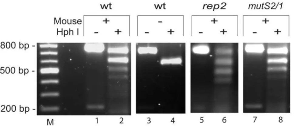
analyzed using RFLP assays (Fig. 2), which detect new restriction
sites resulting from switching at thevlsElocus [16]. As noted in the
upper portion ofTable 2,mutS2,recA,recG,rep,nucA,mag,mfdand
nthmutants all displayed switching at thevlsElocus.
The lower portion of Table 2 shows mutant strains that
displayed ,75% positive cultures from ear biopsies at day 21.
When spirochetes could be cultivated at day 21 (sbcD, sbcC and
BBG32) switching was monitored and shown to occur using the RFLP assay. For the remainder of the mutant strains, infections were allowed to continue until day 35. At this time the mice were euthanized and spirochetes cultivated from heart, bladder, joint
and ear. RFLP switching assays indicated that switching atvlsE
had occurred in all mutant strains from tissues where spirochetes could be recovered at day 35, with the exception of those carrying
theruvAorruvBmutations, whose functional genes encode the two
subunits for a Holliday junction branch migrase [43,44,45].
Although theruvAandruvBmutant strains recovered from organ
harvest at day 35 were negative for switching by RFLP, DNA sequencing analysis revealed that switching had occurred at low efficiency (data not shown).
Table 1.Gene disruption targets and knockout plasmid attributes.
Gene target Locus Gene description Plasmid
E. colistrain number (GCE)
Polarity of gent relative to target
T7 terminator
Gene
disruption Merodiploid
recJ BB0254 ssDNA-specific exonuclease pAD51
pAD26 1563 1538
forward reverse
+
2
+
2
2
+
priA BB0014 helicase pAD94 1908 forward + + 2
sbcD BB0829 exonuclease pAD87
pAD86 1599 1598
forward reverse
+ +
+
2
2
+
ruvA BB0023 Holliday junction helicase pAD78 1590 forward + + 2
mutL BB0211 mismatch repair protein pAD61 1573 forward + + 2
ruvB BB0022 Holliday junction helicase pPOH6 1618 forward 2 + 2
sbcC BB0830 exonuclease pAD65 1577 forward + + 2
BBG32 BBG32 putative helicase pAD88 1900 forward + + 2
mutS1 BB0797 mismatch repair protein pPOH2 1604 forward 2 + 2
mutS2 BB0098 mismatch repair protein pAD24 1536 reverse 2 + 2
recA BB0131 DNA-dependent ATPase
pAD101 pAD92 pAD102
1913 1904 1914
reverse forward forward
2
+
2
+
2 2
2 2 2
recG BB0581 ATP-dependent helicase pAD49 1561 reverse + + 2
rep BB0607 ssDNA-dependent ATPase helicase pAD53 1565 reverse + + 2
nucA BB0411 exonuclease involved in competency pAD63 1575 forward + + 2
mag BB0422 39-methyladenine DNA glycosylase pAD57 1569 forward + + 2
mfd BB0623 transcription-repair coupling factor pAD59 1571 reverse + + 2
nth BB0745 endonuclease III pPOH28-5 1684 reverse 2 + 2
dnaB BB0111 replicative helicase pAD80
pAD81 1592 1593
forward reverse
+ +
2 2
2 2
hbb BB0232 DNA-binding protein
pAD100 pAD99 pAD106
1912 1911 1918
reverse forward reverse
+ +
2
2 2 2
2 2 2
recB BB0633 exonuclease pAD22
pAD48 1534 1560
reverse reverse
2
+
2 2
2 2
recC BB0634 exonuclease
pAD84 pAD104 pAD103
1596 1916 1915
reverse forward reverse
+
2 2
2 2 2
2 2 2
Table 2.Effect of DNA repair and replication mutants onB. burgdorferiinefction and switching atvlsEin C3H/HeN mice. B. burgdorferi genotype Strain (GCB) Total
micea Day 7Bloodb Day 7Infection Day21 Ear Day 21Infection Switching atvlsEday 21c
5A4 wt 933 18 18/18 100.0% 18/18 +
mutS2/1 (BB0098) mutS2/2 1135 1136 4 2/2 2/2 100.0% 2/2
2/2 100.0% +
recA2 (BB0131) recA3 1284 1285 4 2/2 2/2 100.0% 2/2
2/2 100.0% +
recG1 (BB0581) recG2 1155 1156 4 2/2 2/2 100.0% 2/2
2/2 100.0% +
rep1 (BB0607) rep2 1158 1159 4 2/2 2/2 100.0% 2/2
2/2 100.0% +
nucA1 (BB0411) nucA2 1176 1177 4 2/2 1/2 75.0% 2/2
2/2 100.0% +
mag1 (BB0422) mag2 1161 1162 4 2/2 1/2 75.0% 2/2
1/2 75.0% +
mfd1 (BB0623) mfd2 1180 1181 4 2/2 1/2 75.0% 2/2
1/2 75.0% +
nth1 (BB0745)
nth2 525 526 4 2/2 2/2 100.0% 2/2
2/2 100.0% +
Day 35
Persistence at day$35
Switching at vlsEday 35f
Heart Bladder Joint Ear Total sitese
5A4 wtd 933 4/4 4/4 4/4 4/4 16/16 100.0%
+ recJ1 (BB0254) recJ5 1153 1154 4 2/2 2/2 100.0% 0/2
0/2 0% n/a
2/2 1/2 2/2 1/2 0/2 1/2 2/2 2/2 6/8
5/8 68.8% +
ruvB4 (BB0022) ruvB5 513 514 4 2/2 2/2 100.0% 0/2
0/2 0% n/a
0/2 0/2 0/2 1/2 0/2 1/2 0/2 0/2 0/8
2/8 12.5% 2
g ruvA1 (BB0023) ruvA2 1174 1175 4 2/2 2/2 100.0% 0/2
0/2 0% n/a
0/2 1/2 1/2 0/2 0/2 1/2 1/2 0/2 2/8
2/8 25.0% 2
g sbcD1 (BB0829) sbcD2 1251 1252 4 1/2 1/2 50.0% 0/2
1/2 25.0% +
1/2 1/2 1/2 1/2 1/2 1/2 1/2 1/2 4/8
4/8 50.0% +
sbcC2 (BB0830) sbcC3 1248 1249 4 2/2 0/2 50.0% 2/2
0/2 50.0% +
2/2 0/2 2/2 0/2 2/2 0/2 2/2 0/2 8/8
0/8 50.0% +
BBG32/6 BBG32/7 1233 1234 4 0/2 2/2 50.0% 0/2
2/2 50.0% +
0/2 2/2 0/2 2/2 0/2 2/2 0/2 2/2 0/8
8/8 50.0% +
priA2 (BB0014) priA3 1205 1206 4 1/2 0/2 25.0% 0/2
0/2 0% n/a
1/2 2/2 1/2 2/2 1/2 2/2 1/2 2/2 3/8
8/8 68.8% +
Effect ofB. burgdorferigene disruptions on SCID C3H/ HeN mouse infections
Mutants ofB. burgdorferistrains that did not show switching in
wild-type C3H/HeN mice using the RFLP assay (ruvAand ruvB)
and five other mutants that displayed a decreased persistence at
day 21 (recJ,mutL,sbcC,sbcDandBBG32) were used to infect SCID
C3H/HeN mice, which lack an acquired immune response. This effectively removes the selective pressure on antigenic variation
and allowsB. burgdorferimutants with defective switching atvlsEto
persist in the host. Direct analysis ofvlsEswitching beyond 21 days
post-infection can, therefore, be performed in SCID mice, whereas by this time non-switching spirochetes would be cleared in a wild-type mouse [16,17,20,42]. All the mutant strains tested displayed wild-type levels of infectivity and persistence throughout the 35
day course of infection in SCID mice (Table 3). This indicated
that theruvAandruvBmutant strains, which did not switch in
wild-type mice using the RFLP assay and which showed greatly
reduced levels of persistence at day 21 (Table 2, bottom), were
fully competent for the infection process in mice lacking an
acquired immune response. The other mutantB. burgdorferistrains
that displayed reduced infectivity and persistence were also fully rescued in mice lacking an acquired immune response.
Analysis of switching atvlsEby DNA sequencing of mutant strains recovered from SCID mouse infections
The RFLP assay used here provides a quick and convenient
assay method to detect switching atvlsE[16]. The incorporation of
new restriction endonuclease sites from the silent cassettes into the
variable region ofvlsEis a clear indicator of the switching process.
However, the fact that the assay is not quantitative, coupled with the observation that switching is apparently less frequent in SCID mice [17], led us to further analyze switching in SCID mice in a limited set of mutants by DNA sequencing. We chose the two mutants that were negative for switching in wild-type mice by
RFLP analysis (ruvA andruvB) as well as two mutants that were
shown to switch by RFLP at 35 days in wild-type mice, but that
displayed no spirochetes in ear cultures at 21 days (recJandmutL).
For DNA sequencing studies the same PCR product used for
the RFLP assay (a 776 bp fragment containing thevlsEvariable
region) was amplified from the spirochetes recovered from four different tissue types at day 35 for each SCID mouse and gel purified (see Material and Methods). Equimolar amounts of the
vlsEPCR product from each tissue from the 4 mice used in the
infections were combined, providing four pools for each disrupted gene: heart, bladder, joint and ear. The pools were cloned and 10 E. coliclones were chosen for each tissue type for a total of 40vlsE
sequences examined for each of therecJ,mutL,ruvA,ruvBand
wild-type genowild-types. Using a primer specific to the cloning vector, the
plasmid DNA was sequenced and compared to theB. burgdorferi
5A4 parentalvlsEsequence for both templated (present in a silent
cassette) and non-templated nucleotide changes [17,19]. Each
sequenced clone where switching atvlsEhad occurred displayed a
unique sequence; hence, all switch variants from each mouse represented independent switching outcomes.
Sequencing revealed that 10 out of 10 wild-type clones contained nucleotide changes corresponding to sequences found in the silent cassettes in the heart and bladder tissue cultures while 5/10 and 8/10 clones had switched in the joint and ear tissues,
respectively (see Fig. 3). These results are similar to previously
reported data which indicated a greater proportion of switched clones in heart, bladder and skin tissues than in joint and ear tissues [17]. The overall switching frequency that we observed (82.5% at day 35 post-infection in SCID C3H/HeN mice) also
correlates closely with the value of 85% at 28 days post-infection recently observed [17]. For proper analysis, a tissue-specific comparison between mutants and wild-type spirochetes was
undertaken as shown inFig. 3.
The most significant reduction in switching occurred withruvA
andruvBmutants with only one of 40 clones (2.5%) differing from
the wild-type vlsEsequence in ruvAand no changes observed in
any of the 40 ruvB clones. The single clone demonstrating
switching atvlsEin the ruvAmutant was from a clone cultivated
from joint that displayed at least four exchanges with silent cassettes and did not show any features with obvious differences
from switching in wild-typeB. burgdorferi. The P-values indicated a
significant difference (,0.05) in the incidence of switching for all
tissues, with the exception of theruvAmutant in joint. These results
corroborated the negative switching phenotype of the ruvA and
ruvBmutants observed in the RFLP switching assay after infection
of wild-type and SCID mice (Tables 2and 3). The results are
further strengthened by the fact thatruvAandruvBencode the two
subunits of an enzyme known to promote branch migration of Holliday junction recombination intermediates.
Clones carrying a mutL mutation displayed an intermediate
phenotype with a decrease in switching resulting in a total of only 27.5% of the clones exhibiting nucleotide changes, versus 82.5% for wild-type. Significant tissue-specific differences were observed
in the bladder and heart but not the joint and ear (Fig. 3).recJ
showed a slight change in the level of switching with 57.5% of the
clones displaying changes in thevlsEvariable region compared to
85% for wild-type. A significant difference in tissue-specific Figure 2. Restriction fragment length polymorphism assay for switching atvlsE. A portion of thevlsE expression site containing the variable regions was amplified using primers B248 and B249 to give a product of 776 bp. PCR reactions were performed onB. burgdorferigrown from ear biopsies taken at day 21 and the products were digested with HphI and run on a 1.2% agarose gel in TAE buffer at 75V for 1.5 hours and stained with ethidium bromide (see Materials and Methods). Wild-typeB. burgdorferiB31-5A4 recovered following infection of a C3H/HeN mouse was used as a template in lanes 1 and 2. An unswitched template (not exposed to mouse infection) is shown in lanes 3 and 4. PCR products fromrep2andmutS2/ 1 DNA templates are found in lanes 5 & 6, and 7 & 8 respectively. M denotes a 100bp molecular weight marker.
doi:10.1371/journal.ppat.1000680.g002
Table 3.Effect of DNA repair and replication mutations onB. burgdorferiinfection in SCID C3H/HeN mice.
B. burgdorferi genotype
Strain (GCB)
Total micea
Day 7 Bloodb
Day 7 Infection
Day 21 Ear
Day 21
Infection Day 35
Persistence at day 35
Heart Bladder Joint Ear
Total sitesc
5A4 wt 933 6 6/6 100.0% 6/6 100.0% 6/6 6/6 6/6 6/6 24/24 100.0%
recJ1 (BB0254) recJ5
1153
1154 4
2/2
2/2 100.0%
2/2
2/2 100.0%
2/2 2/2
2/2 2/2
2/2 2/2
2/2 2/2
8/8
8/8 100.0%
ruvB4 (BB0022) ruvB5
513
514 4
2/2
2/2 100.0%
2/2
2/2 100.0%
2/2 2/2
2/2 2/2
2/2 2/2
2/2 2/2
8/8
8/8 100.0%
ruvA1 (BB0023) ruvA2
1174
1175 4
2/2
2/2 100.0%
2/2
2/2 100.0%
2/2 2/2
2/2 2/2
2/2 2/2
2/2 2/2
8/8
8/8 100.0%
mutL1 (BB0211) mutL2
1178
1179 4
1/2
1/2 50.0%
2/2
2/2 100.0%
2/2 2/2
2/2 2/2
2/2 2/2
2/2 2/2
8/8
8/8 100.0%
sbcD1 (BB0829) sbcD2
1251
1252 4
2/2
2/2 100.0%
2/2
2/2 100.0%
2/2 2/2
2/2 2/2
2/2 2/2
2/2 2/2
8/8
8/8 100.0%
sbcC2 (BB0830) sbcC3
1248
1249 4
2/2
2/2 100.0%
2/2
2/2 100.0%
2/2 2/2
2/2 2/2
2/2 2/2
2/2 2/2
8/8
8/8 100.0%
BBG32/6 BBG32/7
1233
1234 4
2/2
2/2 100.0%
2/2
2/2 100.0%
2/2 2/2
2/2 2/2
2/2 2/2
2/2 2/2
8/8
8/8 100.0%
aTotal number of mice examined for genotype.
bValues listed correspond to number of cultures positive/number of sites tested.
cNumber of positive tissue sites/number of sites tested.
doi:10.1371/journal.ppat.1000680.t003
Effect of Mutations on Switching atvlsE
switching was only observed in the heart. Switching sequencing data were also analyzed by counting the number of nucleotide
changes in each clone (Table S3) and gave similar results (see also
Discussion). DNA sequencing was also performed on the vlsE
variable region from infections with sbcC, sbcD and BBG32 B.
burgdorferimutants, which displayed wild-type switching levels in all
four (sbcC) tissues or in three of the four four (sbcDand BBG32)
tissue types (data not shown).
In addition to the apparent templated nucleotide changes
observed in switches at vlsE, some non-templated changes
(NTCs), where new sequence atvlsEdid not correspond to the
sequence found in any of the silent cassettes, were also observed
[17,46]. There were no NTCs in the total of 80vlsEsequences
analyzed for wild-type and ruvB clones. In ruvA, four NTCs
were observed in non-switching clones and three in the single
clone that switched. In themutLmutant there were five NTCs
and all of these occurred in clones that did not switch. Finally
the recJ heart sample had six NTCs in three clones, two of
which switched. Taken together there were a total of 18 NTCs
in theruvA,mutL and recJmutants.
Discussion
Attempts were made to generate 21 disruptions inB. burgdorferi
genes believed to be involved in DNA recombination, repair and/ or replication to investigate their possible roles in recombinational
switching at the vlsE locus. Seventeen genes were successfully
disrupted. Three of these gene disruptions (recJ, sbcD and recA)
required either reversing the polarity of the gentamicin resistance cassette or adding (or removing) a T7 transcriptional terminator.
Previous attempts atrecA insertional mutation have either been
unsuccessful [41] or have resulted in a single clone [27]. In this
study multiplerecAdisruptions were obtained only in the absence
of a T7 terminator and with theaacC1in the reverse orientation
relative torecA, underscoring the importance of the transcriptional
features (direction of expression and the presence or absence of a transcriptional terminator) in the drug resistance cassette when
attemptingB. burgdorferigene disruptions. It is also worthy of note
that although our gene disruption mutants carry both an,500 bp
deletion of the target gene as well as an insertion of theaacC1gene,
expression of a partial gene product with some level of functionality cannot be rigorously ruled. Antiserum to the 17 disrupted genes is not currently available and immunoblotting experiments could therefore not be performed.
In addition to the 17 successful gene disruptions, we were
unsuccessful in obtaining disruptions of dnaB (a replicative
helicase), recB or recC (subunits of a recombinational helicase/
nuclease) andhbb(an accessory factor with properties ofE. coliHU
and IHF, which introduces sharp bends in DNA [47,48,49]).
Whether these genes are essential functions forB. burgdorferi, as is
expected fordnaB, or whether our gene disruption conditions for
these loci are still not ideal remains to be established. The primary purpose for the construction of mutants described here was to study the possible role of genes in question on recombinational
switching at the vlsE locus. The effect of the 17 mutations on
generalized recombination and DNA repair is currently under study and will be reported elsewhere.
Candidate genes involved in antigenic variation were identified by mouse infections in C3H/HeN and SCID mice. Initial Figure 3. Number of switchedvlsEclones in SCID C3H/HeN
mice. Sequencing of the cloned PCR product of thevlsEvariable regions using primer pJET1.2/forward was performed on 10 clones from each tissue type culture for each genotype (see Materials and Methods). The y-axis denotes the number of clones out of ten that contained templated nucleotide changes in variable regions 1–6 (switches) and the x-axis denotes the tissue type. The P-values
above the bars indicate the level of significance of the difference between the wild-type and mutant samples, calculated using Fisher’s Exact test.
screening for switching utilized an RFLP assay in wild-type mice followed by DNA sequencing of clones recovered from organ harvest cultures from SCID mice at 35 days post-infection. Analysis of the number of switching events and the source (silent
cassette) of the new sequences atvlsEis often difficult to establish
due to sequence redundancy in different cassettes and the complexity of the resulting switch genotypes (see [17]); hence, an exact method for determining the number of switches in all clones does not exist. Sequencing data was, therefore, analyzed by two independent methods. First, the number of clones that contained switches for each genotype was counted and compared to that of
wild-typeB. burgdorferi(Fig. 3). This approach provides
informa-tion on the number of clones where recombinainforma-tional switching occurs, but the number of switching events or extent of recombinational switching in each clone is not considered. The second method involved counting the number of nucleotide changes as an approximate indicator of the degree of switching (Table S3). Because switching events atvlsEusually involve short stretches of DNA [17], this method is expected to provide a reasonable estimate of the degree of switching. Although the binary alternative outcome for the first method results in a preferred statistical analysis, it is not currently known which of the two methods of estimating the extent of switching is more accurate. As discussed below, the data and conclusions from both methods of analysis were concordant.
Finally, it is noteworthy that other than theruvABmutants, which
are required for switching atvlsE, the remainder of the genes we
disrupted were dispensable for animal infection. The major DNA assault expected by a microbe upon animal infection is oxidative DNA damage originating from the innate immune response.
However, due to a lack of iron inB. burgdorferi, oxidative damage of
DNA appears to be much lower than in other organisms, such asE.
coli[50]. Hence, DNA repair functions may be less important in
protecting the pathogen from exogenous sources of DNA damage. Nonetheless, it is of interest that several of the recombination, repair and replication mutants examined in this study displayed altered infection phenotypes that were not attributable to an obvious deficit
invlsEswitching in C3H/HeN mice (Table 2). Various levels of
decreased infectivity were observed at days 7, 21 and 35 in comparison with the 100% infectivity displayed by all 18 wild-type control mice. Surprisingly, wild-type levels of infectivity for these mutants was restored in SCID C3H/HeN mice lacking an acquired
immune response (Table 3). The mechanism for decreased
infectivity in wild-type that is rescued in SCID mice remains open to speculation at this time.
recAis not required for recombinational switching atvlsE Homologous recombination in bacteria is typically initiated by RecA-mediated pairing and strand invasion [51,52,53]. It has been
previously reported that arecAgene disruption inB. burgdorferidid not
affect switching atvlsE[27]. Because therecAgene inB. burgdorferiis
not easily disrupted [41] and because a single clone with the disrupted
gene was used to assess the role ofrecAin recombinational switching at
vlsE[27], we constructed several recAknockouts and tested two of
them for switching at thevlsElocus. Our results confirm the previous
findings that recA is not required [27]. This raises the interesting
question of how pairing and strand invasion is initiated for
recombinational switching at the vlsE locus. The lack of a
requirement for the RecA protein, and the unidirectional segmental
gene conversion events that characterize switching at thevlslocus in
B. burgdorferiargue for the need of a specialized protein(s) to help mediate the process. The expendability of RecA for antigenic
variation in B. burgdorferi is also a stark difference from antigenic
variation systems in other organisms, as will be discussed below.
A role for theruvABencoded branch migrase in recombinational switching atvlsE
The RuvAB complex is required for homologous recombination and facilitates ATP-dependent branch migration of heteroduplex DNA in Holliday junctions [43,44,45]. RuvA tetramers bind and unfold the heteroduplex DNA and recruit RuvB hexamers, which function as a helicase to move DNA through the RuvAB complex. B. burgdorferiRuvA and RuvB share 32% and 48% identity with
theirE. coliorthologues, respectively.ruvABmutants inE. colihave
only modest defects in homologous recombination. However, these defects become significant when there are also mutations in
other recombination proteins, such as recBC, recG and sbcBC
[54,55]. The role of RuvAB in DNA repair and recombination has
not been previously investigated inB. burgdorferi.
The observed infection phenotype of bothruvAand both ruvB
mutant strains reported here was as previously observed for strains
lacking either lp28-1 [16,17,20,21] or thevlslocus [16,17,20,21],
where switching cannot occur. Infection of wild-type mice was 100% at day 7 with apparent complete clearance at day 21
(Table 2). Complete rescue of the persistence defect forruvmutant
strains was observed in all cases in SCID mice (Table 3). A difference in phenotype between strains lacking either lp28-1 or
thevlslocus, with those carrying aruvmutation is that at 35 days
post-infection spirochetes could be recovered in some organ
harvest cultures from wild-type mice infected with theruvmutants.
DNA sequence analysis revealed that a single switch variant was present in spirochetes from a given tissue, or from both tissues in the two mice where positive cultures were recovered from two sites. These results are indicative of low frequency switching in the mutants, resulting in occasional survival and selection of a single switch variant. This phenotype has also been observed by [28].
The phenotype and the dramatic inhibition of switching atvlsE
(Fig. 3) of the strains carrying theruvAandruvBgene disruptions in this study identify the first protein factors involved in switching and support a mechanism involving branch migration of a
recombination intermediate for antigenic variation inB. burgdorferi.
Although we were unable to complement ourruvmutations (data
not shown), genetic complementation inB. burgdorferiis frequently
difficult to achieve, for reasons not currently understood. We nonetheless argue for the absence of secondary mutations in the
mutantB. burgdorferistrains based upon the following arguments: 1)
Our studies were performed with two independent mutations in
both theruvAandruvBgenes. The four independent mutant strains
demonstrated the same phenotype, making the existence of secondary mutations exceedingly remote. 2) Although not a strict genetic complementation, the rescue of persistent infectivity in all
four ruv mutants following the infection of SCID mice is
compelling evidence for the absence of any secondary mutations
affecting infectivity. 3) Similar results and conclusions with ruv
mutants made by transposon mutagenesis in the accompanying idependent study from the Norris lab [28] corroborate the findings presented here and our combined results provide compelling
evidence for a RuvAB role in switching atvlsE.
A companion protein to RuvAB in most bacteria is the Holliday
junction resolving enzyme RuvC.B. burgdorferidoes not encode a
RuvC orthologue and has no characterized junction resolving enzyme. This leaves unanswered the question of how recombina-tion intermediates involved in homologous recombinarecombina-tion or
switching at vlsE are processed. A series of putative LE family
exonucleases encoded by the cp32 family of circular plasmids has
been proposed as possible substitutes for RuvC in B. burgdorferi
[56]. The actual function of these l exonuclease-type proteins
remains to be established and simultaneous disruption of all of
Effect of Mutations on Switching atvlsE
them (,9) is outside the realm of possibility with current genetic
methods available forB. burgdorferi.
A possible role formutLin recombinational switching at vlsE?
mutL and recJ are both players in bacterial mismatch repair
[57,58,59]. InE. coliMutL acts as a liaison between MutS, which
recognizes the mismatch, and MutH which is responsible for
introducing a nick on either side of the mismatch. BothmutS1 and
mutS2are present in the B. burgdorferigenome, but disruption of either gene did not affect the infectivity phenotype in wild-type mice or switching as assayed by RFLP. There is no identifiable
mutHorthologue inB. burgdorferi[25]. Disruption ofmutLresulted
in a modest decrease in switching at vlsE that was significant
(Fig. 3) or near significant (Table S3) in heart and bladder but not in ear and joint. The results did not allow a clear-cut conclusion on the involvement of MutL in switching as demonstrated for RuvA and RuvB. Further analyses will be required to derive an unambiguous answer to this question. It is possible that MutL plays a role in recombinational switching at vlsE, but that anotherB. burgdorferiprotein can substitute for MutL because of functional redundancy. In such a case a double knockout will be required for further investigation; however, a
functional paralogue of MutL has not been identified in B.
burgdorferi at this time. It is also possible that the reduction in
switching inmutLmutants results from a decreased level of fitness
and a slower growth rate of the mutant in the mouse(Table 2).
RecJ is a 59 to 39 exonuclease that in E. coli can promote
mismatch excision and prepares DNA for strand invasion by
creating the single-stranded 39-overhang [53]. Disruption of recJ
resulted in a decrease in infectivity in wild-type mice at 21 days
and a modest decrease in switching atvlsEthat was significant only
in the heart for both methods of analysis (Fig. 3andTable S3).
Again, the results did not permit an unambiguous conclusion as to
the possible involvement of RecJ in switching atvlsE. The slightly
decreasedvlsEswitching phenotype observed inB. burgdorfericould
be a result of redundancy of function forrecJandrecDas has been
previously reported inE. coli[60,61]. ArecD recJdouble mutant, if
viable, might provide further information regarding the role ofrecJ
in vlsE recombination. Alternatively, the decrease in switching
might simply reflect a decreased level of fitness and infectivity of the mutant spirochete in the mouse and there may be no direct
role of RecJ in switching atvlsE.
Finally, it is noteworthy that mutL and recJ were the only
genotypes sequenced that also contained non-templated nucleotide
changes (NTCs).mutLhad five NTCs whilerecJhad six for a total
of eleven across 7 clones. The NTCs in this study occurred predominantly in invariable regions where nucleotide changes are not normally observed. An explanation for why NTCs were only
observed inmutLandrecJcould be due to their involvement in the
mismatch repair pathway. These data suggest that these repair
proteins are normally operative atvlsEto correct mismatches and
would, therefore, normally be temporally and spatially positioned to play a role in the switching process as well. Recent work on
switching atvlsEhas reported that approximately 15% of wild-type
vlsE variants carry NTCs [17]. This was not observed in our
sequencing data with wild-type, ruvAorruvB mutant clones. We
have no explanation for this discrepancy.
Comparison with other antigenic variation systems While a wide variety of bacterial and protozoan pathogens employ antigenic variation systems driven by gene conversion [5,6], the molecular details of the recombinational events underlying the process remain largely obscure. Information about
some of the protein factors required for gene conversion events are
available only from studies on the bacterial pathogenN. gonorrhoeae
and the protozoan parasite Trypanosoma brucei. Both of these
organisms require either RecA [37] or paralogues of its eukaryotic RecA counterpart, Rad51 [62,63]. In contrast, both this study and a previous one [27] have shown that RecA is not necessary for
switching at vlsE in B. burgdorferi. In N. gonorrhoeae the recFOR
pathway is also involved and disruptions inrecQ,recO,recRorrecJ
result in elimination or fairly dramatic reductions in antigenic
variation [30,32,33]. In contrast,B. burgdorferidoes not carryrecO,
recRorrecQorthologues and disruption ofrecJdid not demonstrate
a clear role for its encoded protein in switching at vlsE. The
Holliday junction resolution pathway (ruvA,ruvB,ruvC and recG) was
also found to be important inN. gonorrhoeae, with disruptions in
these genes resulting in a dramatic decrease in antigenic variation
[31,32]. InB. burgdorferiwe found the RuvAB branch migrase to be
required for switching atvlsE, however, there is no known ruvC
orthologue and disruption ofrecG, which encodes a helicase that
can function in Holliday junction migration, does not affect
switching at vlsE. In summary, the process of recombinational
switching at the vlsE locus shows very dramatic differences in
protein requirements compared to the antigenic variation process inN. gonorrhoeae, with only the RuvAB branch migrase in common. Further studies on the recombinational switching underlying antigenic variation will be required to unravel the elusive molecular details of this fascinating process.
Materials and Methods
Bacterial strains and transformation
InfectiousBorrelia burgdorferi 5A4, derived from the type strain
B31 [40], was cultivated in BSK-II medium prepared in-house [64], supplemented with 6% rabbit serum (Cedarlane
Laborato-ries, Burlington, ON, CA)) and incubated at 35uC (with a 1.5%
CO2 environment for plating). Bacterial density was determined
using a Petroff- Hausser Chamber (Hausser Scientific Partnership) and dark-field phase contrast microscopy with a Nikon Eclipse
E400 microscope.E. coliDH5awas used for all knockout plasmid
construction and maintenance.B. burgdorferi5A4 were transformed
as previously described with 25–50mg of knockout plasmid DNA
[65,66]. Following transformation, the cell suspensions were immediately added to 10ml of pre-warmed BSK II supplemented with 6% rabbit serum. The transformations were allowed to
recover for 24 hours at 35uC with 1.5% CO2. Recovery cultures
were added to BSK II with 6% rabbit serum to a final volume of
50–100 ml and supplemented with 200mg/ml gentamicin after
which 250ml aliquots were distributed into 96-well plates and
incubated at 35uC and 1.5% CO2until some wells with a visible
color change from red to yellow were observed, usually between 8–12 days. Yellow wells were chosen for PCR analysis.
Knockout plasmid construction
Primers for amplifying a centrally located,1500 bp portion in
the gene of interest were designed according to the published sequence information (accession numbers NC_001318 and NC_001852) [25]. The target was amplified by PCR using Phusion High-Fidelity DNA Polymerase (Finnzymes), used for all subsequent PCRs unless otherwise noted using 80ng of genomic
DNA template from B. burgdorferi B31 5A4 [40]. In early
primers (see Table S1) with 59 NheI restriction sites were designed within the target gene such that the amplicon produced did not contain approximately 500bp from the center of the target
gene (seeFig. S1). The inverse PCR product was purified using
the QIAquick PCR Purification Kit (Qiagen). TheflgB
promoter-driven gentamicin resistance cassette (aacC1) was used to disrupt theB. burgdorferi target gene. This cassette was incorporated into
the knockout plasmid by amplifying the flgB promoter-driven
gentamicin resistance gene from the plasmid shuttle vector
pBSV2G [67] using primers B415 and B416 containing 59 Nhe
I sites. A gentamicin resistance cassette with a T7 transcriptional
terminator (59 CTG CTA ACA AAG CCC GAA AGG AAG
CTG AGT TGG CTG CTG CCA CCG CTG AGC AAT AAC TAG CA TAA CCC CTT GGG GCC TCT AAA CGG GTC TTG AGG GGT TTT TTG 39) was also used. This cassette was constructed using overlap extension PCR. The gentamicin resistance gene was amplified from pBSV2G using primers B820 and B1350, and the T7 terminator, originally from the pGEM-T easy vector (Promega), was amplified from a plasmid construct (pTAKanT7t) generously provided by Scott Samuels using primers B1349 and B1345. Following NheI (New England BioLabs) digestion the gentamicin resistance cassette and knockout
plasmid backbone were ligated and used to transform DH5a, with
selection using 10mg/ml gentamicin, with the addition of 50mg/ml kanamycin when the pCR BluntII-TOPO vector was used.
Confirmation ofB. burgdorferigene disruption
Transformants were analyzed by PCR to identify clones with
bona fide gene disruptions and to distinguish them from
merodiploids. PCR was performed using Taq polymerase (New England BioLabs) and a combination of primers to confirm
legitimate allelic exchange (seeFig. 1). Primers B348 and B349
were used to amplify the gentamicin resistance cassette, the knockout primers (KO/f and KO/r) for each mutant were used to confirm gene disruption. The target gene primers (target/f and target/r) for each mutant were used to confirm the correct insertion size upon recombination. Finally, primers B349 and B1281 were used in conjunction with the target primers to verify the correct insertion site and integrity of the recombination boundaries.
Southern hybridization analysis was used for verification of legitimate allelic exchange in the mutants selected for further study. Approximately 600ng of genomic DNA, prepared using the Wizard Genomic DNA Purification Kit (Promega) or mini-genomic DNA preps [68], was digested with HindIII (New England BioLabs) and separated on a 1.2% agarose gel run at 75V for 1.5 hours. After staining with 0.5mg/ml ethidium bromide to confirm complete enzymatic digestion the DNA was depurinated, denatured and neutralized as previously noted [69]. DNA was
transferred to membranes (Hybond-N+ Amersham) and
cross-linked using the UV Stratalinker 1800 (Stratagene). The gent
probes were prepared from the PCR product of pBSV2G using primers B348 and B349. The KO/f and KO/r primers specific to each mutant were used to generate the probes using PCR from the
genomic DNA ofB. burgdorferi5A4. The probes were labeled with
[a-32
P] dCTP by random primer labeling with a Random Primers DNA Labeling System Kit (Invitrogen). Standard procedures were used to pre-hybridize, hybridize and wash the blots [69], after which they were exposed to phosphor screens and analyzed with a Cyclone Phosphoimager (Packard). PCR analysis of plasmid content was performed as previously described to ensure the mutant clones contained the full plasmid complement required for infectivity [40,70].
Mouse infections
All animal infections were carried out in accordance with approved protocols from the University of Calgary Animal Research Centre and were approved by the University of Calgary Animal Care Committee. Three to four week old male C3H/HeN
(wild-type) or three to five week old male C3H.C-PrkdcSCID/
IcrSmnHsd (SCID) mice (Harlan, Indianapolis, IN) were
inocu-lated with 200ml of 16104spirochetes/ml, in two 100ml doses via
dorsal subcutaneous and intraperitoneal injection. At seven days
post-infection, 50ml of blood was taken from the saphenous vein
on the hind leg of the mouse under aseptic conditions. The exposed vein was opened using a needle prick and the pooled blood was drawn with a pipette. The pipette tip used to draw the blood was first coated with 0.5M EDTA to prevent clotting. The blood sample was suspended in 1.7ml BSK II supplemented with
6% rabbit serum and 16Borrelia antibiotic cocktail (20mg/ml
phosphomycin, 50mg/ml rifampicin and 2.5mg/ml amphotericin
B) and cultivated as described above. Ear biopsies were performed at days 14 and 21 and the recovered material was cultivated in
1.5ml of BSK II supplemented with 6% rabbit serum and 16B.
burgdorferiantibiotic cocktail for one to five weeks. The presence or absence of spirochetes was periodically monitored by dark-field microscopy. When necessary, a 35 day organ harvest was performed and the heart, bladder, joint and ear biopsy samples were removed aseptically and cultured in 1.7ml of BSK II supplemented with 6% rabbit serum as noted above.
vlsEswitching assay
Switching at the vlsE locus was determined by a restriction
fragment length polymorphism (RFLP) assay using the 775 bp
product of PCR amplification of the vlsE expression site using
primers B248 and B249 [16]. This crude switching assay was performed on week three ear biopsy cultures when available, or on week 5 organ harvest cultures. Phusion polymerase (Finnzymes) was used for PCR and the product was purified using the QIAquick PCR Purification Kit (Qiagen). Approximately 200ng of PCR product was digested with 2 units of HphI (New England BioLabs) for 1.5 hours. Reaction products were analyzed on a 1.2% agarose gel run at 75 V for 1.5 hours in Tris-acetate buffer,
stained with 0.5mg/ml ethidium bromide and images acquired
using a FluorChem 8900 imaging system.
vlsEcloning and sequencing
For detailed analysis of switching atvlsE, mutantB. burgdorferi
strains were used to infect C3H/HeN SCID mice. Switching in SCID mice was characterized through sequencing of the variable
regions of the vlsE expression site. PCR amplification was
performed using primers B248 and B249 on 1ml of BSK-II
cultures, grown to a density of 16106spirochetes/ml, taken from
glycerol stocks of the heart, bladder, joint and ear organ harvests
for each mouse. Reaction conditions were as follows: 98uC for
2 minutes, 28 cycles of 98uC for 10 seconds and 72uC for
30 seconds, followed by a final extension of 72uC for 5 minutes.
PCR products were visualized and quantified on a 1% agarose gel
run in TAE buffer at 75V for 1.5 hours, stained with 0.5mg/ml
ethidium bromide. Equimolar portions of the PCR product from each of the four mice (two mice for each clone) and each tissue type were combined to give a total of four samples: heart, bladder, joint and ear for each genotype investigated. These PCR products were run on a 1% agarose gel and the 775 bp PCR product was excised and gel purified using the Qiagen Gel Extraction Kit (Quiagen). The PCR fragments were cloned into the pJET1.2/
blunt vector (CloneJet, Fermentas) and used to transform E.coli
DH5a. The transformations were plated on LB agar plates
Effect of Mutations on Switching atvlsE
containing 100mg/ml carbenicillin at 37uC. In preparation for sequencing, 10 colonies of each tissue type for each mutant were
picked and grown in five ml LB supplemented with100mg/ml
carbenicillin for a total of 40 samples from each genotype. These
cultures were grown overnight at 37uC and plasmid DNA was
isolated using the Qiagen 96 Turbo miniprep kit (Qiagen). The University of Calgary Core DNA Services sequenced 500 ng of the plasmid DNA with the pJET1.2forward sequencing primer (CloneJet, Fermentas) in a 96 well format using an Applied Biosystems 3730XL 96 Capillary Sequencer (http://www. ucalgary.ca/dnalab/).
Analysis of sequencing results
Alignments comparing the clonedvlsEsequencing results for each
tissue type in each mutant to the parental vlsE sequence of
B.burgdorferi5A4 were performed using the Seqman
DNASTAR-Lasergene v6 Software. Templated nucleotide changes, those corresponding to the sequence of at least one silent casette, were counted in each variable region as well as the invariable regions and
noted (Table S3). Additionally, non-templated changes were
documented (data not shown). It is important to note that each of the 10 sequences obtained for each tissue type for each mutant were different and, therefore, all sequenced clones represented complete-ly independent switching outcomes. Results were anacomplete-lyzed on a
tissue-specific basis via two methods (Fig. 3). The first method took
into account how many clones from each tissue type switched and
how many retained the parental vlsE sequence. The two-tailed
Fisher’s Exact test was used to determine the P-values of the mutant switch events (GraphPad Prism). The second method used to analyze switching was a comparison of data based on the number of
nucleotide changes in each tissue for each mutant (Table S3). A
two-tailed non-parametric Mann-Whitney student t-test was used to determine the P-values of these data (Graph Pad Prism).
Supporting Information
Figure S1 Strategy for the construction of knockout plasmids. Construction of the knockout plasmids was accomplished by PCR amplification of an approximate 1.5 kb central portion of the target gene (yellow) inserted into a commercial blunt-end cloning vector. Inverse primers (see Materials and Methods and Table S1) with NheI sites were used to amplify the vector and target gene
minus a central ,0.5 kb portion of the target gene. Following
digestion with Nhe I the product of the inverse PCR reaction was ligated to a gentamicin resistance cassette under the control of the
flgB promoter (blue) with Nhe I sticky ends. The knockout
plasmids were propagated inE. colistrain DH5a.
Found at: doi:10.1371/journal.ppat.1000680.s001 (0.24 MB PDF) Figure S2 Gene disruption was demonstrated by PCR analysis (see Fig. 1B) and subsequently confirmed by Southern hybridiza-tion of genomic DNA. In the Southern blot shown, DNA from mutL,sbcDandruvAdisruptions was digested with HindIII and run on a 1.2% agarose gel with a 1kb molecular weight ladder (M). Probes complementary to the gentamicin resistance cassette were used to probe for the gent insertion (left panel). pBSV2G served as
the positive control (c+) and B. burgdorferi 5A4 genomic DNA
served as the negative control (wt).mutL1 and 2,sbcD1 and 2 and
ruvA1 and 2 clones displayed the expected fragments of 5.5kb, 7.4kb, and 2.8kb respectively. In the right hand panel, probes complementary to the deleted portion of the target gene were generated using the knockout primers (see Table S1). The expected size of the hybridization fragments for these blots was determined based upon the nearest flanking HindIII sites to
targeted gene in the B. burgdorferi B31 genomic DNA sequence
[25].mutL+
,sbcD+
andruvA+wild type genomic DNA provided the
expected signals of 4.8kb, 6.4kb and 1.7kb respectively. As
expected, no signals were observed for the mutL, sbcD and ruvA
knockout genotypes indicating the central portion of the target genes was replaced. Equal amounts of DNA were loaded in each lane.
Found at: doi:10.1371/journal.ppat.1000680.s002 (1.23 MB PDF) Table S1 Primers used in this study.
Found at: doi:10.1371/journal.ppat.1000680.s003 (0.03 MB PDF) Table S2 Southern blot analysis.
Found at: doi:10.1371/journal.ppat.1000680.s004 (0.02 MB PDF) Table S3 Expanded C3H/HeN SCIDvlsEaequence data. Found at: doi:10.1371/journal.ppat.1000680.s005 (0.14 MB PDF)
Acknowledgments
We would like to thank Scott Samuels for suggesting the use of a T7 terminator in the construction of gene disruptions and for providing the plasmid pTAKanT7t. We also thank Dr. Steven Norris and his collaborators for communication of unpublished results.
Author Contributions
Conceived and designed the experiments: ARD POH GC. Performed the experiments: ARD POH. Analyzed the data: ARD POH GC. Wrote the paper: ARD POH GC.
References
1. Dzikowski R, Deitsch K (2006) Antigenic variation by protozoan parasites: insights fromBabesia bovis. Mol Microbiol 59: 364–366.
2. Dzikowski R, Templeton TJ, Deitsch K (2006) Variant antigen gene expression in malaria. Cell Microbiol 8: 1371–1381.
3. Taylor JE, Rudenko G (2006) Switching trypanosome coats: what’s in the wardrobe? Trends Genet 22: 614–620.
4. Brayton KA, Palmer GH, Lundgren A, Yi J, Barbet AF (2002) Antigenic variation ofAnaplasma marginalemsp2 occurs by combinatorial gene conversion. Mol Microbiol 43: 1151–1159.
5. Palmer GH, Brayton KA (2007) Gene conversion is a convergent strategy for pathogen antigenic variation. Trends Parasitol 23: 408–413.
6. van der Woude MW, Baumler AJ (2004) Phase and antigenic variation in bacteria. Clin Microbiol Rev 17: 581–611.
7. Criss AK, Kline KA, Seifert HS (2005) The frequency and rate of pilin antigenic variation inNeisseria gonorrhoeae. Mol Microbiol 58: 510–519.
8. Ma L, Jensen JS, Myers L, Burnett J, Welch M, et al. (2007) Mycoplasma genitalium: an efficient strategy to generate genetic variation from a minimal genome. Mol Microbiol 66: 220–236.
9. Zhang JR, Norris SJ (1998) Genetic variation of theBorrelia burgdorferigenevlsE involves cassette-specific, segmental gene conversion. Infect Immun 66: 3698–3704.
10. Dai Q, Restrepo BI, Porcella SF, Raffel SJ, Schwan TG, et al. (2006) Antigenic variation byBorrelia hermsiioccurs through recombination between extragenic repetitive elements on linear plasmids. Mol Microbiol 60: 1329–1343. 11. Centurion-Lara A, LaFond RE, Hevner K, Godornes C, Molini BJ, et al. (2004)
Gene conversion: a mechanism for generation of heterogeneity in thetprKgene ofTreponema pallidumduring infection. Mol Microbiol 52: 1579–1596. 12. Stringer JR (2007) Antigenic variation in pneumocystis. J Eukaryot Microbiol
54: 8–13.
13. Barbour AG (2001) Borrelia: a diverse and ubiquitous genus of tick-borne pathogens. In: Scheld MW, Craig WA, Hughes JM, eds. Emerging Infections 5. Washington, D.C.: American Society for Microbiology. pp 153–173. 14. Steere AC, Coburn J, Glickstein L (2004) The emergence of Lyme disease. J Clin
Invest 113: 1093–1101.
15. Stanek G, Strle F (2003) Lyme borreliosis. Lancet 362: 1639–1647. 16. Bankhead T, Chaconas G (2007) The role of VlsE antigenic variation in the
Lyme disease spirochete: persistence through a mechanism that differs from other pathogens. Mol Microbiol 65: 1547–1558.