Dot1-Dependent Histone H3K79 Methylation Promotes
the Formation of Meiotic Double-Strand Breaks in the
Absence of Histone H3K4 Methylation in Budding Yeast
Mohammad Bani Ismail, Miki Shinohara, Akira Shinohara*
Institute for Protein Research, Graduate School of Science, Osaka University, Suita, Osaka, Japan
Abstract
Epigenetic marks such as histone modifications play roles in various chromosome dynamics in mitosis and meiosis. Methylation of histones H3 at positions K4 and K79 is involved in the initiation of recombination and the recombination checkpoint, respectively, during meiosis in the budding yeast. Set1 promotes H3K4 methylation while Dot1 promotes H3K79 methylation. In this study, we carried out detailed analyses of meiosis in mutants of theSET1andDOT1genes as well as methylation-defective mutants of histone H3. We confirmed the role of Set1-dependent H3K4 methylation in the formation of double-strand breaks (DSBs) in meiosis for the initiation of meiotic recombination, and we showed the involvement of Dot1 (H3K79 methylation) in DSB formation in the absence of Set1-dependent H3K4 methylation. In addition, we showed that the histone H3K4 methylation-defective mutants are defective in SC elongation, although they seem to have moderate reduction of DSBs. This suggests that high levels of DSBs mediated by histone H3K4 methylation promote SC elongation.
Citation:Bani Ismail M, Shinohara M, Shinohara A (2014) Dot1-Dependent Histone H3K79 Methylation Promotes the Formation of Meiotic Double-Strand Breaks in the Absence of Histone H3K4 Methylation in Budding Yeast. PLoS ONE 9(5): e96648. doi:10.1371/journal.pone.0096648
Editor:Dean S. Dawson, Oklahoma Medical Research Foundation, United States of America
ReceivedFebruary 18, 2014;AcceptedApril 9, 2014;PublishedMay 5, 2014
Copyright:ß2014 Bani Ismail et al. This is an open-access article distributed under the terms of the Creative Commons Attribution License, which permits unrestricted use, distribution, and reproduction in any medium, provided the original author and source are credited.
Funding:This work was supported by a Grant-in-Aid from the Ministry of Education, Science, Sport and Culture to A.S. and M.S., as well as grants from the Asahi-Glass Science Foundation, the Uehara Science Foundation, the Mochida Medical Science Foundation and the Takeda Science Foundation to A.S. M.S. was supported by the Japanese Society for the Promotion of Science through the Next Generation World-Leading Researchers program (NEXT). The funders had no role in study design, data collection and analysis, decision to publish, or preparation of the manuscript.
Competing Interests:The authors have declared that no competing interests exist.
* E-mail: [email protected]
Introduction
Germ cells undergo meiosis to generate haploid gametes. Meiosis involves two consecutive chromosome segregations following one round of DNA replication. During meiosis I, homologous chromosomes segregate to opposite poles, and during meiosis II, as in mitosis, sister chromatids are separated [1]. Physical linkages between the homologous chromosomes ensure the proper segregation of the chromosomes during meiosis I. This physical linkage is cytologically visualized as the chiasma. The formation of chiasmata requires exchanges between parental homologous chromosomes, products of homologous recombina-tion during meiosis [2].
Meiotic recombination occurs at distinct regions of the genome, called recombination hotspots [3,4]. The hotspots are distributed non-randomly along chromosomes. The recombination is initiated by the formation of double-strand breaks (DSBs) at the hotspot by a meiosis-specific topoisomerase II-like protein, Spo11, and its associated partner proteins [3,4]. Meiotic DSB formation in yeast often occurs in intergenic regions, which are depleted in nucleosomes [5,6]. Meiotic recombination hotspots are marked with histone post-translational modifications such as histone H3K4 methylation in budding yeast and mammals, and histone H3K9 acetylation in fission yeast [7–9]. Histone H3K4 methylation at the hotspot is catalyzed by Set1 and Prdm9 methyltransferases in budding yeast and in mammals, respectively [8,10]. Deletion of Set1 in the yeast reduces DSB formation and changes its
distribution, and Prdm9 knockout in mouse changes the distribu-tion of DSBs across the genome [11,12]. Indeed, substitudistribu-tion of histone H3K4 modulates DSB formation, as seen in the set1
mutant [12,13]. Moreover, Spp1, a component of the Set1 complex (COMPASS), recognizes H3K4 methylation through its PHD finger and binds to a Spo11 partner, Mer2, by tethering the hotspot located in chromatin loops to the chromosome axis-associated DSB machinery [12,13]. Importantly, theset1mutant of yeast andprdm9 mutant mice still show significant residual DSB formation, and therefore show meiotic recombination [7,11]. The yeastset1 mutant affects DSB distribution with creation of new recombination hotspots [7], suggesting the presence of an alternative pathway for DSB formation. How the formation of these residual DSBs is promoted in the absence of H3K4 methylation remains unsolved.
through an early-branched pathway prior to SEI and dHJ formation [14,19].
During this recombination, a meiotic cell undergoes drastic changes in chromosome structures [20]. One prominent meiosis-specific chromosome structure is the synaptonemal complex (SC), which has a zipper-like morphology. Two sister chromatids are tightly connected to form a chromosome axis. In the SC, 2 chromosome axes from homologous chromosomes pair with each other through transverse filaments between the axes. Chromosome axis structures in SCs are referred to as axial/lateral elements. The formation of SCs is tightly coupled with ongoing recombination in the budding yeast [21].
It has been proposed that meiotic recombination and possibly SC formation are subject to surveillance. One of the surveillance mechanisms is a coupling mechanism of the meiotic events with cell cycle progression, which is often referred as to the pachytene checkpoint or the recombination checkpoint [22]. This surveil-lance mechanism has been studied extensively using mutants defective in meiotic recombination and/or SC formation; e.g., the
dmc1 mutant for meiotic recombination andzip1 mutant for SC formation [15,23]. These mutants show delay or arrest in entry into meiosis I. When recombination is defective, meiotic cells cannot exit the middle of the pachytene phase. This is due to an inability of the mutant cell to express the Ndt80 transcriptional activator [24], which promotes the expression of so-called ‘‘middle sporulation’’ genes such as Cdc5 polo-like kinase and Clb1 cyclin for exit from the pachytene phase [25]. Increased Cdc5 as well as increased Cdk1 activities are key to exiting the mid-pachytene phase for SC disassembly and resolution of dHJs [26,27]. Genetic screens have identified several mutations that suppress meiotic cell progression delay/arrest by thedmc1orzip1mutations. Mutations of theDOT1(PCH1) andPCH2genes have been found to alleviate arrest in the zip1 mutant [28,29]. The PCH2 gene encoding a meiosis-specific AAA+ ATPase is also involved in chromosome
morphogenesis and recombination [28,30,31]. The DOT1 gene encodes a histone H3K79 methyltransferase which is required for gene silencing and control of some DNA damage repair pathways in mitosis [32–36]. Interestingly, both Set1-dependent H3K4 methylation and Dot1-dependent H3K79 methylation are pro-moted by the Rad6/Bre1-dependent ubiquitination of the histone H2BK123 [37,38]. In meiosis, H2BK123 ubiquitination is also important for DSB formation and for timely entry into meiosis I [39].
In this study, we analyzed the role of Set1 and Dot1 histone H3 methyltransferases in DSB formation and SC formation during meiosis. Consistent with previous studies, theset1mutant reduces DSBs on the genome as revealed by immunostaining studies for Rad51 foci. Surprisingly, set1 deletion or H3K4 methylation-defective mutants still retain two-thirds the levels of Rad51 foci, and thus presumably DSBs, compared to those in the wild type. This suggests the presence of additional determinants in hotspots for DSB formation. Indeed, we find that Dot1-dependent H3K79 methylation is critical for the efficient formation of DSBs in the absence of Set1. Therefore, there might be multiple histone
modifications controlling the formation of meiotic DSBs. These studies reinforce the importance of histone posttranslational modifications for chromosome dynamics during meiosis.
Materials and Methods
Strains and plasmids
All strains described here are derivatives of theS. cerevisiaeSK1 diploid strain NKY1551 (MATa/MATa, HO::LYS2/’’, lys2/’’, ura3/’’, leu2::hisG/’’, his4X-LEU2-URA3/his4B-LEU2, arg4-nsp/ arg4-bgl). The genotypes of each strain used in this study are described in Table S1. The hht1-K4R hht2-K4R mutant was constructed by PCR-based mutagenesis. Briefly, wild-typeHHT1
and HHT2 genes were cloned onto pBluescript II KS+ (Stratagene). PCR-based site-directed mutagenesis using mutant primers was carried out and the presence of the mutation was confirmed by DNA sequencing. Thehht1-K4R hht2-K4Rmutant genes were cloned into YIplac211 and pRS406, respectively. After digestion withKpnI, the DNA was integrated by transformation. TheURA3gene was popped-out by counter-selection for theura2
phenotype on a 5-FOA plate. Mutant sequences were verified by DNA sequencing using genomic DNAs for candidates. The hht1-K79R hht2-hht1-K79R strain was a generous gift from Dr. Takehiko Usui. The primers for strain construction are shown in Table S2.
Cytological analysis and antibodies
Immunostaining was conducted as described [40]. Stained samples were observed using an epifluorescence microscope (BL51; Olympus, Tokyo, Japan) with a 1006objective (NA1.4). Images were captured by CCD camera (Cool Snap; Photometrics) at room temperature, and then processed using iVision (Sillicon, California) software. Pseudo-coloring was performed using Photo-shop (Adobe) software. At each timepoint, about 100 spreads were analyzed for counting foci. Primary antibodies directed against Rad51 (guinea pig, 1:500 dilution), Dmc1 (rabbit, 1:500 dilution), Zip1 (rabbit, 1:1000 dilution), Red1 (chicken, 1:400 dilution), and Rec8 (rabbit, 1:1000 dilution) were used. Secondary antibodies (Alexa-fluor-488 and -594, Molecular Probes, Carlsbad, CA) directed against primary antibodies from the different species were used at a 1:2000 dilution. Open-reading frames of Hop1 were PCR-amplified and inserted into a pET21a plasmid (Novagen) in which the C-terminus was tagged with hexahistidine (His6). Fusion proteins with His6 were affinity-purified on nickel/cobalt columns, which was performed by the manufacturers, and used for immunization of guinea pig (MBL Co. Ltd, Nagoya, Japan). The resulting antibody preparation was used at a 1:1000 dilution for western blotting and at a 1:500 dilution for immunostaining. A monoclonal antibody directed against thea-subunit of rat tubulin
was also used (AbD Serotec, Oxford, UK). Meiotic time course analysis for cytology was carried out 3 times and a representative result is shown.
Figure 1. Dot1 plays a meiotic role in the absence of Set1.(A) Schematic representation of events during meiosis. (B, C) Spore viability of various strains was measured by dissecting spores. Spores were incubated at 30uC for 3 days. Each bar indicates percentage of spore viability and actual number of total dissected tetrads (parenthesis). Distribution of viable spores per tetrad is shown in (C). Wild type, NKY1303/1543;set1mutant, MBY015/016;dot1mutant, MBY005/006;set1 dot1double mutant, MBY037/039. (D) Meiotic cell division I was analyzed by DAPI staining of wild type (blue circles; NKY1303/1543),dot1(green circles; MBY005/006),set1(purple triangles; MBY005/006 andset1 dot1(red triangle; MBY037/039) mutant cells. At least 150 cells were counted by DAPI staining for each time point. Plotted values are the mean values with standard deviation (S.D.) from four independent time courses. (E) Expression of various meiotic proteins was verified by western blotting. At each time point, cells were fixed with TCA and cell lysates were subject to the analysis. Representative images are shown. Phosphorylated species of Zip1, Hop1, Rec8, and Clb1 are shown by arrows. Wild type, NKY1303/1543;set1, MBY015/016;dot1, MBY005/006;set1 dot1double mutant, MBY037/039.
doi:10.1371/journal.pone.0096648.g001
Southern and western blotting
For western blotting, cell precipitates were washed twice with 20% (w/v) trichloroacetic acid (TCA) and then disrupted using a bead beater (Yasui Kikai Co. Ltd., Osaka, Japan). Precipitated proteins were recovered by centrifugation and then suspended in sodium dodecyl sulfate polyacrylamide gel electrophoresis (SDS-PAGE) sample buffer. After adjusting the pH to 8.8, samples were
incubated at 95uC for 2 min. Antibodies against Cdc5 (sc-33625, SantaCruz), Clb1 (sc-50440, SantaCruz), Hop1, Zip1, Rec8, Red1, and thea-subunit of rat tubulin (Serotec, UK) were used.
Antibodies against histone H3K4-me3 (ab8580) and H3K79-me3 (ab2621) were from Abcam (Cambridge, UK).
For Pulse Field Gel Electrophoresis (PFGE), DNAs were prepared in agarose plugs as described [41], and run under the
Figure 2. Set1 is necessary for meiotic recombination.(A) Schematic representation of theHIS4-LEU2locus. Sizes of fragments for DSB and recombinant analysis are shown with lines below. (B) DSB formation and repair at theHIS4-LEU2locus in different strains were verified by Southern blotting. The experiments were independently performed several times and representative blots are shown. Genomic DNAs were digested withPstI. (C) Formation of crossovers and no-crossovers was also analyzed. The experiments were independently performed several times and representative blots are shown. Genomic DNAs were digested withMluI andXhoI. (D) The bands of DSB I (top left) and DSB II (top right), R1 (crossovers; CO; bottom right), R2 (CO; bottom middle) and R3 (non-crossovers; NCO; bottom left) and were quantified. The symbols represent the wild type (blue circles; NKY1303/1543),dot1mutant (green circles; MBY005/006),set1mutant (purple triangles; MBY015/016) andset1 dot1mutant (red triangle; MBY037/ 039). Plotted values are the mean values with standard deviation (S.D.) from three independent time courses.
Figure 3. Dot1 promotes Rad51-focus formation in the absence of Set1.(A) Immunostaining analysis of Rad51 (green) and Dmc1 (red) for wild type (NKY1303/1543),dot1(MBY005/006),set1(MBY015/016) andset1 dot1(MBY037/039) mutant strains was carried out. The bar indicates 2mm.
(B) Kinetics of Rad51 (left) or Dmc1 (right)-focus positive cells in various strains. A focus-positive cell was defined as a cell with more than 5 foci. More than 100 nuclei were counted at each time point. The symbols represent the wild type (blue circles; NKY1303/1543),dot1mutant (green circles; MBY005/006),set1mutant (purple triangles; MBY015/016), andset1 dot1mutant (red triangle; MBY037/039). (C) A number of foci of Rad51 were counted in different strains. The symbols represent the wild type (blue circles; NKY1303/1543),dot1mutant (green circles; MBY005/006),set1mutant (purple triangles; MBY015/016), andset1 dot1mutant (red triangle; MBY037/039). The average number of foci per positive nucleus with S.D. is shown on top. (D) Immunostaining analysis of Rad51 (green) for thedmc1mutant (MBY009/010),dmc1 dot1mutant (MBY003/004),dmc1 set1mutant MBY021/022), anddmc1 set1 dot1mutant (MBY282/285) was carried out. The bar indicates 2mm. (E) Meiotic cell division I was analyzed by DAPI
condition (120u, 14Cu, 46 h at 6 V/cm) by CHEF DR-III (BioRad). Switching time was 25 to 125 seconds.
Southern blotting was performed as described previously [42,43]. For the HIS4-LEU2 locus, genomic DNA was digested usingMluI andXhoI (for CO and NCO) orPstI (for meiotic DSB). For theYCR048Wlocus, the DNA was digested withBglII. Probes for Southern blotting were ‘‘Probe 155’’ for CO/NCO, and ‘‘Probe 291’’ for DSB detection at theHIS4-LEU2locus [43]. For DSBs at the YCR047C/048W locus, a probe for theYCR052W
locus (215426-216686) was used. For DSBs along chromosome III and VII, CHA1 and CUP2 were used as a probe, respectively. Image Gauge software (Fujifilm Co. Ltd., Tokyo, Japan) was used to quantify bands.
Results
Set1 and Dot1 play differential roles during meiosis
Previous studies established the role of Set1-mediated histone H3K4 methylation in DSB formation and the role of Dot1-mediated histone H3K79 methylation in signaling for defective SC formation [10,29]. To understand the role of these methyltrans-ferases in events during meiosis, we characterized the meiotic phenotypes of theset1 and dot1single mutants, and the set1 dot1
double mutant in the SK1 background, which confers synchro-nous meiosis (Figure 1A). As shown previously [29], thedot1single mutant exhibits wild-type spore viability. On the other hand, the
set1single mutant shows a slight reduction to 86.8%, compared to 98.4% in the wild type (Figure 1B). This is different from a published result in which spore viability in theset1deletion mutant is not different from that in wild type [12]. Theset1 dot1double mutant shows a synergistic decrease in viability to 46.5% compared to either single mutant, indicating that Set1 and Dot1 work independently in meiosis. Importantly, the distribution of viable spores per tetrad indicated that the double mutant is more biased towards 4-, 2-, and 0-viable spores rather than 3- and 1-viable spores (Figure 1C), suggesting non-disjunction of homolo-gous chromosomes at meiosis I, which is caused by a defect in meiotic prophase-I. However, among 122 2-spore-viable tetrads, we found that only 1 spore was non-mater, which is indicative for non-disjunction of chromosome III, indicating that non-disjunc-tion of chromosome III is not elevated in the mutant (see Discussion).
49,6-Diamidino-2-phenylindole (DAPI) staining reveals that the
dot1mutant shows wild-type like entry into meiosis I (Figure 1D). As reported [10,12], the set1 single mutant delays the entry of meiosis I by 2 h compared to wild type, which is mainly caused by delay in the meiotic S-phase [10]. Theset1 dot1double mutant cells exhibit similar delay to theset1single mutant, although the double mutant is more heterogeneous in synchronous progression of the meiotic division than theset1single mutant.
We also studied the expression of various proteins in the meiotic prophase, including the SC components Zip1, Hop1, Red1, and Rec8, as well as the pachytene marker proteins Clb1 cyclin and Cdc5 polo-like kinase (Figure 1E). Consistent with the DAPI analysis described above, western blot analysis showed that, in wild type, the appearance of Clb1 and Cdc5 is consistent with decrease of Rec8 level, which is roughly consistent with the entry into MI. Thedot1mutant shows similar expression pattern of Hop1, Red1, Cdc5 and Clb1 to wild type with slight delayed disappearance of Rec8. The set1 mutant shows normal appearance of Hop1 and
Zip1, but a,1-h delay in the appearance of phosphorylated Hop1 and phosphorylated Zip1, a,3-h delay in the appearance of Cdc5 and Clb1, a,3-h delay in the disappearance of Rec8 and more than 3-h delay in the disappearance of Zip1, compared to the wild type. Like the set1 mutant, the set1 dot1 double mutant shows normal appearance of Hop1 and Zip1, but a,1-h delay in the appearance of phosphorylated Hop1 and phosphorylated Zip1. Importantly, the double mutant exhibits,1-h delay in appearance of Cdc5 and Clb1 compared to wild type, but about 2 h earlier appearance than theset1mutant. Consistent with this, disappear-ance of Zip1, Rec8, and phospho-Hop1 in the double mutant is earlier than theset1single mutant. These could be explained by the role of Dot1 in coupling of recombination with exist of pachytene in the absence of Set1 (see below). This is consistent with the role of Dot1 in the pachytene checkpoint when the recombination is perturbed [28,29].
In order to know the role of Set1 and Dot1 in meiotic recombination, we studied DSB repair and recombinant forma-tion at a recombinaforma-tion hotspot, theHIS4-LEU2locus (Figure 2A) [44]. In the wild type, DSB starts at 2 h, peaks at 3 h, and then gradually disappears (Figure 2B and 2D). Thedot1mutant exhibits slight delay in the formation of DSBs and delay in the DSB repair relative to the wild type. As reported [10], theset1mutant shows a delay in DSB appearance by,2 h and a peak at 5 h with reduced steady-state levels of DSBs at site I to 18% of the levels seen in the wild type (at 3 h vs. 5 h in theset1). This confirms the role of Set1 in efficient DSB formation [7,10]. The set1 dot1double mutant exhibits similar kinetics to those seen in theset1single mutant. The double mutant shows a similar level of steady-state DSBs as seen in theset1single mutant, suggesting that Dot1 does not play a role in DSB formation at theHIS4-LEU2locus in the absence of Set1.
Using restriction site polymorphisms present on 2 parental DNAs, formation of both CO (R1 and R2) and NCO (R3) was assessed at theHIS4-LEU2locus [43] (Figure 2A). Thedot1mutant exhibits delayed formation of both COs and NCOs by 1–2 h relative to wild type, but the CO and NCO levels in the mutant are almost similar to those in the wild type (Figure 2C and 2D). The set1 single mutant shows a delay in the formation of recombinants by 3 h and decreases COs (R2) to ,35% and NCOs (R3) to ,25% of the levels in the wild type (at 7 h), supporting a role for Set1 in efficient meiotic recombinant formation. The levels of the 2 recombinants in the set1 dot1
double mutant are almost indistinguishable from those in theset1
single mutant (Figure 2D).
Dot1 plays a role in DSB formation in the absence of Set1
To address the role of Set1 and Dot1 in the formation and repair of DSBs across the genome, we carried out immunostaining analysis for Rad51, a RecA homolog [40], involved in both mitotic and meiotic recombination, and the meiosis-specific RecA homolog, Dmc1 [15]. The collaboration of Rad51 and Dmc1 is key to interhomolog recombination [16,45,46]. As shown previ-ously [47], Rad51 shows punctate staining on meiotic chromo-somes (Figure 3A). Rad51 foci correspond with sites of ongoing recombination [40,48]. Counting of nuclei positive for Rad51 foci (more than 5 foci) shows the kinetics of DSB repair (Figure 3B). The dot1 mutant shows the similar kinetics of Rad51-focus appearance as seen in the wild type. However, the disappearance of Rad51 foci occurs earlier in thedot1mutant than the wild type. The appearance of Rad51 foci in the set1mutant is delayed by foci. More than 100 nuclei were counted at each time point. (G) The number of Rad51 foci was counted in different strains as described above. The average numbers of foci per a Rad51-foci positive nucleus with S.D. is shown on top.
about 2 h relative to the wild type, consistent with the delay of the onset of the pre-meiotic S phase in the mutant. On the other hand, disappearance of Rad51 foci in the mutant shows a ,3-h delay compared to the wild type. If the S-phase delay is accounted for [10], the set1 mutant delays Rad51-focus disassembly by about 1 h, suggesting a role for Set1 in DSB repair in meiosis. Theset1 dot1double mutant shows delayed appearance of Rad51 similar to theset1single mutant. However, the disappearance of Rad51 foci in the double mutant is 1 h later than that in the single mutant. This suggests a role for Dot1 in meiotic DSB repair in the absence of Set1.
Numbers of Rad51 foci per focus-positive cell are indicative of the steady-state numbers of DSBs in a cell (Figure 3C). The average numbers of foci in the wild type and thedot1mutant at 4 h are 36610 (n = 78) and 32612 (n = 89), respectively (P value = 0.01; Mann-Whitney’s U-test). The set1 mutant shows a slightly reduced number of foci (3168.9; n = 95; 86% of wild-type level) at 4 h and more reduced number (2168.8; n = 79; 58% of wild-type level at 4 h) at 6 h, consistent with the reduction of DSBs in this mutant (wild type; P value = 0.0041, 2.2 6 10215, respectively, Mann-Whitney’s U-test). Reduced focus number at 6 h compared to that at 4 h may be due to disassembly of Rad51 from chromosomes by DSB repair in the mutant. Moreover, the
set1 dot1 double mutant shows a reduced Rad51-focus number (1766.6; n = 116) at 6 h, which is much lower than that in the
set1single mutant (at 6 h; P value = 5.361025), as well as at 4 h (1967.0; n = 78; versus at 4 h in theset1, P value = 2.2610216), suggesting a significant role for Dot1 in DSB formation in the absence of Set1. Mutations in theSET1and/orDOT1genes show similar effects in the kinetic analysis of Dmc1 as those seen with Rad51 foci (Figure 3B).
Given the critical role of Set1-dependent histone H3K4 tri-methylation in DSB formation across genome [7,12,13], relatively high numbers of Rad51 foci, thus DSBs, in theset1single mutant are a bit surprising. Consistent with significant DSB formation in the set1 mutant, we observed that steady levels of Hop1 phosphorylation, which depends on DSBs through the activation of Mec1/Tel1 kinases [49], in theset1mutant were comparable to those in wild type (shifted bands [arrows] in Figure 1E). To confirm the results, we also counted the number of Rad51 foci in the background of thedmc1mutant (Figure 3D), which is defective in the repair of DSBs and, as a result, accumulates the foci [15]. As reported [29], thedot1 mutation weakly suppressesdmc1-induced cell cycle arrest (Figure 3E). Interestingly, the combination ofset1
anddot1mutations alleviatesdmc1arrest to a greater extent than does thedot1 mutation alone. As expected, all 4 strains with the
dmc1mutation accumulate Rad51-focus positive cells (Figure 3D and 3F). As with Rad51-focus counting, we analyzed early time
points up to 5 h when Rad51 is in the assembly stage. In thedmc1
anddot1 dmc1mutants, the average numbers of Rad51 foci at 3 h are 41613 (n = 41) and 42 617 (n = 30), respectively (Figure 3G). Theset1 dmc1mutant shows a reduced number of foci (34612; n = 42) at 5 h (P value = 1.561024, versus at 3h in
dmc1, Mann-Whitney’sU-test), confirming the role of Set1 in DSB formation, although the effect of theset1mutant is 30% reduction compared to wild type. Again, the set1 dot1 dmc1 triple mutant shows a decreased number of foci, to 1966.9 (n = 52) at 5 h, which is much lower than that in theset1 dmc1mutant (P value = 5.661026, versusdmc1 set1at 5 h, Mann-Whitney’sU-test). This supports a role for Dot1 in DSB formation without Set1.
Dot1 plays a role in DSB formation at some regions of chromosomes
The above cytological analysis of Rad51 foci across the genome indicates a role of Dot1 for DSB formation in the absence of Set1. On the other hand, physical analysis at the artificial recombination hotspot, HIS4-LEU2, did not support this idea (Figure 2D). To know the role of Dot1 in genome-wide DSB formation, we studied the distribution of DSBs on single chromosomes by using pulse-field gel electrophoresis (PFGE) [41]. We analyzed DSB distribu-tion on chromosome III as a representative of small chromosomes of yeast and VII as a long chromosome in the dmc1 mutant background (Figure 4A and 4B). For the mapping, we used the
dmc1 rather than the rad50S, in which Tel1/ATM kinase is activated to down-regulate DSB formation, particularly on long chromosomes [50]. The DSB mapping showed region-specific enrichment of DSBs on chromosome III and VII in the dmc1
mutant. We also chose several regions hot for DSB formation and quantified the amounts of DSBs at the regions (Figure 4C and 4D). The dot1 mutant (with the dmc1) shows similar patterns of DSB distributions on both chromosomes III and VII with similar DSB formation efficiencies to the control wild type. Theset1 mutant greatly reduces DSB formation along chromosomes with variation in its effect. This is consistent with previous observations by ChIP(Chromatin Immunoprecipitation)-chip [7,12]. The set1
mutation not only reduces DSB formation but also increases DSB formation at several loci [7,12]. Increased levels of DSBs in the set1 dmc1 mutant are seen at regions, a, b and d on chromosome III compared to the dmc1 mutant (Figure 4C). Although the dot1 mutation did not affect DSB formation at regions c and e+f of chromosome III in the absence of theSET1, the mutation reduced DSB formation at regions a, b, and d on chromosome III, and g and h on chromosome VII. The positive role of Dot1 in DSB formation is clearly seen in regions in which DSB formation is increased in the absence of Set1. Interestingly, thedot1(andset1 dot1) mutant shows novel DSB hotspots at a late
Figure 4. Dot1 promotes the formation of DSBs in the absence of Set1.(A) Distribution of DSBs along Chromosome III was analyzed by PFGE followed by indirect labeling of one chromosome end using theCHA1. Samples from meiotic time courses of thedmc1mutant (MSY2630/2632),dmc1 dot1mutant (MBY003/004),dmc1 set1mutant (MBY021/022), anddmc1 set1 dot1mutant (MBY282/285) were analyzed. Band positions used for the quantification in (C) are shown on right. On left, approximate size of chromosomes and the position of two recombination hotspots,HIS4-LEU2and THR4are indicated. Green bars on the right side are a possible ‘‘Dot1’’-dependent DSB bands. (B) Distribution of DSBs along Chromosome VII was analyzed by PFGE followed by indirect labeling of one chromosome end using theCUP2. Band positions used for the quantification in (D) are shown on right. (C) Quantification of DSB frequencies at defined positions on chromosome III were carried out for thedmc1mutant (Blue bars, MSY2630/ 2632),dmc1 dot1mutant (Green Bars, MBY003/004),dmc1 set1mutant (purple bars, MBY021/022), anddmc1 set1 dot1mutant (red bars, MBY282/ 285). Total amounts of DSBs along the chromosome are also shown in right. Plotted values are the mean values with standard deviation (S.D.) at 7 h from three independent time courses. (D) Quantification of DSB frequencies at defined positions on chromosome VII were carried out as shown in (C). Plotted values are the mean values standard deviation (S.D.) at 7 h from two independent time courses. (E) Schematic representation of theYCR047C/ CR048Wlocus. Sizes of fragments for DSB are shown with lines below. (F) DSB formation at theYCR047C/CR048Wlocus in different strains was verified by Southern blotting. Genomic DNAs were digested withBglII. (G) The bands of DSBs I (left) and II (right) at theYCR047C/CR048Wlocus were quantified. The experiments were independently performed three times and representative blots are shown. The symbols represent thedmc1(blue circles; MSY2630/2632),dmc1 dot1mutant (green circles; MBY003/004),dmc1 set1mutant (purple triangles; MBY021/022) anddmc1 set1 dot1mutant (red triangle; MBY282/285). Plotted values are the mean values with standard deviation (S.D.) from three independent time courses.
Figure 5. Set1 promotes the formation of synaptonemal complex.(A) Immunostaining analysis of chromosome proteins, Zip1 (red) and Hop1 (green), was carried out for wild type and different mutant strains. Representative images are shown for each strain. Representative images for parallel Hop1 lines in theset1mutants are shown in right. White arrows indicate polycomplexes of Zip1. Wild type, NKY1303/1543;set1mutant, MBY015/016;dot1mutant, MBY005/006;set1 dot1double mutant, MBY037/039. The bar indicates 2mm. (B) Zip1 staining in wild type and mutant
time point (green bars around,150 kb region of chromosome III, right green bar in Figure 4A), supporting a possible role of Dot1 in DSB formation even in normal meiosis.
We also analyzed the role of Dot1 on DSB formation at a single locus. We focused on a hotspot, YCR047C/YCR048W locus on chromosome III (Figure 4E). Thedmc1mutant accumulates DSBs at the YCR047C/YCR048W locus (Figure 4F and 4G). On the other hand, DSBs in thedot1 dmc1double mutant accumulate as in thedmc1mutant but gradually reduce during further incubation. This might be due to either more resection of DSB ends or DNA repair [15]. The set1 dmc1double mutant shows decreased DSB levels (for DSB I) to 23% of wild type (at 6 h). Importantly, theset1 dot1 dmc1triple mutant reduced the DSB levels to 48% (for DSB I) compared to theset1 dmc1double mutant (P = 0.0089, Student’st -test). These results support the idea that Dot1 is involved in DSB formation in the absence of Set1.
Set1 and Dot1 play a role in the formation of synaptonemal complex
Previously, the role of 2 histone H3 methyltransferases in the formation of meiotic chromosome structures had not been described well. We first examined the formation of the synaptonemal complex (SC) by immunolocalization of Zip1, which is a component in the central region of the SC [23]. Zip1 staining was classified into 3 classes: dots, partial lines and full lines, which may roughly correspond with the leptotene, zygotene, and pachytene stages, respectively (Figure 5A). Immunostaining reveals unique contributions of Set1 and Dot1 to SC formation. The dot1 single mutant shows near wild-type kinetics for Zip1 assembly and disassembly, except that dotty staining of Zip1 appears earlier and Zip1 assembly disappears a bit earlier in the
dot1cells relative to wild type (Figure 5B).
The set1 single mutant shows clear defects in SC assembly (Figure 5A and 5B). The appearance of Zip1 dotty staining is delayed by ,1 h, probably due to a delay in the S-phase. strains was classified as follows: dot (dots I, blue), partial linear (short lines, green), full SC (long lines, red). More than 100 nuclei were counted at each time point. Wild type, NKY1303/1543;set1mutant, MBY015/016;dot1mutant, MBY005/006;set1 dot1double mutant, MBY037/039. (C) Kinetics of spreads with Zip1-PCs were analyzed. Wild type, blue circles;set1mutant, green circles;dot1mutant, purple triangles;set1 dot1double mutant, red triangles. (D) Kinetics of spreads positive for Hop1 were verified in different strains. Wild type, blue circles;set1mutant, green circles;dot1mutant, purple triangles;set1 dot1double mutant, red triangles. (E) Hop1-staining in different strains was classified: short lines (green) and long lines (red). Positive cells for each class were counted. More than 100 nuclei were counted at each time point.
doi:10.1371/journal.pone.0096648.g005
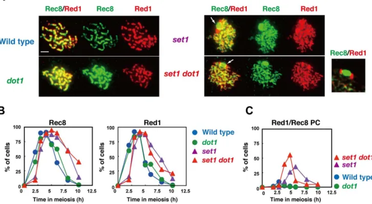
Figure 6. Set1 promotes normal assembly of chromosome axes.(A) Immunostaining analysis of chromosome proteins, Red1 (red) and Rec8 (green), were carried out for wild type and different mutant strains. Representative images for pachytene (wild typedot1) and pseudo-pachytene (set1 andset1 dot1) stages are shown for each strain; wild type 5 h; thedot1, 5h; theset1, 6 h; theset1 dot1, 6 h. White arrows in theset1orset1 dot1 mutants shows Rec8/Red1 aggregates. An image for Rec8/Red1 aggregates in theset1 dot1mutant is enlarged and shown in right. Wild type, NKY1303/1543;set1mutant, MBY015/016;dot1mutant, MBY005/006;set1 dot1double mutant, MBY037/039. The bar indicates 2mm. (B) Kinetics of
spreads positive for Red1 (right) and Rec8 (left) were verified in different strains. The symbols indicate the wild type (blue circles; NKY1303/1543),dot1 mutant (green circles; MBY005/006),set1mutant (purple triangles; MBY015/016), andset1 dot1mutant (red triangle; MBY037/039). (C) Kinetics of spreads with Red1/Rec8-PCs were analyzed. Wild type, blue circles;set1mutant, green circles;dot1mutant, purple triangles;set1 dot1double mutant, red triangles.
Moreover, the mutant shows reduced frequencies of full-length SCs. Furthermore, Zip1 disassembly occurs ,1 h later than in wild type, even after compensating for the delay in assembly. Consistent with the defect in Zip1 assembly in theset1mutant, the mutant accumulates an aggregate of Zip1, referred to as a polycomplex (PC; Figure 5A and 5C). This confirms previous observation that theset1mutant is defective in SC formation [51]. The set1 dot1 double mutant exhibits more defects in Zip1 elongation (reduced pachytene cells) and a greater delay in Zip1-disassembly than does theset1single mutant, suggesting a role for Dot1 in SC formation in the absence of Set1. This SC-defect in the double mutant may be caused by the repair defect and/or reduced DSB formation in the mutant (see above).
To analyze SC defects seen in theset1mutant in more detail, we also examined the localization of Hop1 (Figure 5A), which is a component of the chromosome axis and is required for SC formation as well as meiotic recombination [52]. In the wild type, Hop1 appearance occurs as early as 2 h after the induction of meiosis, and Hop1 disappearance takes place around the pachytene stage; e.g., 5 h (Figure 5D). In wild-type cells, Hop1 shows punctate staining in early meiotic prophase I and reduced staining during late prophase (Figure 5A). Thedot1single mutant shows very similar Hop1 staining patterns to those seen in the wild type, although, as seen for Zip1, Hop1-loading occurs earlier in the mutant than in wild type. Importantly, theset1single mutant shows a 1-h delay in the assembly of Hop1 foci relative to the wild type, and a 3-h delay in the disassembly. Moreover, in addition to dotty staining of Hop1, set1cells show elongated lines of Hop1, which is rarely seen in the wild type (Figure 5A and 5E). In some nuclei, 2 lines of Hop1 are aligned side-by-side (Figure 5A, shown by a pair of arrows), suggesting that the pairing of homologous chromosomes takes place normally, but full synapsis is impaired in theset1mutant. Thedot1 set1double mutant exhibits very similar patterns with the exception of greater proportions of long Hop1 lines and delayed disappearance of Hop1 from chromosomes relative to theset1mutant, consistent with a role for Dot1 in SC formation without Set1.
Consistent with a previous observation [30], double staining of Zip1 and Hop1 clearly shows that Hop1-enriched regions lack strong Zip1 signals in all strains including wild type (Figure 5A), confirming the previous idea that Hop1 in yeast is disassembled along with Zip1 elongation as seen in other eukaryotic organisms, such as Hormad1 in mammals [53] and Asy1 in plants [54]. The accumulated localization of Hop1 along the chromosomes in the
set1mutant is possibly consistent with the fact that Set1 is required for Zip1 elongation.
We also analyzed the localization of another axis protein, Red1 [55], as well as that of the meiosis-specific kleisin, Rec8, which is a
component of the cohesion complex [56](Figure 6). Red1 works together with Hop1 as well as with Mek1/Mre4 in both meiotic recombination and chromosome morphogenesis [57,58]. Red1 initially appears as focal staining like Hop1, but later, unlike Hop1, it forms discontinuous lines as the SC elongates (Figure 6A and 6B) [59]. In theset1andset1 dot1mutants, both of which shows delay in the assembly and disassembly of Red1 (Figure 6B), there are little thick Red1 lines, consistent with defective SC elongation in the mutants. Rec8 localization is similar to that of Red1 in wild type anddot1strains. On the other hand, theset1andset1 dot1mutants rarely form thick lines of Rec8 as seen in the wild-type anddot1
mutant cells, consistent with the lack of full SCs in the mutants (Figure 5A). Notably, we observed aggregates of Red1 and Rec8 in theset1andset1 dot1mutants (Figure 6A). At 6 h, about 35% ofset1
mutant cells contain Red1 and Rec8 aggregates (Figure 6C). This number is increased to 55% at 5 h in theset1 dot1double mutant. In the PC-like structure, Red1 shows bipolar staining on a large Rec8-block (Figure 6A). It is important to point out that these Red1 and Rec8 aggregates are not formed in other SC-deficient mutants such as thedmc1mutant. These results show that Set1 is important for SC elongation and that Dot1 plays a role in SC formation only in the absence of Set1. Set1 may be important for the organization of the chromosome axis containing Red1 and Rec8 for synapsis. On the other hand, recent ChIP-chip study shows that theset1 mutant shows wild-type distribution of Rec8 along chromosomes [12]. Therefore, more higher order structure of chromosome axes might be compromised in the mutant.
The histone H3K4 mutant is defective in SC formation
Set1 is a H3K4 methyltransferase [38]. To confirm the role of Set1 in meiotic chromosome metabolism through this histone modification, we constructed ahht1-K4R hht2-K4Rdouble mutant (hereafter,hht1,2-K4R) at the native chromosomal loci. This strain construct is different from a previous strain, in which bothHHT1
andHHT2were deleted, but anARS-CENplasmid with the hht1-K4R hht2-hht1-K4Rmutations were present [13]. The absence of H3K4 tri-methylation was confirmed by western blotting (Figure 7A). The hht1,2-K4R double mutant shows wild-type spore viability (Figure 7C). This is different from slight reduction of spore viability of the set1 mutant. The hht1,2-K4R cells show a greater delay (,3 h) in the entry into meiosis I than does theset1single mutant with,2 h delay (Figure 7B). Thehht1,2-K4Ralmost recapitulates the meiotic phenotype of theset1single mutant. The hht1,2-K4R
mutant shows reduced DSBs and is defective in SC assembly. Rad51/Dmc1 staining (Figure 7D and 7E) shows that steady state number of Rad51 foci in thehht1,2-K4Rmutant is, on average, 2666.8 (n = 143) at 6 h (Figure 7F; statistically significant from numbers at 3 h in wild type, P value = 1.4 610-5,
Mann-Figure 7. Histone H3K4 is critical for DSB and SC formation.(A) Expression of histone H3K4 trimethylation during meiosis. Western blotting analysis for wild type (NKY1303/1543),hht1,2-K4R(MBY211/218) was carried out using anti-histone H3K4-me3. (B) Meiotic cell division I was analyzed by DAPI staining of wild-type (blue circles; NKY1303/1543),hht1,2-K4R(purple triangles; MBY211/218), andhht1,2-K4R dot1(red triangles; MBY233/ 237) strains. At least 150 cells were counted by DAPI staining for each time point. (C) Distribution of viable spores per tetrad in wild-type and hht1,2-K4R dot1(MBY233/237) strains. For each strain, 100 tetrads were dissected. (D) Immunostaining analysis of Rad51 (green) and Dmc1 (red) for wild type (NKY1303/1543),hht1,2-K4R(MBY211/218), andhht1,2-K4R dot1(MBY233/237) strains was carried out. The bar indicates 2mm. (E) Kinetics of Rad51
focus-positive cells in various strains. A focus-positive cell was defined as a cell with more than 5 foci. More than 100 nuclei were counted at each time point. The symbols indicate the wild type (blue circles; NKY1303/1543),hht1,2-K4R(purple triangles; MBY211/218), andhht1,2-K4R dot1(red triangles; MBY233/237) strains. (F) Rad51 focus numbers per nucleus were counted in different strains. The symbols indicate the wild type (blue circles; NKY1303/1543),hht1,2-K4R(purple triangles; MBY211/218), andhht1,2-K4R dot1(red triangle; MBY233/237) strains. The average number of foci is shown per positive nucleus. (G) Representative images for staining of Zip1(red) and Hop1(green) in wild-type and mutant strains are shown. Hop1 parallel lines thehht1,2-K4Rmutant are shown in a pair of arrows on the right. The bar indicates 2mm. (H) Zip1-staining was classified into 3 classes:
dot (dots, blue), partial linear (short lines, green), and full SC (long lines, red). More than 100 nuclei were counted at each time point Kinetics of spreads (Zip1 polycomplexes) were analyzed. Wild type, NKY1303/1543;hht1,2-K4R, MBY211/218;hht1,2-K4R dot1, MBY233/237. (I) Kinetics of Zip1-PC in different strains. The number of spreads containing Zip1-PC was counted in each strain. The symbols indicate the wild type (blue circles; NKY1303/ 1543),hht1,2-K4R(purple triangles; MBY211/218), andhht1,2-K4R dot1(red triangle; MBY233/237) strains.
Whitney’sU-test), indicating the role of H3K4 in DSB formation. Moreover, thehht1,2-K4Rmutant with thedot1 mutation, with a greater reduction in spore viability (63.5%) compared to the wild type, shows a greater reduction in Rad51-focus number with 1765.6 (n = 140) at 6 h (versus wild type; P value<0, Mann-Whitney’s U-test). Rad51 focus number in the hht1,2-K4R dot1
mutant is 65% of the number in the hht1,2-K4Rmutant. These support the notion that the Dot1 plays a role in DSB formation in the absence of H3K4 methylation.
The hht1,2-K4R double mutant is also defective in Zip1 elongation, and therefore in SC formation like the set1 mutant (Figure 7G and 7I). Thehht1,2-K4R dot1mutant shows more delay in SC disassembly compared tohht1,2-K4R(Figure 7G and 7H). We also found that thehht1,2-K4Rdouble mutant often shows 2
parallel Hop1 lines like theset1mutant (Figure 7G). These strongly suggest a role for Set1 in SC formation as well as DSB formation through the methylation of histone H3K4.
Histone H3K79 is critical for DSB formation in the absence ofSET1
In order to know the involvement of histone H3K79-methyl-ation in DSB formH3K79-methyl-ation, we also used a strain with histone H3K79R mutations at the native chromosomal loci (hht1-K79R hht2-K79R, hereafter hht1,2-K79R; Takehiko Usui and A.S., unpublished). The absence of H3K79 methylation was confirmed by western blot analysis using an anti-histone H3K79 methylation antibody (Figure 8A). Thehht1,2-K79R mutant shows wild-type spore viability (Figure 8B). Importantly, when hht1,2-K79R was
Figure 8. Histone H3K79 is critical for DSB without Set1.(A) Expression of histone H3K79-methylation during meiosis. Western blotting analysis for the wild-type (NKY1303/1543) andhht1,2-K79R(MBY151/152) strains was carried out using anti-H3K79-methylation. (B) Distribution of viable spores per tetrad in thehht1,2-K79R(MBY151/152) andset1 hht1,2-K79R(MBY219/221) strains. For each strain, 100 tetrads were dissected. (C) Immunostaining analysis of Rad51 (green) and Dmc1 (red) in wild-type cells at 3 h (NKY1303/1543) andset1 hht1,2-K79R(MBY219/221) cells at 6 h. The bar indicates 2mm. (D) Rad51 focus numbers per nucleus were counted in different strains. Wild type (blue circles; NKY1303/1543),hht1,2-K79R
(green circles; MBY219/221), theset1(purple triangles; MBY015/016) andhht1,2-K79R set1(red triangle; MBY219/221). Both theset1andset1 hht1,2-K79Rmutants show delayed appearance of Rad51 foci on chromosomes. Thus, focus numbers of Rad51 at 6 h was measured. The number for theset1 is the same as that in Figure 3C. The average number of foci with SD is shown per positive nucleus.
doi:10.1371/journal.pone.0096648.g008
combined with theset1 deletion, the triple mutant shows 47.8% spore viability (Figure 8B), similar to theset1 dot1double mutant (see Figure 1). We found that theset1 hht1,2-K79Rmutant shows a decreased number of Rad51 foci (Figure 8C and 8D; 12.163.5; n = 62), which is more reduced in theset1mutant. This supports the idea that Dot1-dependent histone H3K79 methylation promotes meiotic DSB formation in the absence of Set1-dependent histone H3K4 methylation. The Rad51 focus number in theset1 hht1,2-K79Rmutant is smaller than that in theset1 dot1mutant (Figure 8C and 8D; versus 6 h, P value = 1.561025, Mann-Whitney’sU -test). This may be due to culture-to culture difference.
Discussion
Previous studies have shown that 2 histone-modifications, H3K4 methylation and H2BK123 ubiquitination, play a critical role in the formation of meiotic DSBs [7,12,13,39]. The effect of H2BK123 ubiquitination seems to be indirect since this mark promotes H3K4 methylationin trans[38]. In this study, we have demonstrated the role of Dot1 H3K79 methyltransferase in DSB formation in the absence of Set1.
Cytological analysis of Rad51 foci, which mark sites of ongoing recombination [47], showed that, even in the absence of Set1-dependent H3K4-methylation, meiotic cells form significant numbers of DSBs, about 2/3–3/4 of the levels seen in wild type, consistent with high spore viability of the mutants defective in H3K4 methylation. In contrast, previous studies using whole genome mapping showed a large reduction of DSBs at some regions in the absence of eitherSET1or H3K4 methylation [7]. In addition, the lack of Set1-dependent H3K4 methylation changes the distribution of DSBs along chromosomes [7,12]. However, such studies used ChIP-chip for mapping using dmc1 or rad50S
(sae2) mutants which block recombination; therefore, it is very difficult to quantify/estimate how much DSBs are dependent on the specific histone marks. Our results of Southern blotting for individual loci (Figure 2D and 4G) confirm the previous results. It is reported that the set1 mutant showed increased DSBs at a specific chromosomal locus [7,12], suggesting the presence of a backup system for DSB formation. We used counting of Rad51 foci to get a rough estimate of DSB numbers in a nucleus. Our ongoing research showed that the number of Rad51 foci is roughly proportional to the number of DSBs (M. S., unpublished). We found that the set1 mutant showed a mild reduction of Rad51 focus numbers along the genome. Indeed, DSB mapping on individual chromosomes in the mutant support the idea (Figure 4). Thus, we believe that the contribution of Set1-dependent H3K4 methylation to DSB formation is weaker than expected, at least in the budding yeast. These data suggest the presence of other critical determinants for DSB formation. Indeed, we found that elimina-tion of Dot1-dependent H3K79 methylaelimina-tion reduces DSB levels to about half of that seen in theset1deletion mutant. This indicates the involvement of the histone post-translational modification in DSB formation. In the fission yeast, H3K9 acetylation is known to promote DSB formation, while H3K4 methylation is not involved in DSB formation [9]. Moreover, in mice, meiosis-specific Prdm9-dependent H3K4 methylation shapes hotspot activity for recom-bination. Interestingly, even the absence of Prdm9 methyltrans-ferase changes the distribution of the hotspot by creating new spots [11]. In Prdm9 KO mice, Prdm9-INdependent H3K4 methyla-tion might be responsible for this activity [11]. These studies confirm that multiple histone post-translational modifications determine the site of initiation of meiotic recombination. We want to stress that, even in the absence of both Set1 and Dot1, mutant cells form 40–50% of the wild-type levels of Rad51 foci,
likely DSBs, suggesting the presence of other determinant(s) for hotspot activity. Recently, it is shown that, in a plant,Arabidopsis thaliana, a histone H2A variant, H2A.Z, plays a role in recombination hotspot activity during meiosis [60].
Our results suggest that H3K79 methylation plays a role in DSB formation. H3K79 methylation is recognized by the Tudor domain of Rad9 in yeast [61]. Since therad9mutant is proficient in meiosis [62], it is unlikely that Rad9 plays a role in DSB formation. However, we need to analyze arad9mutant with the
set1 deletion to know the exact role of this protein in DSB formation, since the effect of thedot1is only seen in the absence of the SET1. Alternatively, the other protein involved in DSB formation may recognize this mark. Recent reports suggest a role for Dot1-dependent H3K79-methylation in the recombination checkpoint during meiosis [63]. In the recombination checkpoint, Dot1-dependent H3K79 methylation promotes the efficient binding of the Hop1 protein in thezip1 mutant. This could be interpreted simply as that H3K79 methylation is bound to Hop1. However, this is unlikely, since we showed that thedot1mutant is proficient in Hop1 binding at least in the wild-type background. In wild type meiotic cells, there is another pathway to recruit Hop1 in a Dot1-independent manner. Recently, Dot1 has been shown to play a role in the Tel1/ATM pathway in meiotic recombination [31,63], which somehow controls DSB formation [49,50,64]. If this is true in wild-type meiosis, the role of Dot1 in DSB formation described here is indirect; e.g. signaling. Indeed, recent genome-wide mapping showed that H3K79 methylation is less in promoter regions than coding regions [65]. This suggests that the Dot1-dependent H3K79 methylation play a negative role rather than a positive role in the DSB formation. We suggest that meiotic chromosomes adapt different alternatives to create the recombi-nation hotspot, possibly using different histone marks. This kind of multiple alternatives or flexibility may contribute to the rapid evolution of the recombination hotspots.
The effect of thedot1mutation on DSB formation is clearly seen in the absence of H3K4 methylation. In this line, it is interesting to see subtle effects of thedot1 mutation on DSB formation in the presence of H3K4 methylation. This includes altered kinetics of DSB repair (Figure 2D), slight but significant reduction of Rad51 focus number (Figure 3C) and the appearance of late DSBs formation (Figure 4A) in thedot1mutant. We need further careful evaluation on the role of the Dot1-dependent H3K79 mutation on DSB formation.
Although our studies described here suggest a direct link of Dot1 with DSB formation in the absence of Set1, we cannot exclude the possibility that the effect ofdot1mutation is indirect through the transcription [37]. We also need more careful evaluation on the role of any histone posttranslational modifications in meiotic recombination such as DSB formation.
We also revealed a role for Set1-dependent H3K4 methylation in chromosome morphogenesis in meiosis, such as SC formation. Both theset1andhht1,2-K4Rmutants produce viable spores and retain high levels of DSBs relative to wild type, as judged from the number of Rad51 foci. However, the 2 mutants are almost defective in SC elongation. The SC elongation defect in theset1
promote the assembly of Red1 or Rec8 in the context of meiotic chromosomes.
Alternatively, reduced DSBs might be directly linked with a defect in SC elongation. In this scenario, excess DSBs in wild-type cells are necessary for normal levels of chromosome synapsis rather than recombination. This idea is somehow consistent with previous proposal of 2 types of DSBs; one for synapsis and the other for recombination [66,67]. Indeed, a moderate reduction in DSBs does not affect the frequency of COs due to CO homeostasis [68].
Supporting Information
Table S1 Strain list. (PDF)
Table S2 Primer list. (PDF)
Acknowledgments
We thank Drs. Doug Bishop and Neil Hunter for discussion. We are also indebted to members of the Shinohara lab, particularly Dr. Saori Mori for technical help and Dr. Takehiko Usui for thehht1,2-K79Rstrain.
Author Contributions
Conceived and designed the experiments: MBI MS AS. Performed the experiments: MBI MS. Analyzed the data: MBI MS AS. Contributed reagents/materials/analysis tools: MBI MS AS. Wrote the paper: MBI MS AS.
References
1. Petronczki M, Siomos MF, Nasmyth K (2003) Un menage a quatre: the molecular biology of chromosome segregation in meiosis. Cell 112: 423–440. 2. Kleckner N (2006) Chiasma formation: chromatin/axis interplay and the role(s)
of the synaptonemal complex. Chromosoma 115: 175–194.
3. Borde V, de Massy B (2013) Programmed induction of DNA double strand breaks during meiosis: setting up communication between DNA and the chromosome structure. Curr Opin Genet Dev 23: 147–155.
4. Keeney S (2001) Mechanism and control of meiotic recombination initiation. Curr Top Dev Biol 52: 1–53.
5. Lichten M, de Massy B (2011) The impressionistic landscape of meiotic recombination. Cell 147: 267–270.
6. Yamada T, Ohta K (2013) Initiation of meiotic recombination in chromatin structure. J Biochem.
7. Borde V, Robine N, Lin W, Bonfils S, Geli V, et al. (2009) Histone H3 lysine 4 trimethylation marks meiotic recombination initiation sites. EMBO J 28: 99– 111.
8. Buard J, Barthes P, Grey C, de Massy B (2009) Distinct histone modifications define initiation and repair of meiotic recombination in the mouse. EMBO J 28: 2616–2624.
9. Yamada S, Ohta K, Yamada T (2013) Acetylated Histone H3K9 is associated with meiotic recombination hotspots, and plays a role in recombination redundantly with other factors including the H3K4 methylase Set1 in fission yeast. Nucleic Acids Res 41: 3504–3517.
10. Sollier J, Lin W, Soustelle C, Suhre K, Nicolas A, et al. (2004) Set1 is required for meiotic S-phase onset, double-strand break formation and middle gene expression. EMBO J 23: 1957–1967.
11. Brick K, Smagulova F, Khil P, Camerini-Otero RD, Petukhova GV (2012) Genetic recombination is directed away from functional genomic elements in mice. Nature 485: 642–645.
12. Sommermeyer V, Beneut C, Chaplais E, Serrentino ME, Borde V (2013) Spp1, a member of the Set1 Complex, promotes meiotic DSB formation in promoters by tethering histone H3K4 methylation sites to chromosome axes. Mol Cell 49: 43–54.
13. Acquaviva L, Szekvolgyi L, Dichtl B, Dichtl BS, de La Roche Saint Andre C, et al. (2013) The COMPASS subunit Spp1 links histone methylation to initiation of meiotic recombination. Science 339: 215–218.
14. Hunter N, Kleckner N (2001) The single-end invasion: an asymmetric intermediate at the double-strand break to double-holliday junction transition of meiotic recombination. Cell 106: 59–70.
15. Bishop DK, Park D, Xu L, Kleckner N (1992)DMC1: a meiosis-specific yeast homolog ofE. coli recArequired for recombination, synaptonemal complex formation, and cell cycle progression. Cell 69: 439–456.
16. Cloud V, Chan YL, Grubb J, Budke B, Bishop DK (2012) Rad51 is an accessory factor for Dmc1-mediated joint molecule formation during meiosis. Science 337: 1222–1225.
17. Shinohara A, Ogawa H, Ogawa T (1992) Rad51 protein involved in repair and recombination inS. cerevisiaeis a RecA-like protein. Cell 69: 457–470. 18. Schwacha A, Kleckner N (1994) Identification of joint molecules that form
frequently between homologs but rarely between sister chromatids during yeast meiosis. Cell 76: 51–63.
19. Allers T, Lichten M (2001) Differential timing and control of noncrossover and crossover recombination during meiosis. Cell 106: 47–57.
20. Zickler D, Kleckner N (1999) Meiotic chromosomes: integrating structure and function. Annu Rev Genet 33: 603–754.
21. Borner GV, Kleckner N, Hunter N (2004) Crossover/noncrossover differenti-ation, synaptonemal complex formdifferenti-ation, and regulatory surveillance at the leptotene/zygotene transition of meiosis. Cell 117: 29–45.
22. Hochwagen A, Amon A (2006) Checking your breaks: surveillance mechanisms of meiotic recombination. Curr Biol 16: R217–228.
23. Sym M, Engebrecht JA, Roeder GS (1993) ZIP1 is a synaptonemal complex protein required for meiotic chromosome synapsis. Cell 72: 365–378.
24. Xu L, Ajimura M, Padmore R, Klein C, Kleckner N (1995)NDT80, a meiosis-specific gene required for exit from pachytene inSaccharomyces cerevisiae. Mol Cell Biol 15: 6572–6581.
25. Chu S, Herskowitz I (1998) Gametogenesis in yeast is regulated by a transcriptional cascade dependent on Ndt80. Mol Cell 1: 685–696.
26. Clyne RK, Katis VL, Jessop L, Benjamin KR, Herskowitz I, et al. (2003) Polo-like kinase Cdc5 promotes chiasmata formation and cosegregation of sister centromeres at meiosis I. Nat Cell Biol5: 480–485.
27. Sourirajan A, Lichten M (2008) Polo-like kinase Cdc5 drives exit from pachytene during budding yeast meiosis. Genes Dev 22: 2627–2632.
28. San-Segundo PA, Roeder GS (1999) Pch2 links chromatin silencing to meiotic checkpoint control. Cell 97: 313–324.
29. San-Segundo PA, Roeder GS (2000) Role for the silencing protein Dot1 in meiotic checkpoint control. Mol Biol Cell 11: 3601–3615.
30. Borner GV, Barot A, Kleckner N (2008) Yeast Pch2 promotes domainal axis organization, timely recombination progression, and arrest of defective recombinosomes during meiosis. Proc Natl Acad Sci U S A 105: 3327–3332. 31. Ho HC, Burgess SM (2011) Pch2 acts through Xrs2 and Tel1/ATM to
modulate interhomolog bias and checkpoint function during meiosis. PLoS Genet 7: e1002351.
32. Conde F, Ontoso D, Acosta I, Gallego-Sanchez A, Bueno A, et al. (2010) Regulation of tolerance to DNA alkylating damage by Dot1 and Rad53 in
Saccharomyces cerevisiae. DNA Repair (Amst) 9: 1038–1049.
33. Conde F, Refolio E, Cordon-Preciado V, Cortes-Ledesma F, Aragon L, et al. (2009) The Dot1 histone methyltransferase and the Rad9 checkpoint adaptor contribute to cohesin-dependent double-strand break repair by sister chromatid recombination inSaccharomyces cerevisiae. Genetics 182: 437–446.
34. Conde F, San-Segundo PA (2008) Role of Dot1 in the response to alkylating DNA damage inSaccharomyces cerevisiae: regulation of DNA damage tolerance by the error-prone polymerases Polzeta/Rev1. Genetics 179: 1197–1210. 35. Levesque N, Leung GP, Fok AK, Schmidt TI, Kobor MS (2010) Loss of H3
K79 trimethylation leads to suppression of Rtt107-dependent DNA damage sensitivity through the translesion synthesis pathway. J Biol Chem 285: 35113– 35122.
36. Tatum D, Li S (2011) Evidence that the histone methyltransferase Dot1 mediates global genomic repair by methylating histone H3 on lysine 79. J Biol Chem 286: 17530–17535.
37. Nguyen AT, Zhang Y (2011) The diverse functions of Dot1 and H3K79 methylation. Genes Dev 25: 1345–1358.
38. Shilatifard A (2012) The COMPASS family of histone H3K4 methylases: mechanisms of regulation in development and disease pathogenesis. Annu Rev Biochem 81: 65–95.
39. Yamashita K, Shinohara M, Shinohara A (2004) Rad6-Bre1-mediated histone H2B ubiquitylation modulates the formation of double-strand breaks during meiosis. Proc Natl Acad Sci U S A 101: 11380–11385.
40. Shinohara M, Gasior SL, Bishop DK, Shinohara A (2000) Tid1/Rdh54 promotes colocalization of Rad51 and Dmc1 during meiotic recombination. Proc Natl Acad Sci U S A 97: 10814–10819.
41. Farmer S, Leung WK, Tsubouchi H (2011) Characterization of meiotic recombination initiation sites using pulsed-field gel electrophoresis. Methods Mol Biol 745: 33–45.
42. Shinohara M, Sakai K, Ogawa T, Shinohara A (2003) The mitotic DNA damage checkpoint proteins Rad17 and Rad24 are required for repair of double-strand breaks during meiosis in yeast. Genetics 164: 855–865. 43. Storlazzi A, Xu L, Cao L, Kleckner N (1995) Crossover and noncrossover
recombination during meiosis: timing and pathway relationships. Proc Natl Acad Sci U S A 92: 8512–8516.
44. Cao L, Alani E, Kleckner N (1990) A pathway for generation and processing of double-strand breaks during meiotic recombination inS. cerevisiae. Cell 61: 1089– 1101.
45. Schwacha A, Kleckner N (1997) Interhomolog bias during meiotic recombina-tion: meiotic functions promote a highly differentiated interhomolog-only pathway. Cell 90: 1123–1135.
46. Shinohara A, Gasior S, Ogawa T, Kleckner N, Bishop DK (1997)Saccharomyces cerevisiae recAhomologuesRAD51andDMC1have both distinct and overlapping roles in meiotic recombination. Genes Cells 2: 615–629.
47. Bishop DK (1994) RecA homologs Dmc1 and Rad51 interact to form multiple nuclear complexes prior to meiotic chromosome synapsis. Cell 79: 1081–1092. 48. Miyazaki T, Bressan DA, Shinohara M, Haber JE, Shinohara A (2004) In vivo assembly and disassembly of Rad51 and Rad52 complexes during double-strand break repair. Embo J 23: 939–949.
49. Carballo JA, Panizza S, Serrentino ME, Johnson AL, Geymonat M, et al. (2013) Budding Yeast ATM/ATR Control Meiotic Double-Strand Break (DSB) Levels by Down-Regulating Rec114, an Essential Component of the DSB-machinery. PLoS Genet 9: e1003545.
50. Argunhan B, Farmer S, Leung WK, Terentyev Y, Humphryes N, et al. (2013) Direct and indirect control of the initiation of meiotic recombination by DNA damage checkpoint mechanisms in budding yeast. PLoS One 8: e65875. 51. Trelles-Sticken E, Bonfils S, Sollier J, Geli V, Scherthan H, et al. (2005)
Set1-and Clb5-deficiencies disclose the differential regulation of centromere Set1-and telomere dynamics inSaccharomyces cerevisiaemeiosis. J Cell Sci 118: 4985–4994. 52. Hollingsworth NM, Goetsch L, Byers B (1990) TheHOP1gene encodes a
meiosis-specific component of yeast chromosomes. Cell 61: 73–84.
53. Wojtasz L, Daniel K, Roig I, Bolcun-Filas E, Xu H, et al. (2009) Mouse HORMAD1 and HORMAD2, two conserved meiotic chromosomal proteins, are depleted from synapsed chromosome axes with the help of TRIP13 AAA-ATPase. PLoS Genet 5: e1000702.
54. Armstrong SJ, Caryl AP, Jones GH, Franklin FC (2002) Asy1, a protein required for meiotic chromosome synapsis, localizes to axis-associated chromatin in Arabidopsis and Brassica. J Cell Sci 115: 3645–3655.
55. Rockmill B, Roeder GS (1988)RED1: a yeast gene required for the segregation of chromosomes during the reductional division of meiosis. Proc Natl Acad Sci U S A 85: 6057–6061.
56. Klein F, Mahr P, Galova M, Buonomo SB, Michaelis C, et al. (1999) A central role for cohesins in sister chromatid cohesion, formation of axial elements, and recombination during yeast meiosis. Cell 98: 91–103.
57. Hollingsworth NM, Ponte L (1997) Genetic interactions betweenHOP1,RED1
andMEK1suggest thatMEK1regulates assembly of axial element components during meiosis in the yeastSaccharomyces cerevisiae. Genetics 147: 33–42. 58. Leem SH, Ogawa H (1992) TheMRE4gene encodes a novel protein kinase
homologue required for meiotic recombination inSaccharomyces cerevisiae. Nucleic Acids Res 20: 449–457.
59. Smith AV, Roeder GS (1997) The yeast Red1 protein localizes to the cores of meiotic chromosomes. J Cell Biol 136: 957–967.
60. Choi K, Zhao X, Kelly KA, Venn O, Higgins JD, et al. (2013)Arabidopsismeiotic crossover hot spots overlap with H2A.Z nucleosomes at gene promoters. Nat Genet 45: 1327–1336.
61. Grenon M, Costelloe T, Jimeno S, O’Shaughnessy A, Fitzgerald J, et al. (2007) Docking onto chromatin via theSaccharomyces cerevisiaeRad9 Tudor domain. Yeast 24: 105–119.
62. Lydall D, Nikolsky Y, Bishop DK, Weinert T (1996) A meiotic recombination checkpoint controlled by mitotic checkpoint genes. Nature 383: 840–843. 63. Ontoso D, Acosta I, van Leeuwen F, Freire R, San-Segundo PA (2013)
Dot1-dependent histone H3K79 methylation promotes activation of the Mek1 meiotic checkpoint effector kinase by regulating the Hop1 adaptor. PLoS Genet 9: e1003262.
64. Gray S, Allison RM, Garcia V, Goldman AS, Neale MJ (2013) Positive regulation of meiotic DNA double-strand break formation by activation of the DNA damage checkpoint kinase Mec1(ATR). Open Biol 3: 130019. 65. Zhang L, Ma H, Pugh BF (2011) Stable and dynamic nucleosome states during a
meiotic developmental process. Genome Res 21: 875–884.
66. Stahl FW, Foss HM, Young LS, Borts RH, Abdullah MF, et al. (2004) Does crossover interference count inSaccharomyces cerevisiae? Genetics 168: 35–48. 67. Zalevsky J, MacQueen AJ, Duffy JB, Kemphues KJ, Villeneuve AM (1999)
Crossing over duringCaenorhabditis elegansmeiosis requires a conserved MutS-based pathway that is partially dispensable in budding yeast. Genetics 153: 1271–1283.