Inflammatory Markers of Atherosclerotic Plaque
Stabilization after Acute Coronary Event – Temporal
Trends
Osana Maria Coelho Costa Mouco, José Carlos Nicolau, Tatiana da Rocha e Souza,
Lilia Nigro Maia, José Antônio Franchini Ramires
Hospital de Base da Faculdade de Medicina de São José do Rio Preto – SP e Instituto do Coração do Hospital das Clínicas – FMUSP - São José do Rio Preto, SP - Brazil
Mailing Address: Osana Maria Coelho Costa Mouco • Rua Tiradentes, 2640/122 - 15025-050 – São José do Rio Preto, SP - Brazil E-mail: [email protected] Received on 07/26/05 Accepted on 12/28/05
O
BJECTIVETo evaluate the length of time required for atheros-clerotic plaque stabilization in acute coronary syndromes (ACS), using infl ammatory markers.
M
ETHODSIn this prospective study, C-reactive protein (CRP), fi brinogen, factor VIIIc, interleukin-6 (IL-6), and tumor necrosis factor-α (TNF-α) levels were measured on admission, at discharge, and three and six months post-discharge in 40 patients with non-ST-segment elevation ACS (NSTE-ACS) and 40 healthy subjects.
R
ESULTSC-reactive protein levels were signifi cantly higher on admission and at discharge, but not at three and six months post-discharge, compared with the control group. Fibrinogen levels remained unchanged, except at six months, when they were signifi cantly lower than in the control group. Factor VIII-c did not differ from that of the control group on admission, but it was signifi cantly higher at discharge, with no differences at three and six months. Interleukin-6 levels were signifi cantly higher than in the control group in all time points. However, they declined signifi cantly between discharge and three months. In no time point was TNF-α signifi cantly different from that of the control group. Only IL-6 correlated signifi cantly and independently with future cardiovascular events.
C
ONCLUSIONSWith respect to CRP and factor VIIIc, plaque stabili-zation is suggested in up to three months; IL-6 analysis suggests stabilization as from the third month, although it remained higher than that of the control group for up to six months. Only IL-6 showed prognostic value for further events within a year.
K
EY WORDSAcute coronary syndromes (ACS) – unstable angina and myocardial infarction – are caused mainly by rupture or erosion of atherosclerotic plaque, with subsequent thrombus formation. It is known that plaque instability, among other characteristics, is closely related to the infl ammatory process1-4.
Of the circulating infl ammatory markers, C-reactive protein (CRP) is the most extensively studied; high CRP plasma levels are found in ACS patients and are associated with increased rates of future events5. On the other hand, interleukin-6, which prompts C-reactive protein production, is also elevated in ACS patients and is associated with worse prognosis, thereby reinforcing the role of infl ammation in these syndromes6. Tumor necrosis factor-α (TNF-α), a cytokine produced mostly by activated macrophages, stimulates the synthesis of other cytokines7. Persistently increased TNF-α levels after acute myocardial infarction (AMI) are correlated with worse prognosis; however, little information is available on TNF-α kinetics in non-ST-segment elevation ACS8. Fibrinogen is an acute-phase protein directly involved in the coagulation cascade; increased fi brinogen levels are associated with higher risk of thrombotic events9 and higher risk of events in ACS patients10-14. Finally, factor VIII-c, a procoagulant enzyme cofactor, is also involved in infl amation15 and is a marker of ischemic heart disease16-18.
Little has been published about how long it takes for stabilized coronary lesions to heal completely in patients with non-ST-segment elevation ACS. A study using cell adhesion molecules (CAMs) suggested that the infl ammatory response might persist for up to six months following an acute coronary event19.
The primary focus of this study was to evaluate the length of time required for atherosclerotic plaque to become stabilized in patients with non-ST-segment elevation ACS treated clinically, through the behavior of plasma infl ammatory and coagulation markers levels.
M
ETHODSForty out of the 64 patients admitted to our hospital between September 2000 and May 2001 with unstable angina and no-ST-segment elevation AMI were included in the study (22 men; mean age 61 ± 12). Forty age- and sex-matched healthy volunteers served as the control group. Patients were followed-up over one year.
Inclusion criteria were chest pain lasting 24 hours, suggestive of myocardial ischemia of accelerated pattern, or a prolonged one (> 20 minutes), or with recurrent episodes at rest, or at minimal exertion, in addition to at least one of the following: (a) new or presumed new ECG changes (any of the following three characteristics): ST-segment depression ≥ 0.5 mm, transient ST-segment elevation (< 20 minutes) ≥ 1 mm, T-wave inversion ≥ 3 mm in two or more contiguous leads; (b) and raised levels of cardiac markers (CKMB
≥ 2X the upper limit of normal).
Exclusion criteria were persistent ST-segment elevation; secondary angina; history of percutaneous coronary intervention (PCI) or coronary artery bypass grafting (CABG) within the previous six months; left bundle branch block (LBBB) or pacemaker rhythm; fi brinolytic therapy in the last 48 hours; any infl ammation, infection or neoplasic disease; regular or chronic use of anti-infl ammatory drugs in the previous two months; AMI and stroke within the past six months; severe congestive heart failure (CHF) or cardiogenic shock; severe systemic disease; serum creatinine > 2.5 mg/dL; or surgery or trauma within the past thirty days. Patients who underwent CABG or PCI during the study were also excluded (nine patients were excluded on the basis of this criterion).
Clinical management - In-hospital medications were used according to the medical approach (see Table 1). The assistant doctor also made decisions regarding invasive or conservative strategy and the need of coronary revascularization.
Blood samples - Peripheral venous blood samples were collected on admission, at hospital discharge, and three and six months after discharge. A single measurement was performed in healthy controls. All samples (patients and controls) were centrifuged, stored at -70 °C, and tested for high-sensitivity CRP, IL-6, TNF-α, fi brinogen and factor VIII-c in a single series at the end of the study.
Laboratory assessment - High-sensitivity CRP (hs-CRP) was determined by particle-enhanced immunoturbidimetric assay (COBAS INTEGRA 700; Roche Diagnostics). IL-6 and TNF-α were evaluated by ELISA (Enzyme-Linked Immunosorbent Assay- R&D Systems). Fibrinogen was measured using the automated Clauss method (Fibriquik, manufactured by Organon Teknika). Factor VIII activity was measured by determining the ability of the tested sample to correct the clotting time of factor VIII-defi cient plasma (Organon Teknika).
Purpose - The primary purpose of this study was
to indirectly evaluate the length of time required for atherosclerotic plaque stabilization in ACS patients (unstable angina and non-ST-segment elevation AMI), by measuring infl ammatory markers (CRP, fi brinogen, factor VIII, IL-6, and TNF-α) during six months after hospital discharge. The secondary purpose was to correlate the plasma concentrations of these markers with the prognosis (risk of future cardiovascular events) in patients with non-ST-segment elevation ACS in up to one year of follow-up.
± standard deviation.
To compare both groups, the Mann-Whitney test (non-parametric) was used for non-normally distributed values, while Student’s unpaired t-test, Welch-corrected, was used for normally distributed values.
Categorical variables were expressed as number and percentage. Fisher’s exact test was performed to compare these variables.
When the patient was his own control and thus acted as paired groups do, Wilcoxon’s non-parametric paired test was used to compare the different time points analyzed. The same comparisons in the different time points were performed for normally-distributed markers using the paired t-test.
Correlation between cytokines and acute phase protein values was performed using a Spearman non-parametric correlation.
Every marker was compared with the composite endpoint of recurrent angina, readmission for angina, reinfarction or cardiovascular death after a one-year follow-up. In these analyses, the Mann-Whitney test (univariate analysis) and logistic regression (multivariate analysis) were used.
Group differences were considered statistically signifi cant when p ≤ 0.05 (two-tailed).
Ethical considerations - The project was approved by the Institutional Research Ethics Committee of the Faculdade de Medicina de São José do Rio Preto, São Paulo (Famerp) and the Faculdade de Medicina da Universidade de São Paulo (FMUSP). Informed consent was obtained from all patients.
R
ESULTSThe characteristics of the study population are shown in Table 1. As may be noted, groups were paired according to age and gender. As expected, the case group showed a larger proportion of patients with specifi c risk factors and pre-treatments.
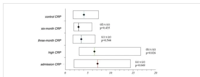
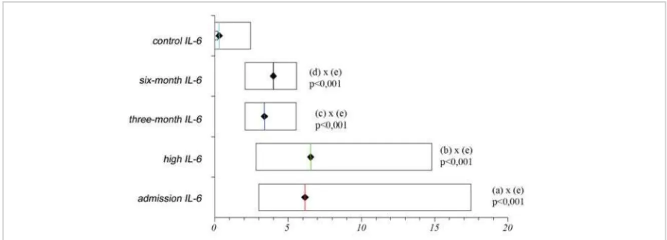
Figure 1 shows that median CRP levels were signifi -cantly higher in ACS patients, compared to control subjects, both on admission and at discharge, but not at three and six months. Median IL-6 levels declined significantly between discharge and three months. However, they remained signifi cantly higher than those of the control group for up to six months (Fig. 2). Median TNF-α levels decreased signifi cantly between three and six months after discharge. Yet, the levels compared with the controls did not reach statistical signifi cance at any time of follow-up (Fig. 3). Change in patterns over time were also found for fi brinogen (Fig. 4), although no signifi cant increase was found during follow-up compared with controls. Factor VIII-c levels were signifi cantly higher at discharge, compared with controls, but not at three and six months (Fig. 5).
Correlations between acute-phase proteins and cytokines- There were signifi cant correlations between CRP and IL-6 levels measured on admission (r = 0.5; p < 0.001), at discharge (r = 0.6; p < 0.001), and after three months (r = 0.4; p < 0.007); between CRP and fi brinogen levels measured on admission (r = 0.4; p = 0.003) and at discharge (r = 0.4; p = 0.003); and between CRP and TNF-α levels measured on admission (r = 0.3; p = 0.024) and at discharge (r = 0.4; p = 0.009).
No signifi cant correlations between CRP and factor VIIIc were found at any time during follow-up.
Clinical events and inflammatory marker levels during a one-year follow-up- At the end of a follow-up period of one year, there were 45 cardiovascular events (32 recurrent cases of angina without hospitalization, four deaths, eight readmissions for angina, and one non-fatal AMI). Of the infl ammatory markers analyzed, only IL-6 levels on admission correlated signifi cantly and independently with the composite endpoint, as shown in Tables 2 and 3.
D
ISCUSSIONCRP - This study showed that CRP levels were high in patients with unstable angina or non-ST-segment elevation AMI. Liuzzo et al.20 demonstrated that 65% of the patients had CRP levels ≥ 3 mg/L on admission, similar therefore to our fi ndings, in which 62.5% of the patients had CRP levels ≥ 4 mg/L (median CRP for controls) at the time of admission. On the other hand, elevated CRP plasma levels on admission were found in 65% of the patients, higher than those described by Biasucci et al21 (49%), which may suggest a higher risk population in the present study.
Following hospital discharge, O’Malley et al22 did not fi nd signifi cant differences in the CRP levels of patients and controls after three months of follow-up, which is similar to our fi ndings. To our knowledge, no comparative data is available in the literature that analyzes CRP levels after three months of follow-up in patients with non-ST-segment elevation ACS.
Different studies have emphasized the usefulness of CRP measurement during acute coronary syndrome as a marker for future cardiac events12,20,23,24. However, this correlation has not been found by others22,25-28, which is again similar to our results.
Table 1 – Characteristics of the Study Population
Cases Controls p value
Number of patients 40 40
Age (years, mean ± SD) 61 ± 12 65 ± 10 0.11
Gender (M/F) 22/18 22/18 1.0
Risk factors; number of patients (%):
-Family history of CAD 18 (45) 0 <0.001
-Dyslipidemia 14 (35) 9 (22.5) 0.069
-Diabetes 10 (25) 3 (7.5) 0.033
-Hypertension 38 (95) 14 (35) <0.001
-Cigarette smoking 14 (35) 2 (5) <0.001
Past history; number of patients (%):
-Previous MI > six months 11 (27.5) 0
-Stable angina 2 (5) 0
-Unstable angina 2 (5) 0
-CABG > six months 10 (25) 0
-PTCA > six months 4 (10) 0
Previous medication (%):
-Beta-blocker 14 (35) 1 (2.5) <0.001
-ACEI 15 (37.5) 4 (10) 0.007
-ASA 16 (40) 1 (2.5) <0.001
-Calcium antagonist 11 (27.5) 1 (2.5) 0.003
-Vastatin 6 (15) 1 (2.5) 0.108
-Digitalis 5 (12.5) 0 0.054
-Diuretics 12 (30) 7 (17.5) 0.293
-Nitrate 6 (15) 0 0.025
In-hospital medication (%):
-Beta-blocker 39 (97.5) 0
-ACEI 39 (97.5) 0
-ASA 40 (100) 0
-Calcium antagonist 22 (55) 0
-Vastatin 19 (47.5) 0
-Diuretics 16 (40) 0
-Nitrate 29 (72.5) 0
Admission diagnosis (%):
-Unstable angina 34 (85) 0
-Non-ST-segment elevation AMI 6 (15) 0
CAD - coronary artery disease; AMI - acute myocardial infarction; MR - myocardial revascularization; PTCA - percutaneous transluminal coronary angioplasty; ACEI - angiotensin-converting enzyme inhibitor; ASA - aspirin; ST - standard deviation; n - number; CABG - coronary artery bypass graft.
Fig. 2 – Median (25th-75th percentile) IL-6 levels (pg/mL) of the case group compared with the control group at the four time points analyzed during follow-up.
Fig. 3 – Median (25th-75th percentile) TNF-α levels (pg/mL) of the case group compared with the control group at the four time points analyzed during follow-up.
also below detection limit. These fi ndings support the hypothesis that detectable levels of IL-6 are related to atherosclerotic plaque instability, suggesting that this is not independent of myocardial necrosis, since this cytokine is also detectable in the absence of raised levels of creatine kinase or troponin T6.
That study6 found a signifi cant correlation between IL-6
and CRP levels (p = 0.013) measured on admission. In the present study, a signifi cant correlation between both markers was obtained not only on admission (p = 0.001) but also at discharge and at three months of follow-up. These fi ndings corroborate the hypothesis that, in the presence of ACS, increased levels of acute-phase proteins are a result of cytokine infl ammatory pathways.
Fig. 4 – Mean ± SD fi brinogen levels (mg/dL) of the case group compared with the control group at the four time points analyzed during follow-up.
With regard to IL-6 evolution after discharge, O’Malley et al22 found that, compared to controls, unstable patients showed signifi cantly higher IL-6 levels on admission, followed by a drop in three months, at which time IL-6 levels in both groups became similar. In our study, although the number of patients with elevated IL-6 levels in the case group decreased signifi cantly between discharge and at three months, this number remained signifi cantly higher than that of the control group in up to six months of follow-up. These differences may be explained by the fact that, in the O’Malley and et al study, the unstable angina population was at lower risk if we take into account that median IL-6 level on admission (3±3 pg/ml-1) was half of that obtained on admission in our study. It could be also speculated that the persistently elevated levels observed in our patients may be related to reactivation of the infl ammatory process.
As for the prognosis, it was recently demonstrated that high IL-6 levels on admission in patients with non-ST-segment elevation ACS is a good marker of future events7,29,30. Accordingly, in the present study no signifi cant and independent correlation was found between IL-6 levels on admission and cardiovascular events up to one year of follow-up.
TNF-α - Little data is available regarding TNF-α levels in non-ST-segment elevation ACS. Simon et al31 compared patients with unstable angina with healthy subjects and found no signifi cant difference between both groups. Similarly, in the present study TNF-α levels in the case group were not signifi cantly different from those of the control group in any of the periods studied. This might be justifi ed by Bazaran et al fi ndings that demonstrated that
Table 2 – Infl ammatory Markers (admission) Correlation with Events* in a One-year Follow-up – Univariate Analysis
Infl ammatory marker with event(n=25) without event(n=55) p-value 95% CI
CRP (median, 25th-75th percentile) 7.82, 1.73-12.21 4.74, 1.71-7.84 0.288 –1.21-5.26
IL-6 (median, 25th-75th percentile) 5.87, 2.37-13.90 1.32, 0-4.55 < 0.001 1.8- 5.87
TNF-α (median, 25th-75th percentile) 0, 0-1.24 0, 0-0 0.644 0-0
Fibrinogen (mean + SD) 272.1+ 67.9 288.6 + 73.1 0.337 –50.6-17.5
Factor VIII (mean + SD) 73.8 + 42.8 66.9 + 23.7 0.354 –7.9-21.7
*Events = recurrent angina, readmission, myocardial infarction, death; CRP = C-reactive protein; IL-6 = interleukin-6; TNF = tumor necrosis factor.
Table 3 – Infl ammatory Markers (admission) Correlation with Events* in a One-year Follow-up
– Multivariate Analysis
Infl ammatory marker OR p value
CRP -0.570 0.569
IL-6 2.190 0.029
TNF-α -0.757 0.449
Fibrinogen -1.431 0.152
Factor VIII-c 0.520 0.603
*Events = recurrent angina, readmission, myocardial infarction, death; CRP = C-reactive protein; IL-6 = interleukin-6; TNF = tumor necrosis factor.
plasma TNF-α levels in patients with AMI and unstable angina peaked within six hours and disappeared after 24 hours from symptoms onset. In our analysis, TNF-α levels on admission were higher compared with the control group, although not signifi cantly. Indeed, TNF-α plasma half-life is short, and baseline levels are low in most patients32.
Just as TNF-α levels did not rise signifi cantly compared with the control group at any time point of follow-up, neither was a correlation found between TNF-α levels and future events by the present study. Cusack et al30 also found no difference regarding infl ammatory marker levels among patients with unstable angina who subsequently experienced a major coronary event .
On the other hand, Ridker et al33, analyzing a sample of patients included in the CARE trial (Cholesterol And Recurrent Events), demonstrated that plasma TNF-α levels were elevated several months after AMI among subjects at high risk for recurrent coronary events. Blood samples were collected on an average of nine months after AMI, suggesting that the higher risk for recurrent coronary events associated with TNF-α is not merely the result of a transient marker increase following coronary occlusion. These fi ndings also suggest that subclinical persistent instability can be detected by presence of infl ammatory markers, such as TNF-α. Nevertheless, it is worth noting that the origin of persistently elevated TNF-α levels among high risk subjects (post-AMI) remains unknown33.
Fibrinogen - Becker et al34 demonstrated an initial drop in fi brinogen levels within the fi rst 12 to 24 hours, exceeding baseline levels in 96 hours. This result is consistent with fi brinogen levels measured on admission in the present study, when lower levels were found, compared to those of the control group, which increased up to discharge. In sum, fi brinogen levels during the acute phase seem to peak between three to fi ve days, returning gradually to baseline values after resolution of the infl ammation35.
which may be related to an exacerbation of fi brinogen uptake after the acute episode. To our knowledge, that is no study that analyzes fi brinogen profi le up to six months of follow-up.
With regard to the prognostic value of fi brinogen levels after an acute coronary event, some authors describe this infl ammatory marker as useful10-14,34, while others do not36-38, as is the case of this study.
Factor VIII-c - Olinic et al17 found factor VIIIc activity to be signifi cantly higher (p < 0.01) in 17 patients with unstable angina than in 10 healthy control subjects. In our analysis, factor VIIIc activity in the case group was similar to that of the control group on admission and signifi cantly higher at discharge, returning to normal levels at three months of follow-up. Similarly, al-Nozha et al16 demonstrated progressive and signifi cant elevation in factor VIIIc activity in patients with unstable angina between eight hours and fi ve days of evolution.
In the present analysis, no correlation was found between factor VIIIc and future events in ACS. In fact, there is no evidence in the literature regarding the prognostic value of increased factor VIIIc in ACS.
Study limitations - Firstly, the lack of correlation between the infl ammatory markers analyzed and future events, with the exception of IL-6, should be interpreted cautiously, due to the small number of patients studied. Furthermore, in this study, patients who underwent revascularization procedures at any time during the follow-up period were excluded so that the natural course of infl ammation could be documented in non-ST-segment elevation ACS. In most studies, such patients are included in this population, which might affect the results. Secondly, routine laboratory measurements of infl ammatory markers, especially cytokines, present a host of technical diffi culties, partly due to their short
plasma half-lives and the presence of blocking factors39, resulting in differences when comparing several studies. Additionally, thus far no standard and widely used methodology is available, and there are several kits to measure a single product, such as high-sensitivity CRP. The relevance of this issue was underscored by Roberts et al40, who analyzed four high-sensitivity CRP methods and demonstrated differences among then in assessing a healthy population. Finally, troponin T was not measured in these patients, because this infl ammatory marker was not available at the time the study was made.
C
ONCLUSIONIndirect temporal analysis of plaque stabilization following acute coronary event varies according to the infl ammatory markers analyzed:
1. When taking CRP and factor VIII into account, plaque stabilization is suggested in up to three months.
2. IL-6 analysis suggests stabilization as from the third month of follow-up, remaining at these levels, though elevated compared to controls in up to six months of follow-up.
3. Fibrinogen and TNF-α were not useful markers in evaluation of plaque stabilization, since they did not increase signifi cantly compared with the control group.
As for the prognosis, among the inflammatory markers analyzed, only IL-6 correlated significantly and independently with the development of future cardiovascular events.
Potencial Confl ict of Interest
No potential confl ict of interest relevant to this article was reported.
R
EFERENCES1. Kinlay S, Selwyn AP, Libby P, Ganz P. Infl ammation, the endotelium, and the acute coronary syndromes. J Cardiovasc Pharmacol. 1998; 32 (suppl.3): S62-6.
2. Lettino M, Dailey-Sterling F, Badimon L, Chesebro JH, Badimon JJ. Wall passivation for unstable angina. Semin Interv Cardiol. 1996; 1: 60-6.
3. Van der Wal AC, Becker AE. Atherosclerotic plaque rupture-pathologic basis of plaque stability and instability. Cardiovasc Res. 1999; 41: 334-44.
4. Shah PK. Plaque disruption and thrombosis – potential role of infl ammation and infection. Cardiol Clin. 1999; 17: 271-81.
5. Ridker PM. Evaluating novel cardiovascular risk factors: can we do better predict heart attacks? Ann Intern Med. 1999; 130: 933-7.
6. Biasucci LM, Vitelli A, Liuzzo G, et al. Elevated levels of interleukin-6 in unstable angina. Circulation. 1996; 94: 874-7.
7. Koukkunen H, Penttila K, Kemppainen A, et al. C-reactive protein, fi brinogen, interkeukin-6 and tumour necrosis factor-alpha in the prognostic classifi cation of unstable angina pectoris. Ann Med. 2001; 33: 37-47.
8. Irwin MW, Mak S, Mann DL, et al. Tissue expression and immunolo-calization of tumor necrosis factor-α in postinfarction dysfunctional myocardium. Circulation. 1999; 99: 1492-98.
9. Ridker PM. Role of infl ammatory biomarkers in prediction of coronary heart disease. Lancet. 2001; 358: 946-8.
10. ECAT Angina Pectoris Study Group. ECAT Angina Pectoris Study: baseline associations of haemostatic factors with extent of coronary arteriosclerosis and other coronary arteriosclerosis and other coronary risk factors in 3000 patients with angina pectoris undergoing coronary angiography. Eur Heart J. 1993; 14: 8-17.
11. Bolibar I, Kienast J, Thompson SG, Matthias R, Niessner H, Fechtrup C. Relation of fi brinogen to presence and severity of coronary artery disease is independent of other coexisting heart disease. The ECAT Angina Study Group. Am Heart J. 1993; 125: 1601-5.
12. Meade TW, Mellow S, Brozovic M, et al. Haemostatic function and ischaemic heart disease: principal results of the Northwick Park Heart Study. Lancet. 1986; 2: 533-37.
angina pectoris. Am J Cardiol. 1998; 82: 845-50.
28. Choussat R, Montalescot G, Collet J, et al. Effect of prior exposure to Chlamydia pneumoniae, Helicobacter pylori, or Cytomegalovirus on the degree of infl ammation and one-year prognosis of patients with unstable angina pectoris or non-Q-wave acute myocardial infarction. Am J Cardiol. 2000; 8: 379-84.
29. Passoni F, Morelli B, Seveso G, et al. Comparative short-term prognostic value of hemostatic and infl ammatory markers in patients with non-ST elevation acute coronary syndromes. Ital Heart J. 2002; 3: 28-33.
30. Cusack MR, Marber MS, Lambiase PD, Bucknall CA, Redwood SR. Systemic infl ammation in unstable angina is the result of myocardial necrosis. J Am Coll Cardiol. 2002; 39: 1917-23.
31. Simon AD, Yazdani S, Wang W, Schwartz A, Rabbani LE. Circulating levels of IL-1beta, a prothrombotic cytokine, are elevated in unstable angina versus stable angina. J Thromb Thrombolysis. 2000; 9: 217-22.
32. Basaran Y, Basaran MM, Babacan KF, et al. Serum tumor necrosis factor levels in acute myocardial infarction and unstable angina pectoris. Angiology. 1993; 44: 332-7.
33. Ridker PM, Rifai N, Pfeffer M, Sacks F, Lepage S, Braunwald E. Elevation of tumor necrosis factor-α and increased risk of recurrent coronary events after myocardial infarction. Circulation. 2000; 101: 2149-53.
34. Becker RC, Cannon CP, Bovill EG, et al. Prognostic value of plasma fi brinogen concentration in patients with unstable angina and non-Q-wave myocardial infarction (TIMI IIIB Trial). Am J Cardiol. 1996; 78: 142-7.
35. Rosenson RS. Myocardial injury: the acute phase response and lipoprotein metabolism. J Am Coll Cardiol. 1993; 22: 933-40.
36. Friesewinkel O, Marbet GA, Ritz R. Factor VII and protein-C markers are no prognostic indicators in acute coronary heart disease. Schweiz Med Wochenschr. 1993; 123: 82-4.
37. Montalescot G, Philippe F, Ankri A, et al. Early increase of von Willebrand factor predicts adverse outcome in unstable coronary artery disease: benefi cial effects enoxaparin. French investigators of the ESSENCE Trial. Circulation. 1998; 98: 294-9.
38. Saitoh M, Matsuo K, Nomoto S, Uchiyama T, Kondoh T, Yanagawa T. Prognostic signifi cance of electrocardiographic change during anginal attack in patients with unstable angina. Intern Med. 2000; 39: 369-74.
39. Gabay C, Kushner I. Acute-phase proteins and other systemic responses to infl ammation. N Engl J Med. 1999; 340: 448-54.
40. Roberts WL, Sedrick R, Moulton L, Spencer A, Rifai N. Evaluation of four automated high-sensitivity C -reactive protein methods: implications for clinical and epidemiological applications. Clin Chem. 2000; 46: 461-8.
14. Keshavamurthy CB, Kane GR, Magdum AP, Sahoo PK. Serum fi brinogen and C-reactive protein levels predict major adverse cardiac events in unstable angina. Indian Heart J. 2000; 52: 36-9.
15. Meade TW. Haemostatic function and arterial disease. Br Med Bull. 1994; 50: 755-75.
16. Al-Nozha M, Gader AM, Al-Momen AK, Noah MS, Jawaid M, Arafa M. Haemostatic variables in patients with unstable angina. Int J Cardiol. 1994; 43: 269-77.
17. Olinic D, Brudasca I, Colhon D, Stoia M, Cucuianu M. Increased plasma factor VIII:c activity in patients with unstable angina pectoris. Rom J Intern Med. 1996; 34: 65-71.
18. Tracy RP, Bovill EG, Yanez D, Psaty BM, Fried LP, Heiss G, et al. Fibrinogen and factor VIII, but not factor VII, are associated with measures of subclinical cardiovascular disease in the elderly. Results from The Cardiovascular Health Study. Arterioscler Thromb Vasc Biol. 1995; 15: 1269-79.
19. Mulvihill NT, Foley JB, Murphy R, Crean P, Walsh M. Evidence of prolonged infl ammation in unstable angina and non-q wave myocardial infarction. J Am Coll Cardiol. 2000; 36: 1210-16.
20. Liuzzo G, Biasucci LM, Gallimore JR, et al. The prognostic value of C-reactive protein and serum amyloid A protein in severe unstable angina. N Engl J Med. 1994; 331: 417-24.
21. Biasucci LM, Liuzzo G, Grillo RL, et al. Elevated levels of C-reactive protein at discharge predicts recurrent instability in patients with unstable angina. Circulation. 1999; 99: 855-60.
22. O’Malley T, Ludlam CA, Riememrsa RA, Fox KAA. Early increase in levels of soluble inter-cellular adhesion molecule-1 (sICAM-1): potential risk factor for the acute coronary syndromes. Eur Heart J. 2001; 22: 1226-34.
23. Morrow DA, Rifai N, Antman EM, et al. C-reactive protein is a potent predictor of mortality independently or in combination with troponin T in acute coronary syndromes: a TIMI 11A substudy. J Am Coll Cardiol. 1998; 31: 1460-5.
24. Ferreirós ER, Boissonnet CP, Pizarro R, et al. Independent prognostic value of elevated C-reactive protein in unstable angina. Circulation. 1999; 100: 1958-63.
25. Borras PS, Gomez ME, Romero RA, Campos FC, Molina E, Valentin SV. Infl ammation study in unstable angina and myocardial infarction without ST segment elevation. Value of ultra-sensitive C-reactive protein. An Med Interna. 2002; 19: 283-8.
26. Curzen NP, Patel DJ, Kemp M, et al. Can C reactive protein or troponins T and I predict outcome in patients with intractable unstable angina? Heart. 1998; 80: 23-7.