Copyright © 2008 by Sociedade Brasileira de Pediatria
O
RIGINALA
RTICLEAccuracy of white blood cell count, C-reactive protein,
interleukin-6 and tumor necrosis factor alpha for
diagnosing late neonatal sepsis
Jamil P. S. Caldas,1 Sérgio T. M. Marba,2 Maria H. S. L. Blotta,3 Roseli Calil,4
Sirlei S. Morais,5 Rômulo T. D. Oliveira6
Abstract
Objective:To evaluate the diagnostic value for late neonatal sepsis of white blood cell count (WBC) and assays for C-reactive protein (CRP), interleukin-6 (IL-6) and tumor necrosis factor alpha (TNF-α), in isolation and in conjunction.
Methods:This was a diagnostic test validation study. Chemiluminescence was used to assay CRP, IL-6 and TNF-α at the time of clinical suspicion and again after 24 and 48 hours, whereas the WBC was performed only once, at the time of suspicion. Patients were classified into three groups based on clinical progress and culture results: confirmed sepsis (CS), probable sepsis (PS), and not infected (NI). Statistical analysis was performed using the Wilcoxon and chi-square tests and Friedman analysis of variance; cutoffs were defined by plotting receiver operator characteristic curves.
Results:The total study sample comprised 82 children, 42 of whom were classed as CS, 16 as PS and 24 as NI. At all three test times, the medians for CRP and IL-6 were significantly more elevated in the CS and PS groups, while the medians for TNF-αwere abnormal only in the CS group. The CRP test had elevated indices of diagnostic utility at all three test times, better accuracy than the WBC and similar accuracy to the first IL-6 and TNF-αassays. There was no statistical difference between the cytokines, nor between them and the WBC. Combining tests did not increase diagnostic power, with the exception of the combination of WBC with CRP2 and when the sequential CRP assays were combined.
Conclusions:Both CRP and WBC were useful for the diagnosis of late neonatal sepsis and comparable with IL-6 and TNF-α. Accuracy increased when CRP and WBC were combined and when sequential CRP assay results were used.
J Pediatr (Rio J). 2008;84(6):536-542:Neonate, sepsis, C-reactive protein, interleukin-6, tumor necrosis factor, cytokine, inflammatory mediators.
Introduction
Sepsis remains one of the most common diseases of the neonatal period and is still a significant cause of mortality and
morbidity.1,2Factors linked to its prevalence among new-borns (NB), especially preterm infants, include the need for invasive procedures combined with immunological immaturity.3-5
1. Mestre, Faculdade de Ciências Médicas, Universidade Estadual de Campinas (UNICAMP), Campinas, SP, Brazil. Médico assistente, Centro de Atenção Integral à Saúde da Mulher, UNICAMP, Campinas, SP, Brazil.
2. Professor livre-docente, Departamento de Pediatria, Faculdade de Ciências Médicas, UNICAMP, Campinas, SP, Brazil.
3. Doutora. Professora assistente, Departamento de Patologia Clínica, Faculdade de Ciências Médicas, UNICAMP, Campinas, SP, Brazil.
4. Doutora, Faculdade de Ciências Médicas, UNICAMP, Campinas, SP, Brazil. Médica assistente, Centro de Atenção Integral à Saúde da Mulher, UNICAMP, Campinas, SP, Brazil.
5. Estatística, Centro de Atenção Integral à Saúde da Mulher, UNICAMP, Campinas, SP, Brazil. 6. Biólogo, Mestre, Ciências Médicas, Faculdade de Ciências Médicas, UNICAMP, Campinas, SP, Brazil.
Article derived from a masters dissertation entitled “A utilidade do leucograma, proteína C-reativa, interleucina-6 e fator de necrose tumoral-alfa no diagnóstico da sepse neonatal tardia”, presented by Jamil P. S. Caldas at Universidade Estadual de Campinas (UNICAMP), Campinas, SP, on February 21 2006.
Financial support: Fundo de Amparo à Pesquisa do Estado de São Paulo (FAPESP), process no. 01/028361.
No conflicts of interest declared concerning the publication of this article.
Suggested citation:Caldas JP, Marba ST, Blotta MH, Calil R, Morais SS, Oliveira RT. Accuracy of white blood cell count, C-reactive protein, interleukin-6 and tumor necrosis factor alpha for diagnosing late neonatal sepsis. J Pediatr (Rio J). 2008;84(6):536-542.
Manuscript received May 16 2008, accepted for publication Aug 11 2008.
doi:10.2223/JPED.1838
Neonatal sepsis generally exhibits an insidious onset, with signs and symptoms the majority of which are highly nonspe-cific and easily confused with conditions to be expected based on the age and clinical progress of very low weight NB, which is sometimes unstable.5-8
Blood cultures are considered the gold standard for diag-nosis of neonatal sepsis. Nevertheless, their positivity varies widely (50 to 87%) and the results are not available rapidly for use in defining therapeutic management. For this reason, other, faster, laboratory tests are used. The tests used include the white blood cell count (WBC) and assays for markers of inflammatory reaction in serum, such as interleukin-8, C-reactive protein (CRP), interleukin-6 (IL-6), tumor necro-sis factor alpha (TNF-α) and procalcitonin.5,8-10
These biological markers, combined with clinical assess-ment, increase the probability of correct diagnosis and offer physicians greater confidence in promptly initiating antimi-crobial therapy, in parallel with supportive care. On the other hand, they can also avoid the indiscriminate use of antibiotic treatment, thereby reducing the risk of multi-resistant patho-gens developing and helping reduce hospital costs.4,8,11,12
Thus, the objective of this study was to investigate the value of the WBC, CRP, IL-6 and TNF-α, in isolation and in com-bination, for detecting late neonatal sepsis at three different times during the first 48 hours of the disease.
Methods
This is a diagnostic test validation study. The research was approved by the Research Ethics Committee at the institu-tion, and the blood samples for the tests were authorized by parents by signing a free and informed consent form.
Patients chosen for the study were all NB with a clinical suspicion of late neonatal sepsis admitted to a neonatal inten-sive care unit (ICU) between September of 2001 and May of 2003. Late neonatal sepsis was defined as sepsis in which the first symptoms emerge after more than 48 hours of life.13 Chil-dren were excluded if they had a confirmed congenital chronic infection, if they had been given immunoglobulin, if free and informed consent form was not signed by parents or if it was not possible to take blood for the sequential assays for the markers (at least two of the three samples).
The following signs and symptoms, in varying combina-tions, were considered clinical indicators of sepsis: hypother-mia or hypertherhypother-mia, apnea, bradycardia, tachycardia, hypoactivity, pallor, changes to peripheral perfusion, moan-ing, tachypnea, worsening of breathmoan-ing, cyanosis, vomitmoan-ing, gastric residues, abdominal distension, enterorrhagia, bleed-ing in general or acute changes in tone or behavior (hypoto-nia or hyperto(hypoto-nia, irritability, lethargy, convulsions).
In accordance with the ICU’s routine procedure, those NB with a clinical suspicion of late sepsis had blood samples taken for a full blood test, CRP assay and blood cultures, in addition
to cerebrospinal fluid and urine samples. After the cultures had been taken, antibiotic therapy was initiated in all cases. Chest and/or abdominal X rays were taken as necessary.
The WBC results were considered abnormal if both the total number of neutrophils and the immature/total neutro-phil ratio were abnormal simultaneously.14The routine CRP assay normally used when treating NB is performed by nephelometry (with a cutoff of 1 mg/dL) and the results of those tests were not included in this analysis. For the pur-poses of this study, CRP was assayed later, at the same time as IL-6 and TNF-α.
The blood samples for blood cultures were taken sequen-tially from distinct sites, always of peripheral blood, using aseptic techniques, and the blood culture were read automati-cally (BacT/ALERT®PF, BioMerieux Inc., Durham, NC, United States). Cerebrospinal fluid was taken by lumbar puncture, and urine samples were taken by suprapubic puncture. Where there was significant clinical instability, making it difficult to collect cerebrospinal fluid, and/or there was oligoanuria, mak-ing the urine sample difficult to obtain, the respective samples were not taken.
Blood cultures were defined as positive when the same microorganism with the same antimicrobial sensitivity grew in both samples within 72 hours of collection.
The series of blood samples for the sequential CRP, IL-6 and TNF-αassays were taken at the time of clinical suspicion and 24 and 48 hours after this time. The WBC was only per-formed at the time the condition was first suspected. The sample was preferably taken at the same time that samples were being taken for routine ICU tests, by venipuncture, and was the first sample taken before administration of the anti-microbials. A 1 mL sample of blood was stored in a plastic tube and promptly transported to the laboratory where it was pro-cessed and the plasma frozen at -70 ºC.
All samples were assayed together at a later date by a technician who was blind to the clinical history of the NB. Mea-surements were taken using an automated analyzer (IMMU-LITE®PILK6P-4, Euro/DPC Ltd, United Kingdom) by enzyme immunometric assay-chemiluminescence, in accordance with the manufacturer’s instructions. The analytical sensitivity of this method for CRP is 0.01 mg/dL; for IL-6 it is 5 pg/mL; and for TNF-αit is 1.7 pg/mL. The calibration range is 15 mg/dL for CRP, up to 1,000 pg/mL for IL-6 and up to 1,000 pg/mL for TNF-α. Values greater and smaller than these were obtained by extrapolation on the basis of the tests of linearity described by the manufacturer of the reagents.
A - Sepsis confirmed by culture (CS): clinical status compat-ible with sepsis and positive culture, whether from blood, urine or cerebrospinal fluid samples;
B - Probable sepsis (PS): clinical status compatible with sep-sis, WBC and/or CRP abnormal and/or chest X ray compat-ible with pneumonia, but with negative cultures;
C - Not infected (NI): initial clinical status provoking suspi-cion of sepsis, but clinical developments not compatible, WBC and CRP normal and negative cultures.
After 72 hours’ administration, antimicrobials were sus-pended for group C, but maintained for groups A and B.
The sample size was determined based on the sensitivity of each diagnostic test, with satisfactory precision, and on a study published by Silveira & Procianoy,15who found sensi-tivities of 90 and 87.9%, for IL-6 and TNF-α, respectively. The level of significance was set at 5%, and it was assumed that blood culture positivity would be 60% and that the precision of sensitivity would be 10 and 11%, respectively, meaning that the sensitivity of IL-6 could vary around 90±10% and that of TNF-αaround 87.9±11%. Based on these estimations, the sample size was estimated at n = 69 patients.
The chi-square test, or the Fisher test when necessary, was used to compare groups in terms of birth weight, sex, nutritional status at birth, gestational age, age at suspicion of sepsis and 1 and 5 minute Apgar scores.
Friedman analysis of variance and the Wilcoxon test were applied to the medians of the CRP, IL-6 and TNF-αresults in order to compare groups and sample collection times. Receiver operator characteristic (ROC) curves were plotted in order to determine the cutoffs for the two cytokines and CRP, based on data from analysis of the figures for the group with confirmed sepsis and the uninfected NB. Sensitivity, specificity, positive and negative predictive values and accu-racy were calculated using the cutoffs obtained from the ROC curve and 95% confidence intervals were calculated. The sig-nificance level adopted was p < 0.05. Statistical calculations were performed using the statistical package SPSS 7.5 for Windows.
Results
From a total of 96 NB with suspected late sepsis, 14 were rejected by the exclusion criteria. Therefore, a sample of 82 children was studied, being 42 NB with episodes of sepsis con-firmed by culture, 16 with probable sepsis and 24 children who were not infected.
These three groups of NB were comparable in terms of birth weight, sex, gestational age, age at onset of symptoms and Apgar scores. The median birth weight in the CS group was 1603± 827.82 g; in the PS group it was 1760.94±878.86 g; and in the NI group it was 1970.21±1009.21 g. Male sex was more common in all three groups (47.6% in the CS group, 81.3% in the PS group and 54.2% in the NI group). Median
gestational age was 31 weeks in the PS group, 33 weeks in the PS group and 32 weeks in the NI group. Age at clinical suspicion of sepsis was 14 days in the PS group, 12 in the PS group and 10 in the NI group, and the proportion of children with 5 minute Apgar scores ≥ 7 was 88, 100 and 96%, for groups PS, CS and NI respectively.
In the confirmed sepsis group, primary bloodstream infec-tions predominated (67% of cases), with a predominance of
S. epidermidis(16 cases) andS. aureus(12 cases). In 18 cases (43%) there was a local focus, being eight cases of pneumonia, three of meningitis, five urinary tract infections and two cases of necrotizing enterocolitis. Still with relation to the same group, blood cultures were positive in 38 cases (90%), and in just four cases the agent was only isolated from urine samples. In all three cases of meningitis the blood cul-tures were also positive and in one case of systemic candidi-asis the blood and urine cultures were positive.
The WBC results in the CS group were considered abnor-mal in 27 cases (64.3%) and norabnor-mal in 15 (35.7%). The pat-tern in the PS group was similar, with abnormal results in 11/16 patients (68.8%). In the NI group, results were abnor-mal in just six cases (25%).
With relation to the serum markers, the cutoffs for CRP, taken as the maximum point on the ROC curve, were as follows: first measure (CRP1), 1.73 mg/dL; second measure (CRP2), 1.16mg/dL; third measure (CRP3), 1.32 mg/dL. For IL-6 the cutoffs were: first measure (IL-6 1), 25.8 pg/mL; sec-ond measure (IL-6 2), 14 pg/mL; third measure (IL-6 3), 5.6 pg/mL. For TNF-α: the cutoffs were first (TNF1) and third (TNF3) measures, 12.5 pg/mL; second measure (TNF2), 11pg/mL. The areas under the curves for these tests were: CRP1, 0.92; CRP2, 0.94; CRP3, 0.93; IL-6 1, 0.85; IL-6 2, 0.72; IL-3, 0.66; TNF1, 0.84; TNF2, 0.80; and TNF3, 0.73.
The median CRP results were elevated at all three mea-surement times in both septic groups, and with statistically significant difference at all three points with relation to the NI group (p < 0.0001). There were significant increases in median from CRP1 to CRP2 in the CS group (p = 0.0019) and the PS group (p = 0.0155). There was no statistical difference between the three measures in the NI group (Table 1).
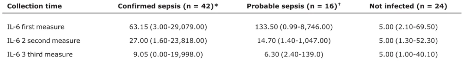
The median IL-6 values were statistically higher in the CS group than in the NI group at all three measurement times (p < 0.0001, p = 0.0024 and p = 0.0317, respectively). There was a progressive and significant reduction from the first to the second median (p = 0.0036) and from the second to the third (p = 0.00174). In the PS group, the medians were also statistically significant at all times (p = 0.0016, p = 0.0219 and p = 0.0373), and there was a significant drop from the first to the second measure (p = 0.0052). The medians in the NI group remained constant (Table 2).
< 0.0001; p = 0.0002 and p = 0.0038, respectively), with a reduction in median values from TNF1 to TNF2 (p = 0.0145) and from TNF2 to TNF3 (p = 0.0320). In the PS group there was no significant difference (p = 0.1668, p = 0.5209 and p = 0.9019) or reduction over time (p = 0.0830 and p = 0.1465). The median in the NI group remained almost unaltered for all three measurements (Table 3).
The CRP test exhibited high values for diagnostic proper-ties at all three measurement points, with no significant varia-tion over time for sensitivity (p = 0.1353), specificity (p = 1.0) or accuracy (p = 0.1062), and with elevated values for nega-tive predicnega-tive value at all three measurement points. The accuracy of this test was superior to the WBC at all three mea-surement times (p = 0.0335, p = 0.010 and p = 0.0024). The
values for sensitivity, specificity and accuracy for CRP1 were similar to those for IL-6 1 (p = 0.9070, p = 0.9557 and p = 0.5502, respectively) and for TNF1 (p = 0.6594, p = 0.5947 and p = 0.7366, respectively). In the comparison between CRP2 and IL-6 1 and TNF1, there were no statistical differ-ences between values for sensitivity (p = 0.1118 for IL-6 1 and p = 0.0628 for TNF1) or for specificity (p = 0.9557 for IL-6 1 and p = 0.5947 for TNF1), although accuracy was supe-rior to TNF-α (p = 0.0349).
The sensitivity and specificity of the WBC were similar to IL-6 1 (p = 01924 and p = 0.3033, respectively) and TNF1 (p = 0.3485 and p = 0.5783, respectively), although with infe-rior accuracy to IL-6 1 (p = 0.0459).
The IL-6 test was similar to the TNF-αtest in terms of lev-els of sensitivity (p = 0.7460), specificity (p = 0.6369) and
Table 1- Median CRP results for the confirmed sepsis, probable sepsis and not infected groups for the three collection times
Collection time Confirmed sepsis (n = 42) Probable sepsis (n = 16) Not infected (n = 24) p*
CRP first measure 5.09 (0.56-30.60) 4.14 (0.19-31.30) 0.28 (0.01-3.61) < 0.0001
CRP second measure 9.90 (0.29-83.20) 9.93 (0.70-31.90) 0.51 (0.01-2.38) < 0.0001
CRP third measure 5.74 (0.11-144.00) 6.21 (0.46-60.90) 0.33 (0.02-3.20) < 0.0001
CRP = C-reactive protein.
Values expressed in mg/dL – median (variation).
* Statistically significant difference between the medians for the confirmed sepsis or probable sepsis groups with the not infected group.
Table 2- Median IL-6 results for the confirmed sepsis, probable sepsis and not infected groups for the three sample collection times
Collection time Confirmed sepsis (n = 42)* Probable sepsis (n = 16)† Not infected (n = 24)
IL-6 first measure 63.15 (3.00-29,079.00) 133.50 (0.99-8,746.00) 5.00 (2.10-69.50)
IL-6 2 second measure 27.00 (1.60-23,818.00) 14.70 (1.40-1,047.00) 5.00 (1.30-52.30)
IL-6 3 third measure 9.05 (0.00-19,998.0) 6.30 (2.40-139.0) 5.00 (1.00-40.10)
IL-6 = interleukin-6.
Values expressed in pg/mL – median (variation).
* Statistically significant difference between medians for groups CS and NI in all three measurements (p < 0.0001; p = 0.0024 and p = 0.0317, respec-tively).
†Statistically significant difference between medians for groups PS and NI in all three measurements (p < 0.0016; p = 0.0219 and p = 0.0373,
respec-tively).
Table 3- Median TNF-αresults for the confirmed sepsis, probable sepsis and not infected groups for the three collection times
Collection time Confirmed sepsis (n = 42)* Probable sepsis (n = 16)† Not infected (n = 24)
TNF-αfirst measure* 24.70 (4.80-397.00) 10.05 (0.02-47.70) 6.40 (2.10-29.70)
TNF-αsecond measure† 19.20 (4.30-376.00) 8.10 (2.00-37.20) 6.60 (0.75-30.40)
TNF-αthird measure 15.00 (1.40-238.00) 6.7 (2.30-28.50) 7.00 (3.70-17.30)
TNF-α= tumor necrosis factor alpha.
Values expressed in pg/mL – median (variation).
* Statistically significant difference between medians for groups CS and NI in all three measurements p < 0.0001; p = 0.0002 and p = 0.0038, respec-tively).
†No statistically significant difference between medians for groups PS and NI in any of the three measurements (p = 0.1668; p = 0.5209 and p = 0.9019,
accuracy (p = 0.3067) at the first measurement. There was no significant difference between the first and second IL-6 measurements or between the first and third IL-6 measure-ments in sensitivity (p = 0.1924), specificity, (p = 0.7289 and 0.3033) or accuracy (p = 0.9044 and 0.950). The TNF-α results exhibited the same pattern, with no statistical differ-ence in sensitivity (p = 0.273), specificity (p = 0.1775) or accuracy (p = 0.6486) between TNF1 and TNF2.
In general, combining tests did not prove superior to each marker assayed in isolation. However, combining WBC with CRP2 offered a sensitivity superior to WBC in isolation (p = 0.0018), and taking the series of CRP assays offered a signifi-cant increase in sensitivity when compared with the initial assay alone (p = 0.0321). The values of these diagnostic test properties are listed in Table 4.
Discussion
The sample studied here is representative of the popula-tion normally seen at neonatal intensive care centers, with a
predominance of very low weight preterm NB and a predomi-nance of gram-positive cocci as the agents of late neonatal sepsis. Coagulase-negative staphylococcus have been the agents most commonly isolated from blood cultures in late neonatal sepsis,12,16although they are the most common contaminating agents. The fact that these agents grew in two blood culture samples and had the same antimicrobial resis-tance profile helped to define them as the agents responsible for the sepsis.
This study has shown that the WBC offers comparable diagnostic accuracy to that of IL-6 and TNF-αassays, although it is not as accurate as sequential CRP tests. The values for sensitivity and specificity in the literature vary widely, since there are significant differences in definitions used to count total neutrophils and sub-fractions; with sensitivity varying from 17 to 100% and specificity from 31 to 100%.7,17-19The elevated negative predictive value of the WBC has been con-sidered a valuable attribute, allowing clinicians greater confi-dence when ruling out a suspicion of late neonatal sepsis.7,19
Table 4- Figures for sensitivity, specificity, positive and negative predictive values and accuracy at the three different measurement times for CRP, IL-6, TNF-αand WBC
Test Sensitivity (95%CI) Specificity (95%CI) PPV NPV Accuracy (95%CI)
CRP
First measure 78.6 (66.2-100.0) 87.5 (74.3-100.0) 91.7 70.0 81.8 (72.5-91.1)
Second measure 90.5 (81.6-99.4) 87.5 (74.3-100.0) 92.7 84.0 89.4 (82.0-96.8)
Third measure 85.7 (75.1-96.3) 91.7 (80.6-100.0) 94.7 78.6 87.9 (80.0-95.8)
IL-6
First measure 77.5 (64.9-90.4) 87.0 (73.2-100.0) 91.2 69.0 81.0 (71.3-90.6)
Second measure 64.3 (49.8-78.8) 83.3 (68.4-98.2) 87.1 67.1 71.2 (60.3-82.1)
Third measure 64.3 (49.8-78.8) 75.0 (57,7-92.3) 81.8 54.5 68.2 (56.9-79.4)
TNF-α
First measure 74.3 (59.8-86.8) 81.8 (65.7-97.9) 86.7 66.7 77.2 (66.3-88.1)
Second measure 71.8 (57.7-85.9) 86.4 (72.0-100.0) 90.3 63.3 77.0 (66.5-87.6)
Third measure 62.2 (46.5-77.8) 95.2 (86.1-100.0) 95.8 58.8 74.1 (62.9-85.4)
hite blood cell count 64.3 (49.8-78.8) 75.0 (58.7-92.3) 81.8 54.5 68.2 (56.9-79.4)
Combinations
WBC + CRP1 87.5 (74.3-100.0) 90.9 (78.9-100.0) 91.3 87.0 89.1 (80.1-98.1)
WBC + CRP2* 100.0 (100.0-100.0) 94.1 (82.9-100.0) 95.8 100.0 97.5 (92.7-100.0)
CRP1 + CRP2† 95.0 (88.2-100.0) 84.2 (67.8-100.0) 92.7 88.9 91.5 (84.4-98.6)
CRP1 + IL-6 1 88.0 (75.3-100.0) 90.0 (76.9-100.0) 91.7 85.7 88.9 (79.7-98.1)
CRP1 + TNF1 95.2 (86.1-100.0) 94.1 (82.0-100.0) 95.2 94.1 94.7 (87.6-100.0)
CRP2 + IL-6 1 93.5 984.9-100.0) 90.5 (77.9-100.0) 93.5 90.5 92.3 (85.1-99.6)
CRP2 + TNF1 95.8 (87.8-100.0) 94.1 (82.9-100.0) 95.8 94.1 95.1 (86.5-100.0)
IL-6 1 + TNF1 87.0 (73.2-100.0) 94.1 (82.9-100.0) 95.2 90.0 90.0 (80.7-99.30)
95%CI = 95% confidence interval; CRP = C-reactive protein; IL-6 = interleukin-6; NPV = negative predictive value; PPV = positive predictive value; TNF-α= tumor necrosis factor alpha; WBC = white blood cell count. The number of test indicates the collection time.
* Statistically significant difference between the sensitivity with this combination and with the WBC in isolation (p = 0.018).
The CRP test proved to be a good diagnostic marker, supe-rior to the WBC and with comparable performance to the two cytokines, even for the first measurement and even though this has traditionally been considered a later marker since there is an interval between the infectious stimulus and the increase in CRP production and serum concentration. On the other hand, its elevated negative predictive value has also been shown to be clinically important, since sequential serum assay results that remain normal are an indicator, with a rea-sonable degree of certainty, that the child is not infected. Ben-itz et al.20found that, during episodes of late sepsis, using sequential samples increased sensitivity from 61.5 to 84.4% and specificity from 68.9% to 74.6%, comparing an initial sample with one taken 24 hours later.
The diagnostic test properties given in studies of late neo-natal sepsis also vary widely, with sensitivity oscillating between 39 and 97.5% and specificity between 47 and 100%. These oscillations are the result of differences in collection (single or a sequence), assay method, variations in the cut-offs defined and differences in the definition of sepsis itself (which is not always confirmed by culture).19-23
The diagnostic accuracy of IL-6 also proved to be good, and, although a significant reduction was observed in the median level of this marker over time, the sensitivity, speci-ficity and accuracy did not alter significantly. Procianoy & Sil-veira24also report that the timing of sample collection is important to detecting high levels of this cytokine in plasma. In the majority of sepsis cases serum levels return to normal in less than 48 hours.25-27The principal diagnostic utility of this cytokine is as an early marker; later assays can be dis-pensed with since they could result in interpretation errors.15,28
Tumor necrosis factor alpha exhibited a similar perfor-mance to IL-6 for diagnosis of late sepsis at all three collec-tion times. Roman et al.29found higher figures for sensitivity and specificity (91.3 and 100%, respectively). However, this difference can be explained by the smaller number of chil-dren in the study and the significant proportion of chilchil-dren who suffered shock and/or death. Ng et al. (1997),30performed a study of late sepsis in 68 very low birth weight NB and dem-onstrated elevated values for sensitivity (82%), specificity (86%), positive predictive value (82%) and negative predic-tive value (85%), maintaining an elevated value 24-48 hours after the first test.
When combinations of the tests were analyzed, the per-formance of WBC combined with CRP was significantly better than when used in isolation, and sequential CRP assays also had better sensitivity, and a very high negative predictive value. This being so, since both tests are routine in neonatal intensive care units, the correct use and interpretation of these tests are of great value.
Adding the two cytokine assays at the time of clinical sus-picion to these two routine tests did not improve upon the
assays in isolation, even though many authors have found evi-dence that diagnostic power is increased by combining CRP with IL-6 and TNF-α.15,30
The group of probable sepsis cases had WBC abnormali-ties and elevated levels of CRP and of the two cytokines, mak-ing a very similar profile to the group of sepsis cases confirmed by culture. This shows that these cases most probably were indeed sepsis, despite the negative cultures. Other studies have also described similar findings and some have included these children with those with confirmed sepsis.7,21,26
In conclusion, it was possible to demonstrate that using WBC and CRP was comparable to the IL-6 and TNF-αtests, allowing earlier diagnosis of late neonatal sepsis. Taking into consideration that these tests offer the advantages of being more often available, of having more standardized methods and having more accessible costs, they can be of aid in taking the difficult medical decision of whether or not to initiate anti-microbial treatment when faced with an NB with a suspicion of sepsis.
References
1. Klein J, Marcy MS. Bacterial sepsis and meningitis. In: Remington JS, Klein JO, editors. Infectious diseases of the fetus & newborn infant. 6th ed. Philadelphia: WB Saunders; 2005. p. 835-90.
2. Lemons JA, Bauer CR, Oh W, Korones SB, Papile LA, Stoll BJ, et al.Very low birth weigth outcomes of the National Institute of Child Health and Human Development Neonatal Research Network, January 1995 through December 1996.Pediatrics. 2001;107:E1.
3. Mussi-Pinhata MM, Rego MA.Particularidades imunológicas do pré-termo extremo: um desafio para a prevenção da sepse hospitalar. J Pediatr (Rio J). 2005;81:S59-S68.
4. Calil R, Marba ST, von Nowakonski A, Tresoldi AT.Reduction in colonization and nosocomial infection by multiresistant bacteria in a neonatal unit after institution of educational measures and restriction in the use of cephalosporins. Am J Infect Control. 2001;29:133-8.
5. Baltimore RS.Neonatal sepsis: epidemiology and management.
Paediatr Drugs. 2003;5:723-40.
6. da Silva O, Ohlsson A, Kenyon C.Accuracy of leukocyte indices and C-reactive protein for diagnosis of neonatal sepsis: a critical review. Pediatr Infect Dis J. 1995;14:362-6.
7. Jackson GL, Engle WD, Sendelbach DM, Vedro DA, Josey S, Vinson J, et al.Are complete blood cell counts useful in the evaluation of asymptomatic neonates exposed to suspected chorioamnionitis?Pediatrics. 2004;113:1173-80.
8. Ng PC. Diagnostic markers of infection in neonates. Arch Dis Child Fetal Neonatal Ed. 2004;89:F229-F35.
9. Gerdes JS.Diagnosis and management of bacterial infections in the neonate.Pediatr Clin North Am. 2004;51:939-59.
11. Stover BH, Shulman ST, Bratcher DF, Brady MT, Levine GL, Jarvis WR. Pediatric Prevention Network.Nosocomial infection rates in US children’ s hospitals’ neonatal and pediatric intensive care units. Am J Infect Control. 2001;29:152-7.
12. Stoll BJ, Hansen N.Infections in VLBW infants: studies from the NICHD neonatal research network. Semin Perinatol. 2003; 27: 293-301.
13. Gaynes RP, Edwards JR, Jarvis WR, Culver DH, Tolson JS, Martone WJ.Nosocomial infections among neonates in high-risk nurseries in the United States. National Nosocomial Infections Surveillance System.Pediatrics. 1996;98:357-61.
14. Manroe BL, Weinberg AG, Rosenfeld CR, Browne R.The neonatal blood count in health and disease I. Reference values for neutrophilic cells.J Pediatr. 1979;95:89-98.
15. Silveira RC, Procianoy RS.Evaluation of interleukin-6, tumour necrosis factor-alpha and interleukin-1beta for early diagnosis of neonatal sepsis.Acta Paediatr. 1999;88:647-50.
16. Fanaroff AA, Korones SB, Wright LL, Verter J, Poland RL, Bauer CR, et al.Incidence, presenting features, risk factors and significance of late onset septicemia in very low birth weight infants. The National Institute of Child Health and Human Development Neonatal Research Network.Pediatr Infect Dis J. 1998;17:593-8.
17. Rodwell RL, Taylor KM, Tudehope DI, Gray PH.Hematologic scoring system in early diagnosis of sepsis in neutropenic newborns. Pediatr Infect Dis J.1993;12:372-6.
18. Guillois B, Donnou MD, Sizun J, Bendaoud B, Youinou P.
Comparative study of four tests of bacterial infection in the neonate. Total neutrophil count, CRP, fibrinogen and C3d. Biol Neonate. 1994; 66:175-81.
19. Berger C, Uehlinger J, Ghelfi D, Blau N, Fanconi S.Comparison of C-reactive protein and white blood cell count with differential in neonates at risk for septicemia.Eur J Pediatr. 1995;154: 138-44.
20. Benitz WE, Han MY, Madan A, Ramachranda P.Serial serum C-reactive levels in the diagnosis of neonatal infection. Pediatrics. 1998;102:E41.
21. Ehl S, Gering B, Bartmann P, Högel J, Pohlandt F.C-reactive protein is a useful marker for guiding duration of antibiotic therapy in suspected neonatal bacterial infection. Pediatrics. 1997;99:216-21.
22. Chiesa C, Pellegrini G, Panero A, Osborn JF, Signore F, Assumma M, Pacifico L.C-reactive protein, interleukin-6, and procalcitonin in the immediate postnatal period: influence of illness severity, risk status, antenatal and perinatal complications, and infection.Clin Chem. 2003;49:60-8.
23. Franz AR, Bauer K, Schalk A, Garland SM, Bowman ED, Rex K, et al.; International IL-8 Study Group.Measurement of interleukin 8 in combination with C-reactive protein reduced unnecessary antibiotic therapy in newborn infants: a multicenter, randomized, controlled trial. Pediatrics. 2004;114:1-8.
24. Procianoy RS, Silveira RC.A influência do tempo de coleta sobre os níveis de interleucina-6 na sepse neonatal precoce.J Pediatr (Rio J). 2004;80:407-10.
25. Buck C, Bundschu J, Gallati H, Bartmann P, Pohlandt F.
Interleukin-6: a sensitive parameter for the early diagnosis of neonatal bacterial infection. Pediatrics. 1994; 93:54-8.
26. de Bont ES, Martens A, van Raan J, Samson G, Fetter WP, Okken A, et al.Tumor necrosis factor-alpha, interleukin-1 beta, and interleukin-6 plasma levels in neonatal sepsis. Pediatr Res.1993; 33:380-3.
27. Ng PC, Li K, Wong RP, Chui K, Wong E, Li G, et al.Proinflammatory and anti-inflammatory cytokine responses in preterm infants with systemic infections. Arch Dis Child Fetal Neonatal Ed. 2003; 88:F209-13.
28. Panero A, Pacifico L, Rossi N, Mancuso G, Stegagno M, Chiesa C.
Interleukin 6 in neonates with early and late onset infection.
Pediatr Infect Dis J. 1997;16:370-5.
29. Roman J, Fernandez F, Velasco F, Rojas R, Roldan MR, Torres A.
Serum TNF levels in neonatal sepsis and septic shock.Acta Paediatr. 1993;82:352-4.
30. Ng PC, Cheng SH, Chui KM, Fok TF, Wong MY, Wong W, et al.
Diagnosis of late onset neonatal sepsis with cytokines, adhesion molecule, and C-reactive protein in preterm very low birthweight infants. Arch Dis Child Fetal Neonatal Ed. 1997;77: F221-7.
Correspondence:
Sérgio Tadeu Martins Marba Rua Paulo Setúbal, 335/64
CEP 13020-240 - Campinas, SP - Brazil Tel.: +55 (19) 3521.9307, +55 (19) 9187.7727 Fax: +55 (19) 3521.9307