DYSFUNCTION OF THE TEMPORALIS MUSCLE AFTER
PTERIONAL CRANIOTOMY FOR INTRACRANIAL ANEURYSMS
COMPARATIVE, PROSPECTIVE AND RANDOMIZED
STUDY OF ONE FLAP VERSUS TWO FLAPS DIERESIS
FRANCISCO CARLOS DE ANDRADE JR.*, FRANCISCO CARLOS DE ANDRADE*, CELSO MACHADO DE ARAUJO FILHO**, JOSÉ CARCAGNOLO FILHO***
ABSTRACT - Patients with intracranial aneurysm(s) of the carotid artery territory, treated with pterional craniotomy, were prospectively and randomly addressed to one layer flap (n=36) or myocutaneous (MC) versus two layers’ dieresis (n=32) or interfascial (IF). The study protocol included the patient’s sex, age, area of craniotomy, time of flap dieresis and synthesis, time of bone dieresis and synthesis, the intracranial time, including dura mater dieresis and synthesis and time of flap retraction. Before and after surgery, the patients were evaluated with examination specially oriented to V and VII cranial nerves, bi-temporal diameter measurement, the symmetry of the temporal region, tempora-mandibularis joint (TMJ) movements and cranial CT scan. The evaluations of the TMJ dysfunctions were postoperative pain, movement limitations at mastication, occlusion, mouth aperture and lateral movements of the jaw. The statistical analysis showed that the incidence of pain at TMJ and moderate and severe temporalis muscle atrophy was observed, comparing MC and IF, and there were significant differences among these ones, being greater in IF group. We concluded that both techniques permit equivalent access to the studied intracranial aneurysm(s), and the atrophy of temporalis muscle, pain and movement limitations of the temporomandibularis joint were prevalent, worse and more long-lasting in two-layers flap dieresis than in one-layer flap dieresis.
KEY WORDS: atrophy temporalis muscle, intracranial aneurysm, postoperative complications, pterional craniotomy, surgical technique, temporalis muscle, temporomandibularis joint.
Disfunção do músculo temporal após craniotomia pterional para tratamento de aneurismas intracranianos: estudo comparativo, prospectivo e aleatório da diérese em camada única versus camada dupla
RESUMO - Pacientes com aneurisma(s) intracraniano(s) foram tratados pela via pterional e de modo prospectivo e aleatório submetidos às variantes técnicas, miocutânea (MC)(n=36) ou em retalho único e interfascial (IF) (n=32) ou em retalho duplo. No protocolo foram anotados o sexo, idade, área da craniotomia, período de diérese e síntese do retalho, período de diérese e síntese óssea, o período do procedimento intracraniano, incluindo o tempo de diérese e síntese da dura-máter e o período de retração do retalho. Os pacientes foram avaliados antes e após cirurgia, com exame neurológico especialmente orientado para o V e VII pares de nervos cranianos, medida do diâmetro bi-temporal, simetria das regiões temporais, movimentação da articulação temporomandibular (ATM) e tomografia computadorizada de crânio. A ATM foi avaliada em relação a dor articular pós-operatória, limitação dos movimentos a mastigação, oclusão, abertura da boca e movimentos laterais da mandíbula. A análise estatística mostrou que a incidência de dor na ATM e atrofia temporal moderada e grave, comparando MC com IF, houve diferenças significativas entre estes, sendo maior no grupo IF. Concluimos que ambas técnicas permitem acessos equivalentes aos aneurismas estudados, mas a atrofia do músculo temporal, a dor e limitações dos movimentos da ATM foi prevalente, pior e mais duradoura na diérese IF que na diérese MC.
PALAVRAS-CHAVE: aneurismas intracranianos, articulação temporomandibular, atrofia músculo temporal, complicações pós-operatórias, craniotomia pterional, músculo temporal, técnica cirúrgica.
Department of Neurological Surgery and Bucomaxillofacial Surgery Service, Pontifical Catholic University of São Paulo Medical Sciences School, Sorocaba (SP), Brazil: *MD; **MD, DS; ***DS. Aceite: 15-janeiro-1998.
The temporalis muscle atrophy that follows the pterional approach is a concern for patients and neurosurgeons. For the pterional craniotomy, different incisions were employed in the scalp and temporalis muscle1-6. To improve the anterior basal exposition of the pterional approach, the
one-layer flap or myocutaneous (MC) may be splitted in two one-layers. This maneuver permits the sliding and translation of the temporalis muscle to posteroinferior position of the exposure and opens wide the anteroinferior pterional access by decreasing the flap width in this corner.5 Following the
two-layers flap dieresis it is frequent the palsy of frontotemporal branches of the facial nerve (30%)2-4,
temporalis muscle atrophy and temporomandibularis joint dysfunction1-3,7-9. The technical interfascial
variant (IF) 4-6 drastically decreased the incidence of the frontotemporal branches’ palsy of facial
nerve, but not at all.10. Meanwhile, there are few reports on the temporalis muscle atrophy that
follows this procedure2,3.
On the other hand, the sulcus lateralis (Sylvian) access to the anterior supratentorial basal midline does not need a very low level of the flap7, and the inconvenience created by the flap width
can be easily turned through hook traction below the sphenoid ridge2,3.
To study the dilemma, there was a greater risk of flap complications with IF dieresis and technical difficulties to access the anterior supratentorial basal midline through MC dieresis, so we decided to compare them.
METHODS
Study Design
Patients with intracranial aneurysm(s) were consecutively treated with the pterional unilateral approach and randomly directed to MC (born in even years) and IF (born in odd years) flap dieresis (and synthesis), to investigate the incidence of temporalis muscle atrophy, its function and cosmetic consequences.
The study protocol included the sex and age of the patients, area of craniotomy, time of flap dieresis and synthesis (TFDS), time of the bone dieresis and synthesis (TBDS), the intracranial time, including durotomy and synthesis (TIC). The surgical period the flap was retracted in, without alleviation, was named time of flap retraction (TFR). The craniotomy areas and the necessary flap retraction according to the number and location of aneurysms were always variable, and they were calculated by measuring the area of the elevated free bone flap. The biggest bi-temporal diameter was taken pre- and postoperatively to calculate the degree of temporalis muscle atrophy with outside caliper (Vernier).
Selection Criteria
Patients with intracranial aneurysm(s) of the carotid artery territory (except distal portion of anterior cerebral artery), treated between October 1994 and December 1995 at the Medical Sciences School Center of the Pontifical Catholic University of São Paulo (Brazil).
Exclusion Criteria
Patients with follow-up period under 6 months, infection of the surgical wound, absorption of bone flap, previous craniotomy or re-operation, severe transoperative arterial hypotension, antecedents of temporomandibularis joint (TMJ) dysfunction, mobile tooth prosthesis, and craniofacial trauma.
General Characteristics of Patients
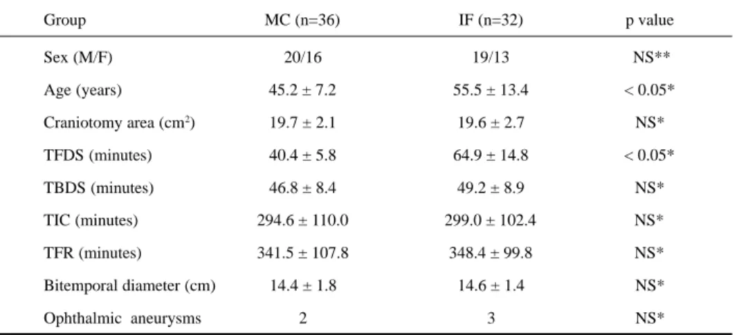
General characteristics of patients submitted to MC and IF pterional dieresis (and synthesis) are in Table 1.
Surgical Technique
In the MC group the the technique of flap’s dieresis (and synthesis) was employed, as detailed in the technical note by Spetzler & Lee3, and in IF group the technique detailed by Yasargil et al.4-6 was employed. The
pre-Table 1. General characteristics of patients submitted to MC and IF variants.
Group MC (n=36) IF (n=32) p value
Sex (M/F) 20/16 19/13 NS**
Age (years) 45.2 ± 7.2 55.5 ± 13.4 < 0.05*
Craniotomy area (cm2) 19.7 ± 2.1 19.6 ± 2.7 NS*
TFDS (minutes) 40.4 ± 5.8 64.9 ± 14.8 < 0.05*
TBDS (minutes) 46.8 ± 8.4 49.2 ± 8.9 NS*
TIC (minutes) 294.6 ± 110.0 299.0 ± 102.4 NS*
TFR (minutes) 341.5 ± 107.8 348.4 ± 99.8 NS*
Bitemporal diameter (cm) 14.4 ± 1.8 14.6 ± 1.4 NS*
Ophthalmic aneurysms 2 3 NS*
Caption: M/F, male/female; n, number of cases; NS, no significant; *, Fisher; **, χ2; media ± standard deviation; TBDS, time of
the bone dieresis and synthesis; TFDS, time of flap dieresis and synthesis;TFR, time of flap retraction;TIC, intracranial time.
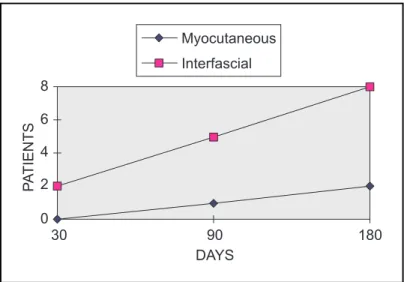
Fig 1. Incidence and evolution of TMJ’s disorders.
and postoperative wound care. The ophthalmic segment aneurysm(s) were treated with extradural removal of orbital posterior roof, lesser sphenoid bone covering the superior and medial surface of superior orbital fissure. The anterior clinoid process was removed intradurally.
Evaluation and Results Criteria
Before and after surgery (7-10, 30, 90, and 180 days), the patients were evaluated with examination specially oriented to V and VII cranial nerves, bi-temporal diameter measurement, the symmetry of the temporal region, TMJ’s movements and cranial CT scan. All patients were evaluated by the bucomaxillofacial team. They were not informed on the surgical procedures. The treatments of TMJ dysfunctions was made with non-hormonal anti-inflammatory drugs, orthodontics and physiotherapy.
The evaluations of the TMJ dysfunctions were postoperative pain, movement limitations at mastication, occlusion, aperture and lateral movements of the jaw. The classifications of these dysfunctions’ degrees were: mild, when regressed before 3 months; moderate with impairment betwen 3 and 6 months; and severe when persisted after 6 months.
The degrees of temporalis muscle atrophy were given through the percentile reduction of postoperative bi-temporal diameter: mild when bellow 5%; moderate between 5 and 10%; and severe over 10%.
Statistical Methods
Data from the experiments were evaluated according to the qui-square (χ2) or Fisher test. An alpha of 5%
was considered statistically significant.
RESULTS
Of the 80 original patients, 12 were excluded due to study criteria. The remaining 68 patients’ data, were submitted to the χ2 test for sex and Fisher exact test (F) to determine the differences’
distribution of the MC and IF groups, (Table 1) and to analyze study results (Fig 1).
Figure 1 presents the incidence and evolution of the TMJ’s studied complications in both groups.
The statistical analysis showed no differences in both randomized groups, related to the following data (Table 1): sex (χ2=0.10; p=0.75), craniotomy area (F=0.03; p=0.86), TBDS (F=1.31;
p=0.26), TIC (F=0.03; p=0.86) and TFR (F=0.07; p=0.78).
On the other hand, there were significant differences in the age (F=16.07; p=0.0009) and TFDS (F=97.02; p=0,00000…) data among groups, being greater in IF group.
The incidence of pain at TMJ, after 10 days (χ2= 4.83; p=0.03), 30 days (χ2=5.33; p=0.02)
and 90 days (χ2=7.29; p=0.007) of the evolution was significantly longer in the IF group, but in 180
days (F; p=0.22) there were no differences. In the MC group, the pain reduced precociously up to 90 days (χ2=3.96;p=0.04), and in the IF group the pain significantly reduced later, between 90 and 180
days (χ2=4.27; p=0.04). (Fig 1a)
The proportion of patients with movements limitation at TMJ, after 10 (χ2=1.24;p=0,26), 30
(χ2=1.35;p=0.25), 90 (χ2=0,87;p=0.35) and 180 (χ2=1.86;p=0.17) days was similar in both groups.
There were no statistically significant differences in evolution of this alteration in MC and IF series (Fig 1b).
There was no difference in postoperative evolution of the moderate temporalis muscle atrophy, in 30 (F; p= 0.21), 90 (χ2=1.25; p=0.25) and 180 (χ2=0.36; p=0.55) days, in both groups (Fig 1d).
The severe temporalis muscle atrophy showed us significant difference in evolution starting after 90 (F; p=0.02) and 180 (F; p=0.02) days, comparing MC and IF. And more, the incidence of such complication increased significantly in IF group between 30 and 90 (F; p=0.03) days. (Fig 1e). When analyzed in conjunction, moderate and severe temporalis muscle atrophy was observed, comparing MC and IF, after 90 (χ2=7.29; p=0.007) and 180 (χ2=5.11; p=0.02) days, and there were
significant differences of these kinds of atrophy in the groups (Fig1f).
DISCUSSION
In both analyzed techniques, myotomy (and synthesis) of the pars horizontalis of the temporalis muscle occurred, which was also responsible for final occlusion of the jaw and subperiostal disassemble of the temporal insertion of the temporalis muscle. In the study, both groups showed that the function of mandible final occlusion had been altered and always followed by mild temporalis muscle atrophy (Fig 1c). Since the skeletal muscle was cut (and sutured), and there was no remarkable regeneration of its fibers and its healing and the muscle repair was made by fibrous tissue11, it is
likely that this temporalis muscle mild atrophy and the final weakness of the jaw occlusion observed in the study can be imputed to such maneuvers.
In these procedures, traction, compression and flexion of the base flap occurred, which were maintained without alleviation during TFR. Without intermittent retraction, the flap was submited to ischemia8. In the study, there was not significant difference in the TFR between the groups, but
the IF technique permits major traction, torsion and kinking of the flap pedicle12. In this way, the flap
irrigation (and drainage) in IF technique is worse than MC technique, with direct or indirect (kinky) compromise of the deep temporal arteries12. In the study, there was a severity progressive pattern of
temporalis muscle atrophy in both groups (Fig 2). But the severity profile of the temporalis muscle atrophy was worse in the IF group.
So, the IF variant promotes more damage to the temporalis muscle irrigation (and drainage) responsible for postoperative temporalis muscle ischemic atrophy.
The pain and movement limitation of the mouth aperture and lateral deviation of the jaw to the side of the craniotomy are a consequence of the ischemic contracture, due to temporalis muscle antagonistic movements9. So, the TMJ function and balance are more compromised in intensity and
duration with the IF technique when compared to MC.
In the study bias, the average age distribution significantly bigger in the IF group (Table 1) does not change our conclusion, because the age is not a major influence on the inflammatory-reparative response, and there are no controlled experimental data to support such fact11. The bigger
TFDS observed at IF series explains itself by the greater technical complex in the making and flap synthesis of this variant, aiming the protection of the facial temporofrontalis limbs4-6.
There were no significant statistical differences in the remaining data analyzed in the study.
As for the additional space given by the muscular in two-layer flap making4-6, this one can be
surrounded by the thicker flap retraction proper to the single flap to the sphenoidal level given by several hooks2,3. Since the access of the studied intracranial aneurysm(s) was always made through
the lateralis fissure and basal cisterns, the limitation to the access of these aneurysm(s) is a function of the craniotomy area and of the cerebral compliance at surgical moment.
In the study, the pterional approach to the intracranial aneurysm(s) studied, were through the sulcus lateralis and basal cisterns.4,7,12 The limitation to the intracranial access of these aneurysm(s)
is only the widen opening of the Sylviam fissure and basal cisterns and cerebral compliance.
In conclusions, the MC and IF techniques of flap dieresis (and synthesis) for pterional craniotomy permit equivalent access to the intracranial aneurysm(s) studied. The risk of moderate and severe temporalis muscle atrophy and TMJ imbalance (pain and movement limitation) was prevalent, worse and long-lasting in IF dieresis than in MC. The MC technique is easier, faster, and has better TMJ function and cosmetic results than IF technique.
Acknowledgment - We thank Reinaldo José Gianini, MD, for statistical assistance.
REFERENCES
1. Ransohoff J. Lateral sphenoidal wing meningeoma. In Rengachary MD, Wilkins RH (eds). Neurosurgical operative atlas. Baltimore: Williams & Wilkins, 1991,1:43-49.
2. Spetzler RF. Two technical notes for neurosurgery. Barrow Neurol Instit Q 4, 1988;38-39.
3. Spetzler RF, Lee KS. Reconstruction of temporalis muscle for the pterional craniotomy: technical note. J Neurosurg 1990;73:636.
4. Yasargil MG: Interfacial pterional (frontotemporosphenoidal) craniotomy. In Yasargil MG, Smith RD, Young PH, Teddy PJ, Roth P (eds). Microneurosurgery. Stuttgart: Thieme Verlag, 1984,1:217-220.
5. Yasargil MG, Fox JL. The microsurgical approach to intracranial aneurysms. Surg Neurol 1975;3:7-14.
6. Yasargil MG, Reichmann MV, Kubik S. Preservation of the frontotemporal branch of the facial nerve using the interfascial temporalis flap for pterional craniotomy: technical note. J Neurosurg 1987;67:463-466.
7. Hamby WB. Remarks concerning intracranial aneurysmal surgery. In Ojemann RG, Tindal GT, Cantu RC, Keener EB, Wilkins RH (eds). Clinical neurosurgery. Baltimore: Williams & Wilkins, 1970;17:1-15.
8. Horwitz NH, Rizzoli HV. Intracranial neoplasm. In Horwitz NH, Rizzoli HV (eds). Postoperative complications in neurosurgical practice. Baltimore: Williams & Wilkins, 1967:5.
9. Rhoton AL Jr. Anatomic foundations of aneurysms surgery: operative approaches. In Loftus CM, Awad IA, Keith LB, et al. (eds). Clinical neurosurgery. Baltimore: Williams & Wilkins, 1993,41:317-324.
10. Aoki N. Incision of the facial nerve branch at aneurysm surgery (Letter). J Neurosurg 1987;66:482.
11. Cotran RS, Kumar V, Robbins SL. Healing and repair. In Cotran RS, Kumar V, Robbins SL (eds). Robbin’s pathologic basis of disease. 4 Ed. Philadelphia: WB Saunders, 1989:72-86.