*Correspondence: F. B. de Araujo Paula. Departamento de Análises Clínicas. Faculdade de Ciências Farmacêuticas. Universidade Federal de Alfenas. Rua Gabriel Monteiro da Silva, 700. Centro, 37130-000 - Alfenas - MG, Brasil. E-mail addresses: [email protected]; [email protected]
A
vol. 51, n. 4, oct./dec., 2015 http://dx.doi.org/10.1590/S1984-82502015000400013
Ethanolic extract of
Passiflora edulis
Sims leaves inhibits
protein glycation and restores the oxidative burst in diabetic rat
macrophages after
Candida albicans
exposure
Carolina Fernandes Ribas Martins
1, Bruno César Corrêa Salles
1, Maisa Ribeiro Pereira Lima
Brigagão
2, Maria Rita Rodrigues
1, Eric Batista Ferreira
3, Stella Maris da Silveira Duarte
1,
Fernanda Borges de Araujo Paula
1,*1Faculty of Pharmaceutical Sciences, Departament of Clinical and Toxicology Analysis, Federal University of Alfenas, MG, Brazil, 2Institute of Biomedical Sciences, Department of Biochemistry, Federal University of Alfenas, MG, Brazil, 3Insitute of
Exact Sciences, Federal University of Alfenas, Federal University of Alfenas, MG, Brazil
This study was conducted to evaluate the efects of the ethanolic extract of Passilora edulis leaves on
blood glucose, protein glycation, NADPH oxidase activity and macrophage phagocytic capacity after
Candida albicans exposure in diabetic rats. The Passilora edulis Sims leaves were dried to 40°C,
powdered, extracted by maceration in 70% ethanol, evaporated under reduced pressure and lyophilised.
The biochemical tests performed were total phenolic content (TP) as determined by the Folin-Ciocalteu assay, trapping potential DPPH assay and total iron-reducing potential.Diabetes was induced by alloxan
injection. Protein glycation was determined by AGE and fructosamine serum concentrations.
Extract-treated diabetic animals demonstrated lower fructosamine concentrations compared with the diabetic group. Our results suggest that ethanolic Passilora edulis Sims leaf extraction may have beneicial
efects on diabetes and may improve glycaemic control in diabetic rats.
Uniterms: Passilora edulis Sims/pharmacognosy. Passilora edulis Sims/ethanolic extract/efects. Blood glucose. Protein glycation. Reactive oxygen species. Diabetes/treatment/experimental study. Medicinal plants.
O objetivo deste estudo foi avaliar os efeitos do extrato etanólico de folhas de Passilora edulis sobre
os níveis de glicose sanguínea, glicação protéica, produção de espécies reativas de oxigênio (ERO) e capacidade fagocítica de macrófagos de ratos diabéticos. As folhas de Passilora edulis Sims foram
secas a 40 °C, trituradas e o extrato preparado por maceração em solução hidroetanólica 70% (v/v) etanol foi evaporado sob pressão reduzida e lioilizado. Os testes químicos realizados demonstraram que além da presença de compostos fenólicos, determinada pelo método de Folin-Ciocalteu, o extrato apresentou potencial sequestrante de radicais DPPH e redutor de ferro.Nos animais diabéticos foi observado aumento na glicação protéica, avaliada pela concentração de frutosaminas e de produtos de
glicação avançada (AGE), e redução na produção de ERO por macrófagos frente à Candida albicans,
quando comparados ao grupo controle. O tratamento dos animais diabéticos com o extrato reduziu as
concentrações de frutosaminas e manteve a produção de ERO em níveis semelhantes aos observados no
grupo controle. Nossos resultados sugerem que o extrato etanólico de folhas de Passilora edulis Sims
pode apresentar efeitos benéicos sobre o diabetes e melhorar o controle glicêmico em ratos diabéticos.
Unitermos: Passilora edulis Sims/farmacognosia. Passilora edulis Sims/extrato etanólico/efeitos. Glicose sanguínea. Glicação protéica. Espécies reativas de oxigênio. Diabetes/tratamento/estudo
INTRODUCTION
Diabetes mellitus is a serious public health problem due to the high incidence, morbidity and mortality rates that are mainly caused by the complications of this chronic disease (Shaw, Sicree, Zimmet, 2010).
Among these complications, diabetic individuals usually present with increased susceptibility to infections, which causes health problems. Studies have demonstrated
that approximately 20% of septicaemia patients are diabetic. Furthermore, the high frequency of infection may aggravate a pre-existing medical condition (Koh et al., 2012).
Many factors contribute to an increased susceptibility to infections; however, deficiency in innate immune function is the major cause of such manifestations (Geerlings, Hoepelman, 1999).
Some studies have suggested that increased susceptibility to infections might be associated with alterations in immune function and the phagocyte
inflammatory response (chemotaxis, phagocytosis and
killing), which could reduce the phagocytic and microbicidal capacity of these cells (Panneerselvam, Govindasamy, 2003; Ferreira et al., 2012; McNelis, Olefsky, 2014).
Alterations in the production of reactive oxygen
species (ROS) by phagocytes have been implicated as a cause of dysfunction in diabetes; however, the data
describing the inluence of diabetes on mononuclear cell NADPH oxidase activity are still divergent (Banerjee,
Sharma, 2012).
The increase in advanced glycation end product (AGE) formation because of interactions between glucose and protein molecules may also influence immune function either by binding to immunoglobulins or by
interacting with speciic receptors (RAGE) that are present on macrophages, which alters proinlammatory cytokine expression (Barbosa, Oliveira, Seara, 2008).
Brazil is the major producer of Passilora edulis, also known as yellow passion fruit. This fruit has great nutritional value, and its leaves have been widely used
in traditional medicine with beneicial efects in various
diseases. P. edulis leaf extract has sedating, anxiolytic,
anti-inlammatory and antibacterial properties (Dhawan
Dhawan, Sharma, 2004; Li et al., 2011).
Experimental studies have suggested that leaf extracts of diferent Passilora species may have hypoglycaemic, antioxidant and anti-glycation properties. These properties
have been attributed mainly to the presence of phenolic compounds, among which the flavonoid C-glycosides
isoorientin, orientin, vitexin, apigenin, and others stand
out. However, many of these studies were performed
in vitro and were simplified to be extrapolated to the
body. Furthermore, scientiic information regarding the
use of passion fruit leaves in diabetes treatment is still controversial (Doyama et al., 2005; Rudnicki et al, 2007).
Thus, the aim of the present study was to evaluate
the effects of the ethanolic extract of Passiflora edulis
Simsleaves on blood glucose, protein glycation, NADPH
oxidase activity and phagocytic capacity of diabetic rats’ macrophages exposed to Candida albicans.
MATERIAL AND METHODS
Obtaining passion fruit leaf samples
The passion fruit leaf samples were classified as
Passilora edulis Sims species. The leaves were collected from passion fruit plant cultivated in sandy soil with organic matter, in March 2012, in the city of Alfenas, Minas Gerais, Brazil, which has the following GPS
coordinates of latitude 21°27’33’ S, longitude 46°01’59’
W to 789 m. The voucher specimen of this sample was
preserved in our department with reference number 22356,
which was obtained by Federal University of Lavras, Department of Biology, ESAL herbarium.
Passiflora edulis Sims lead ethanolic extract
preparation
The Passiflora edulis Sims leaves were dried at
40 °C, powdered in an industrial blender, extracted by 70%
ethanol maceration, evaporated under reduced pressure and lyophilised (Rudinicki et al., 2007).
Total phenolic content determination
Total phenolic content (TP) was determined using the Folin-Ciocalteu assay (Singleton, Orthofer, Lamunela, 1999). Test sample TP content was standardised
against gallic acid and expressed as “mg gallic acid equivalents/100 g extract (GAE)”. Each determination
was performed in triplicate.
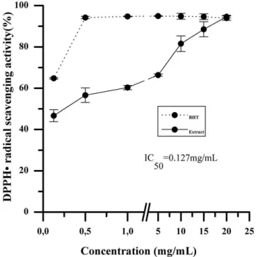
Trapping Potential DPPH assay
This assay has been widely used to evaluate the
antioxidant activity of vegetables extracts in vitro, based
on electron-transfer. The 2,2′-diphenyl-1-picrylhydrazyl
(DPPH.) is a stable free radical which may be reduced in
the presence of antioxidant substances, giving rise to the reduced form with the loss of this violet colour (Molyneux,
The measurement of DPPH radical scavenging activity was performed according to the methodology described by Dudonné et al. (2009). The different concentrations of Passiflora edulis ethanolic extract
samples (0.125, 0.5, 1.0, 5.0, 10.0, 15.0 or 20.0 mg/ mL) were reacted with the DPPH. in a methanolic
solution (0.02%, w/v). The absorbance recorded at 517 nm against an aliquot blank. DPPH radical-scavenging
activity of the Passilora edulis leaf ethanolic extract
was expressed in percentage compared with the control that only contained 0.02% DPPH solution; these data
were compared to a standard curve that was made with BHT ethanolic solution at the same concentration as the
Passilora edulis ethanolic extract. Each determination
was performed in triplicate.
Total iron-reducing potential
The iron-reducing potential of the Passilora edulis
leaf ethanolic extract was determined by adding diferent Passilora edulis extract concentrations (0.5, 10, 20, 40, 80, 120 or 160 mg/mL) into phosphate bufer with 1%
potassium ferricyanide, which was placed in a water bath
at 50°C for 30 minutes. Trichloroacetic acid (10%) was
then added, and the solution was centrifuged at 3,000 rpm
for 10 minutes. After centrifugation, 0.1% ferric chloride was added and the solution was vortexed. The absorbance
was read at 700 nm in a spectrophotometer (Biospectro SP - 220). The iron-reducing properties of the test sample
were standardised against butylated hydroxytoluene (BHT), and the results were expressed as a percentage compared with 1% BHT (Dudonné et al., 2009).
Animals
The study was conducted in accordance with
the ethical principles for animal experimentation that
have been adopted by the Brazilian College of Animal
Experimentation, and the study was approved by the ethics
committee on animal research at the Federal University of Alfenas. In total, 24 adult male Wistar rats (Rattus norvegicus) weighing 350±25 g that were obtained from the Unifal-MG vivarium were used in this study. The rats were housed in a temperature-controlled room on a 12 h light/dark schedule with food and water available ad libitum.
Alloxan-induced diabetic rats
Alloxan (2% solution) was administered
intraperitoneally (150 mg.kg–1) (Etuk, 2010). In this
study, diabetic rats were deined as those with glycaemia
over 250 mg.dl–1 after 7 days of alloxan injection. Blood
glucose was monitored using test strips with blood taken from the tail. At the time of sacrifice, blood was collected by cardiac puncture, and blood glucose levels were measured using an enzymatic method based on the Trinder reaction.
Animal experimental protocol
The animals were allocated into 4 groups of 6
animals: Control (C) – non-diabetic animals that were not
treated with ethanolic extract; Diabetics (D) – alloxan-treated animals; Extract (E) – non-diabetic animals
treated with Passilora edulis leaf ethanolic extract; and
Diabetic Extract (DE) – alloxan-treated animals also
treated with Passilora edulis leaf ethanolic extract. The
ethanolic extract was diluted in water and administered
to the animals by gavage for 8 weeks, after the induction of diabetes. The dose administered was 200 mg dried
ethanolic extract per kilogram of body weight per day.
The final volume administered to each animal was 0.5 mL. The control group received 0.5 mL water (Dhanabal
et al., 2004). After 55 days of treatment with the extract
and water, the animals received 3% sodium caseinate
intraperitoneally for macrophage recruitment. The animals were anaesthetised on the 57th day, and blood was collected
by cardiac puncture after a 12-hour fast. The animals were euthanised, and the macrophages were isolated from the
peritoneal luid with the addition of sterile 4 °C phosphate bufer, pH 7.4.
Obtaining serum and whole blood
To obtain serum, the second aliquot of blood was
distributed into siliconised glass tubes (without additive) and maintained at rest until blood clotting was complete. The samples were centrifuged at 1500 ×g for 10 minutes and stored at -80 °C. These samples were used for blood glucose evaluation and for AGE dosage (Advanced glycation end-product).
Cell suspension preparation
The cell suspension was obtained by peritoneal lavage and centrifuged at 2000 g for 15 minutes. The supernatant was resuspended in PBS plus glucose and kept on ice until the assay. These cell samples were used to
evaluate reactive oxygen species production as well as the
phagocytic and fungicidal ability of the macrophage when
exposed to yeast solution. Cell viability was evaluated by trypan blue exclusion (0.1%). The cell number obtained
Newbauer’s chamber, the different cell types present
were evaluated by morphological analysis, subjected to cytocentrifugation and stained with May-Grunwald-Giemsa (Daniel et al., 1971).
Yeast preparation
Candida albicans yeast (ATCC 69548) was
opsonised at a 1:5 ratio in diabetic or non-diabetic rat serum in 0.01 mol.l–1 phosphate bufer, pH 7.4, containing
0.15 mol.l–1 NaCl (PBS) for 30 minutes at 37 °C with
50 rpm orbital shaking. Viability was determined by
0.05% methylene blue exclusion (99.9%), and the number
of Candida albicans was determined in a Neubauer’s
chamber.
Phagocytic and fungicide capacity assessment
Macrophages (3.106 cells/mL) were incubated in sterile plastic tubes (to avoid cell adherence) at 37 °C with opsonised C. albicans (3.107 cells/mL). After 30, 60,
and 90 min incubation, a 50 µl aliquot of this suspension
was adhered to glass cover slips by cytocentrifugation (Sorocito FANEM). After centrifugation, cover slips were stained with Wright and May–Giemsa stains. The percentage of phagocytic cells was determined by counting the percentage of macrophages that had phagocytosed one or more C. albicans particles. The percentage of phagocytic cells and the number of yeast cells attached per 100 randomly chosen macrophages were counted
by examining at least 200 macrophages per preparation
(Ferreira et al., 2012).
Luminol-amplified chemiluminescence
For this assay, macrophages (1.106 per millilitre) were activated with C. albicans (1.107 per millilitre) in
a reaction mixture containing luminol (1 mmol.L–1) and
PBS, pH 7.4. Chemiluminescence intensity was followed
in a Geomax luminometer W 20/20, and the inal volumes were 0.3 mL. Chemiluminescence is expressed as relative
light units per second (RLU/s). The reaction was followed
for 60 min. All of the measurements were made at 37 ºC.
As controls, the reactions were carried out without cells, without luminol or without C. albicans.
Assessment of protein glycation
Protein glycation was assessed by determining AGE and fructosamine serum concentration. Fructosamine concentration was determined using the 2-point colorimetric kinetic method with a commercial brand
Analisa Gold test. The absorbance was read at 530 nm
in a semi-automatic BioPlus. AGEs were quantified
by measuring fluorescence, as described by Zilin et al. (2001). Previously, we determined serum protein concentration using the Biuret method. Afterwards,
AGEs were quantiied by adding 70 μL serum in 330 μL
chloroform in plastic tubes. Proteins were then precipitated
by adding 1.6 mL 0.15 M trichloroacetic acid followed by vortexing and centrifugation at 12000 ×g for 15 minutes.
The fluorescence was read at wavelengths of 350 nm
(excitation) and 440 nm (emission) with a gap of 5 nm. The results were expressed as arbitrary units divided by
the serum protein concentration (AU/g protein) (Zilin et al., 2001).
Statistical Analysis
The resultswere expressed as the mean ± standard deviation; they were also submitted to one-way analysis
of variance and compared using the Scott-Knott test at 5% signiicance.
RESULTS AND DISCUSSION
For the preliminary evaluation of the antioxidant
properties of Passilora edulis leaf ethanolic extract, some tests were performed to evaluate their reducing potential and free radical scavenging capacity. The FRAP method is based on electron transfer from compounds present in
the extract to iron ions, while the DPPH method assesses the ability of the extract to donate hydrogen atoms to
DPPH and stabilise it. The results demonstrated that the
P. edulis leaf ethanolic extract had antioxidant activity in vitro and was able to donate hydrogen atoms to DPPH and transfer electrons to iron ions (Figures 1 and 2). EC50
values observed for the two methods used in this study, and our results, demonstrate that the in vitro antioxidant
potential presented by the P. edulis ethanolic extract can
be attributed to the ability of scavenging free radicals rather than the FRAP method. This difference could be attributed to the ability of the chemical compounds
present in the extract to donate hydrogen or reduce iron
ions. These results reinforce the importance of using two
or more techniques for antioxidant activity evaluation as
recommended by Wootton-Beard, Moran, Ryan (2011). The results represent the average ± standard deviation performed in triplicate.
human health, such as oxidative stress inhibition and the prevention or treatment of diabetes mellitus, inlammation
and infectious processes (Rodrigo, Miranda, Vergara,
2011). Thus, as a preliminary analysis of the P. edulis
ethanolic extract chemical composition, the total phenolic content was determined for this extract.
The results suggest that the extract has phenolic
compounds. The literature has demonstrated the presence
of phenolic compounds in diferent Passilora species, mostly in the form of flavonoid C-glycosides such as
isoorientin, which is one of the main lavonoids present
in passion fruit juice. However, data on polyphenol concentration in P. edulis leaf extracts are still controversial
because there is great variation in this species, the method
of extract preparation and expression of the results (Li et al., 2011; Rudnick et al., 2007).
The phenolic content of P. edulis (4.67 ± 0.115 g
GAE/100 g extract) observed in the ethanolic extract was
similar to the concentrations reported by Oliveira et al.
(2009), when passion fruit methanol extract waste was examined. Furthermore, the P. edulis ethanolic extract
phenolic content was higher than the concentrations
that were observed in diferent studies with other herbal extracts that have potential anti-inflammatory and
hypoglycaemic properties (Caia et al., 2004; Souza et al., 2008).
The association between phenolic content and
antioxidant activity have been observed in different studies with medicinal plant extracts, including the genus Passilora (Caia et al., 2004; Rudnicki et al., 2007; Souza
et al., 2008). Caia et al. (2004) evaluated the in vitro
antioxidant activity and phenolic content in approximately
112 medicinal plants used in Chinese folk medicine. The authors reported that phenolic compounds were the main
components with antioxidant activity in these plants.
Furthermore, positive correlations have been observed
in diferent studies between phenolic content and extract antioxidant activity from plants that originated from the Amazon Rainforest with potential anti-inlammatory and hypoglycaemic efects (Souza et al., 2008). Some authors
have also reported an association between antioxidant activity and phenolic content in Passiloraceae genus fruit extracts or plant leaves (Oliveira et al., 2009; Rudnick et al., 2007).
Thus, our results suggest that the presence of phenolic compounds in the P. edulis ethanolic extract
could contribute to the iron reduction power and free radical scavenging that was observed in this study.
Antioxidants from plant extracts have an important role in oxidative processes and also in preventing or
treating various diseases including diabetes mellitus (Rodrigo, Miranda, Vergara, 2011). Considering the results obtained from the evaluation of P. edulis ethanolic
extract antioxidant activity in vitro and several studies
FIGURE 1 - Trapping Potential DPPH Assay of the Passilora edulis leaf ethanolic extract compared with the standard BHT.
FIGURE 2 - Total iron-reducing power assay of the Passilora edulis leaf ethanolic extract. The results represent the average
demonstrating the importance of medicinal plants with
antioxidant properties presented by the active principles
present in these plants (Rodrigo, Miranda, Vergara, 2011; Rudinicki et al., 2007), this study evaluated the efects of P. edulis leaf ethanolic extract ingestion on glycaemic control
and serum protein glycation in diabetic rats (Table I). Several studies have used glycation end product measurement in the diabetic patient serum to assess the risk of disease progression, demonstrating a positive correlation between glycated proteins levels in blood samples and the prevalence of diabetes mellitus-induced chronic complications (Brownlee, 1995; Rondeau, Boundon, 2011).
In this study, alloxan-treated animals demonstrated
severe weight loss, hyperglycaemia, elevated serum fructosamines and AGE, demonstrating that the protocol
was efective for diabetes induction (Table I). Among the
strategies adopted to reduce or maintain glycated blood protein levels within the desirable limits, glycaemic control is undoubtedly the main concern. Ethanolic
extract administration failed to induce signiicant changes in blood glucose and luorescent AGE serum levels, but
fructosamine concentration was significantly lower in
the extract-treated diabetic animal serum compared with
untreated diabetic animals, demonstrating that the P. edulis
ethanolic extract prevented an increase in serum protein
glycation in diabetes mellitus.
Fructosamine formation involves an interaction between the nucleophilic group of glucose molecules and the amine group of serum proteins, mainly albumin.
Therefore, fructosamine levels relect glycaemic control
during two to three weeks prior to blood collection, while fasting glucose levels reflects momentary glycaemic
control. These efects can be attributed to time between extract administration and blood collection (12 hours).
Thus, these results suggest that despite the ethanolic
extract did not inluence fasting glucose levels, it could
have reduced the blood glucose level shortly after its administration and contributed to a reduction of the
average glycaemic level during the day, which is beneicial
for glycaemic control.
The hypoglycaemic efects of P. edulis, P. mollisima
and P. quadrangular leaf alcoholic extracts were observed
after oral administration in diabetic mice for a period of 8 days. However, the administration of P. alata tea leaves orally for a period of 15 days was not able to reduce blood glucose levels in normoglycaemic rats (Doyama et al.,
2005).
Arya et al. (2012) demonstrated that the Centratherum anthelminticum seed methanolic fraction exhibited potential
anti-diabetic efects in pancreatic beta cell culture and in streptozotocin-induced diabetic rats. These efects were
associated with polyphenols that were identified in the
methanolic fraction of the extract. Furthermore, several studies have associated the anti-diabetic efects presented
by some medicinal plants to the presence of polyphenols (Rodrigo, Miranda, Vergara, 2011). Therefore, the results observed in this study could be attributed to the presence of phenolic compounds in the P. edulis ethanolic extract,
which could consequently contribute to glycaemic control
either by increasing insulin sensitivity or by decreasing the intestinal absorption of carbohydrates.
Another way of assessing protein glycation is by determining AGE concentration. These products are
formed from Amadori products by means of a complex
cascade of reactions, which can take weeks or months to complete. This determination has been adopted to assess protein glycation and long-term glycaemic control (Barbosa, Oliveira, Seara, 2008).
In the present study, an increase in the AGE concentration was observed in the serum of diabetic rats compared with non-diabetic animals (Table I).
I n v i t ro s t u d i e s h a v e d e m o n s t r a t e d t h a t
hydroethanolic extracts from P. alata leaves inhibited
TABLE I - Efect of P. edulis leaf ethanolic extract on glucose levels, fructosamine concentrations and advanced glycation
end-product (AGE) quantiication in non-diabetic and diabetic animals
Experimental group Glycaemia (mg/dl)
Fructosamines (mmol/L)
AGE (AU/g prot)
Body weight (g)
1st week 8st week
C 113,78 ± 13,52a 1,63 ± 0,18a 0,11 ± 0,02a 354,50 ± 20,87a 417,00 ± 17,03a
D 285,78 ± 69,35b 2,38 ± 0,41b 0,17 ± 0,06b 351,00 ± 19,34a 264,67 ± 38,05b
E 90,95 ± 10,57a 1,61 ± 0,07a 0,10 ± 0,02a 344,17 ± 17,05a 401,83 ± 22,69a
DE 269,98 ± 115,58a 1,85 ± 0,49a 0,13 ± 0,04a 348,50 ± 15,75a 280,66 ± 36,35b
The results represent the average ± standard deviation of 6-8 determinations per treatment. Diferent letters in the same column
AGE formation from albumin and glucose interaction.
The authors attributed these effects to the antioxidant activity in the analysed extracts (Rudnicki et al., 2007). Although other mechanisms may be involved in the anti-glycation activity of natural and synthetic compounds, the actions of most of these compounds has been linked
to their antioxidant properties, particularly the ability to
chelate metals and scavenge carbonyl radicals (Edeas et al., 2010). Moreover, many studies have attributed these
efects to the presence of phenolic compounds such as lavonoids (Edeas et al., 2010).
However, in the present study, there was no
signiicant diference in the concentration of serum AGE
among diabetic animals that were treated or not with the
extract (Table I).
Considering that the ethanolic extract of P. edulis
presented free radical scavenging activity as well as iron-reducing potential and that there was decrease in
the serum fructosamine concentration in the extract-treated diabetic animals, the absence of efects on AGE
concentration could be attributed to fact of that not all
serum AGE have the ability to luoresce between 370 and
440 nm (Schleicher et al., 2001). Thus, ethanolic extract
treatment may exert its efect only on the non-luorescent AGE. Our group should perform future experiments to assess the efects of the extract on non-luorescent AGE concentrations such as that of carboxymethylcysteine
(CML), which is largely found in the serum.
Hyperglycaemia can negatively modulate the phagocytic cell response to infectious agents, which could
explain the increased susceptibility to infections that is
usually noted in diabetic patients (Banerjee, Sharma, 2012; Souza et al., 2007). Moreover, macrophages are central components of the innate immune response to C. albicans, and ROS production by these cells is reportedly an important mechanism for their activation and the destruction of these pathogens (Slauch, 2011).
Our results have demonstrated that diabetes mellitus and P. edulis ethanolic extract treatment were not able
to induce changes in macrophage phagocytic capacity
(Figure 3), but ROS production was signiicantly lower in
diabetic rat peritoneal macrophages compared with
non-diabetic animals (Figure 4). Furthermore, extract treatment
restored macrophage ROS production in diabetic animals. Phagocytosis occurs after pathogen recognition by macrophages via specific receptors that are present on the cell membrane. During phagocytosis, ROS are
generated by NADPH oxidase activation in a process called an oxidative burst. In this process, superoxide
anions are produced, and although this molecule presents
low microbicide activity, it is required to generate more
FIGURE 3 - Efect of P. edulis leaf ethanolic extract on the ability of diabetic and non-diabetic rat peritoneal macrophages to phagocyte Candida albicans. Macrophages were incubated with opsonised yeast C. albicans for 0, 30, 60, 90 or 120 minutes, and the percentage of macrophages that phagocytised one or more yeast molecules (% phagocytosis) was determined. The results
represent the average ± standard deviation of 4-5 determinations
per treatment. C=control group; D=diabetic animals; E=P. edulis ethanolic extract-treated non-diabetic animals; DE=P. edulis ethanolic extract-treated diabetic animals.
FIGURE 4 - Efect of the Passilora edulis leaf ethanolic extract
on Reactive Oxygen Species (ROS) production in diabetic and non-diabetic rats. The values are represented as the means ± SD of the four-ive experiments of the integrated light emission area that was obtained for luminol oxidation (1 mmol.L–1)
promoted by Candida albicans (1x10⁷ cells per test)-stimulated
rat peritoneal macrophages (1x10⁶ cells per test) from control
animals (C), diabetic animals (D), Passilora edulis ethanolic
extract-treated non-diabetic animals (E) and Passilora edulis
potent molecules such as HOCl (Babior, 2002; Slauch, 2011). When macrophages are activated, the generated
ROS can oxidise molecules such as luminol to its excited
and unstable intermediates, which can be measured by chemiluminescence (Vilim, Wilhelm, 1999).
Several studies have demonstrated impaired phagocytic cell activity in diabetes mellitus (Alba-Loureiro et al., 2006). However, the results regarding the
efects of diabetes mellitus on ROS production in these
cells are still divergent. Some authors have observed increased ROS production in phagocytic cells of diabetic animals or humans (Willians et al., 2011); however, Souza et al. (2007) determined that ROS production
by lipopolysaccharide (LPS)-stimulated RAW 264.7
macrophages was lower in the presence of high glucose concentrations (500 mg/dL) when compared with cells that were incubated with normal glucose concentrations (200 mg/dL). Banerjee, Sharma (2012) reported that
the NADPH oxidase activity of macrophages that were maintained in diferent glucose concentrations decreased
as glucose concentrations increased. Furthermore, the authors observed an association between increased cell membrane glycation and reduced macrophage NADPH
oxidase activity.
Thus, our results suggest that inhibition of the
macrophage oxidative burst in diabetic rats could be one
of the mechanisms that is responsible for the susceptibility to infections in diabetic patients and the administration of
P. edulis ethanolic extract could contribute to preventing
this disorder.
Hyperglycaemia can lead to the glycation of proteins that are present in the macrophage membrane including
NADPH oxidase subunits, thus compromising its activity. Therefore, the efect of P. edulis ethanolic extract treatment
on diabetic rat macrophage ROS production could be related to its action on protein glycation, as evidenced by the fructosamine results.
Thus, our results suggest that the ethanolic extract
may have beneficial effects on the diabetic state by contributing to glycaemic control and the modulation of
macrophage oxidative burst in diabetic rats.
CONCLUSIONS
The ethanolic extract demonstrated higher phenolic
content than the various medicinal plants that are used in
folk medicine and demonstrated antioxidant activity in vitro, as evidenced by DPPH radical scavenging capacity
and iron-reducing power. Ethanolic extract administration
in the animals prevented an increase in diabetic animal serum protein glycation and contributes to maintaining
diabetic rat macrophage ROS production at levels that are similar to those of non-diabetic animals.
ACKNOWLEDGMENTS
The authors thank CAPES and FAPEMIG.
REFERENCES
ALBA-LOUREIRO, T.C.; HIRABARA, S.M.; MENDONÇA, J.R.; CURI, R.; PITHON-CURI, T.C. Diabetes causes marked changes in function and metabolism of rat neutrophils. J. Endocrinol., v.188, n.2, p.295-303, 2006.
ARYA, A.; LOOI C.Y.; CHEAH, S.C.; MUSTAFA, M.R.; MOHD, M.A. Anti-diabetic effects of Centratherum anthelminticum seeds methanolic fraction on pancreatic cells, b-TC6 and its alleviating role in type 2 diabetic rats. J. Ethnopharmacol., v.144, n.1, p.22-32, 2012.
BABIOR, B.M. The leukocyte NADPH oxidase. Israel Med. Am. J., v.4, p.1023-1024, 2002.
BARBOSA, J.H.; OLIVEIRA, S.L.; SEARA, L.T. O papel dos produtos finais da glicação avançada (AGEs) no desencadeamento das complicações vasculares do diabetes. Arq. Bras. Endocrinol. Metab., v.52, n.6, p.940-950, 2008.
BANERJEE, D.; SHARMA, P. Dual effect of glucose on macrophage NADPH oxidase activity: a possible link between diabetes and tuberculosis. Oxid. Antioxid. Med. Sci., v.1, n.1, p.91-96, 2012.
BROWNLEE, M. The pathological implications of protein glycation. Clin. Invest. Med., v.18, p.275-281, 1995.
CAIA, Y.; LUOB, Q.; SUNC, M.; CORKE, H. Antioxidant activity and phenolic compounds of 112 traditional Chinese medicinal plants associated with anti-cancer. Life Sci., v.74, n.17, p.2157-2184, 2004.
DANIEL, M.T.; FLANDRIN, G.; LE’VEUNE, F.; LISO, P.; CORTHOLARY, P.F. Les estérases spécifiques monocytaires: utilisacion dans la classiicación de leucemies agués. Nov. Rev. Franc. Hemat., v.11, p.233-240, 1971.
DHANABAL, S.P.; JERALD, E.E.; KUMAR, E.P.; SURES, B.
DHAWAN, K.; DHAWAN, S.; SHARMA, A. Passiflora: a review update. J. Ethnopharmacol., v.94, n.1, p.1-23, 2004.
DOYAMA, J.T.; RODRIGUES, H.G.; NOVELLI, E.L.; CEREDA, E.; VILEGAS, W. Chemical investigation and efects of the tea of Passilora alata on biochemical parameters in rats. J. Ethnopharmacol., v.96, n.3, p.371-374, 2005.
DUDONNÉ, S.; VITRAC, X.; COUTIERE, P.; WOILLEZ, M.; MÉRILLON, J. Comparative study of antioxidant properties and total phenolic content of 30 plant extracts of industrial interest using DPPH, ABTS, FRAP, SOD, and ORAC assays. J. Agric. Food Chem., v.57, n.5, p.1768-1774, 2009.
EDEAS, M.; ATTAF, D.; MAILFERT, A.; NASU, M.; JOUBET, R. Maillard reaction, mitochondria and oxidative stress: potential role of antioxidants. Pathol. Biol., v.58, n.3, p.220-225, 2010.
ETUK, E.U. Animals models for studying diabetes mellitus. Agric. Biol. J. N. Am., v.1, n.2, p.130-134, 2010.
FERREIRA, C.S.; ARAÚJO, T.H.; ÂNGELO, M.L.; PENNACCHI, P.C.; OKADA, S.S.; PAULA, F.B.A.; MIGLIORINI, S.; RODRIGUES, M.R. Neutrophil dysfunction induced by hyperglycaemia: modulation of myeloperoxidase activity. Cell Biochem. Funct., v.30, n.7, p.604-610, 2012.
G E E R L I N G S , S . E . ; H O E P E L M A N , A . I . M . I m m u n e dysfunction in patients with diabetes mellitus (DM). FEMS Immunol. Med. Microbiol., v.26, n.3-4, p.259-265, 1999.
JAMIR, T.T.; SHARMA, H.K.; DOLUI, A.K. Folklore medicinal plants of Nagaland. Fitoterapia, v.70, n.4, p.395-401, 1999.
KOH, G.C.K.W.; PEACOCK, S.J.; VAN DER POLL, S.J.; WIERSINGA, W.J. The impact of diabetes on the pathogenesis of sepsis. Eur. J. Clin. Microbiol. Infect. Dis., v.31, n.4, p.379-388, 2012.
LI, H.W.; ZHOU, P.; YANG, Q.Q.; SHEN, Y.; DENG, J.; LI, L. Comparative studies on anxiolytic activities and lavonoid
compositions of Passilora edulis ‘edulis’ and Passilora
edulis ‘lavicarpa’. J. Ethnopharmacol., v.133, n.3, p.1085-1090, 2011.
MCNELIS, J.C.; OLEFSKY, J.M. Macrophages, immunity, and metabolic disease. Immunity, v.41, n.1, p.36-47, 2014.
M O LY N E U X , P. T h e u s e o f t h e s t a b l e f r e e r a d i c a l diphenylpicrylhydrazyl (DPPH) for estimating antioxidant activity. Songklanakarin J. Sci. Technol., v.26, n.2, p.211-219, 2004.
OLIVEIRA, A.C.; VALENTIM, I.B.; SILVA, C.A.; BECHARA, E.J.H.; BARROS, M.P.; MANO, C.M.; GOULART, M.O.F. Total phenolic content and free radical scavenging activities of methanolic extract powders of tropical fruit residues. Food Chem., v.115, n.2, p.469-475, 2009.
PANNEERSELVAM, S.; GOVINDASAMY, S. Sodium molybdate improves the phagocytic function in alloxan-induced diabetic rats. Chem. Biol. Interact., v.145, n.2, p.159-163, 2003.
RODRIGO, R.; MIRANDA, A.; VERGARA, L. Modulation of endogenous antioxidant system by wine polyphenols in human disease. Clin. Chim. Acta., v.412, n.5-6, p.410-424, 2011.
RONDEAU, P.; BOUNDON, E. The glycation of albumin: Structural and functional impacts. Biochimie, v.93, n.4, p.645-658, 2011.
RUDNICKI, M.; OLIVEIRA, M.R.; PEREIRA, T.V.; REGINATTO, F.H.; DAL-PIZZOL, F.; MOREIRA, J.C.F. Antioxidant and anti-glycation properties of Passiflora alata and Passilora edulis extracts. Food Chem., v.100, n.2, p.719-724, 2007.
SHAW, J.E.; SICREE, R.A.; ZIMMET, P.Z. Diabetes atlas. Global estimates of the prevalence of diabetes for 2010 and 2030. Diabetes Res. Clin. Pract., v.87, n.1, p.4-14, 2010.
SCHLEICHER, E.D.; BIERHAUS, A.; HARING, H.U.; NAWROTH, P.P.; LEHMANN, R. Chemistry and pathobiology of advanced glycation end products. Contrib. Nephrol., v.131, p.1-9, 2001.
SINGLETON, V.L.; ORTHOFER, R.; LAMUNELA, R.M. Analysis of total phenols and other oxidation substrates by means of Folin-Ciocalteau reagent. Meth. Enzymol., v.299, p.152-178, 1999.
SOUZA, L.F.; BARRETO, F.; DA SILVA, E.G.; ANDRADES, M.E.; GUIMARÃES, E.L.; BEHRGA, M.J.C.; BERNARD, E.A. Regulation of LPS stimulated ROS production in peritoneal macrophages from alloxan-induced diabetic rats: Involvement of high glucose and PPARγ. Life Sci., v.81, n.2, p.153-159, 2007.
SOUZA, J.N.; SILVA, E.; LOIR, A.; REES, J.; ROGEZ, H.; LARONDELLE, Y. Antioxidant capacity of four polyphenol-rich Amazonian plant extracts: A correlation study using chemical and biological in vitro assays. Food Chem., v.106, n.1, p.331-339, 2008.
VILIM, V.; WILHELM, J. What do we measure by a luminol-dependent chemiluminescence of phagocytes? Free Radic. Biol. Med., v.6, n.6, p.623-629, 1999.
WILLIANS, N.L.; MORRIS, J.L.; RUSH, C.; GOVAN, B.L.; KETHEESAN, N. Impact of streptozotocin-induced diabetes on functional responses of dendritic cells and macrophages towards Burkholderia pseudomallei. FEMS Immunol. Med. Microbiol., v.61, n.2, p.218-227, 2011.
WOOTTON-BEARD, P.C.; MORAN, A.; RYAN, L. Stability of the total antioxidant capacity and total polyphenol content of 23 commercially available vegetable juices before and after in vitro digestion measured by FRAP, DPPH, ABTS and Folin–Ciocalteu methods. Food Res. Inter., v.44, n.1, p.217-224, 2011.
ZILIN, S.; NAIFENG, L.; BICHENG, L.; JIPING, W. The determination of AGE peptides by low injection assay, a practical marker of diabetic nephropathy. Clin. Chim. Acta., v.313, n.1-2, p.69-75, 2001.