*Correspondence: K. Segala. Departamento de Engenharia de Materiais e Bioprocessos. Faculdade de Engenharia Química. Universidade Estadual de Campinas. Avenida Albert Einstein, 500, 13083-970 - Campinas - SP, Brazil. E-mail: [email protected]
A
vol. 51, n. 4, oct./dec., 2015 http://dx.doi.org/10.1590/S1984-82502015000400017
Silver nanoparticles incorporated into nanostructured biopolymer
membranes produced by electrospinning: a study of antimicrobial
activity
Karen Segala
1,*, Silvia Vaz Guerra Nista
1, Lívia Cordi
2,3, Maria Trindade Marques Bizarria
1, José
de Ávila Júnior
4, Sirlene Adriana Kleinubing
1, Deborah Cristina Cruz
1, Marcelo Brocchi
2, Liliane
Maria Ferrareso Lona
1, Nelson Eduardo Durán Caballero
3,5,6, Lucia Helena Innocentini Mei
11Department of Materials Engineering and Bioprocess, School of Chemical Engineering, University of Campinas, UNICAMP,
Campinas, SP, Brazil, 2Department of Genetics, Evolution and Bioagents, Institute of Biology, University of Campinas,
UNICAMP, Campinas, SP, Brazil, 3Chemistry Institute, University of Campinas, UNICAMP, Campinas, SP, Brazil, 4Mechanical
Engineering Department, Federal University of São João del-Rey, São João del-Rei, MG, Brazil, 5NanoBioss, Institute of
Chemistry, UNICAMP, Campinas, SP, Brazil, 6Brazilian Network on Nanotoxicology, MCTI/CNPq, Institute of Chemistry,
UNICAMP, SP, Brazil
This study examines the antimicrobial activity of silver nanoparticles incorporated into nanostructured membranes made of cellulose acetate (CA) and blends of chitosan/poly-(ethylene oxide, CTS/PEO) and prepared by electrospinning. The formation of chemically synthesized Ag nanoparticles (AgNPs) was monitored by UV-visible spectroscopy (UV-Vis) and characterized by transmission electron microscopy (TEM). The size distribution of the AgNPs was measured by dynamic light scattering (DLS), with an
average size of approximately 20 nm. The presence of AgNPs on the surface of electrospun nanoibers was observed by ield emission electron microscopy (FEG) and conirmed by TEM. The antimicrobial
activity of AgNPs incorporated into nanostructured membranes made of CA and CTS/PEO electrospun
nanoibers was evaluated in the presence of both Gram-positivebacteria, such as Staphylococcus aureus ATCC 29213 and Propionibacterium acnes ATCC 6919, and Gram-negativebacteria, such as Escherichia coli ATCC 25992 and Pseudomonas aeruginosa ATCC 17933. Microbiological results showed that the
presence of AgNPs in CA and CTS/PEO nanostructured membranes has signiicant antimicrobial activity for the Gram-positivebacteria Escherichia coli and Propionibacterium acnes.
Uniterms: Electrospinning. Silver nanoparticles/antibacterial activity. Cellulose acetate. Chitosan.
Neste trabalho avaliou-se a atividade antimicrobiana das nanopartículas de prata (AgNPs) incorporadas em membranas de acetato celulose (AC) e blendas de quitosana/poli-óxido de etileno (CTS/PEO) preparadas
pelo método de eletroiação. A formação das AgNPs previamente sintetizadas foi monitorada por UV-Vis e caracterizada por microscopia eletrônica de transmissão (MET). A distribuição de tamanho das AgNPs foi mensurada por espalhamento de luz dinâmico, com tamanho médio em torno de 20 nm. A presença das NPs na superfície das nanoibras eletroiadas foi observada por microscopia eletrônica com emissão de campo (FEG) e conirmada por MET. A atividade antimicrobiana das membranas nanoestruturadas de AC e CTS/PEO foi avaliada pelo uso de bactérias Gram-positivas, tais como Staphylococcus aureus ATCC 29213 e Propionibacterium acnes ATCC 6919, e Gram-negativas, como Escherichia coli ATCC 25992 e Pseudomonas aeruginosa ATCC 17933. Os resultados microbiológicos mostraram a presença das
AgNPs nas membranas de AC e CTS/PEO com signiicativa atividade antimicrobiana para Escherichia coli e Propionibacterium acnes, respectivamente.
INTRODUCTION
The use of metal nanoparticles in various medical and biotechnological applications is one of the most investigated areas in materials science. These applications require appropriate chemical functionalization of the nanoparticles with organic molecules or their incorporation into polymer matrices (Dallas, Sharma, Zboril, 2011). Among the numerous types of nanoparticles that have been used to decorate polymers, silver nanoparticles (AgNPs) are the most researched, due to their electronic properties (Líu et al., 2010; Tricoli, Pratsinis, 2009; Chen et al., 2010), and optical (Zeng et al., 2007), catalytic (Severin et al., 2007; Signori et al., 2010) and antimicrobial activities (Panacek et al., 2006; Kvitek et al., 2008; Prema, Raju, 2009; Chaloupka, Malam, Seifalian, 2010).
Recently, there has been increasing interest in studying nanostructured biopolymers with antimicrobial characteristics for their immense potential use in devices that require a high antiseptic character. Recent studies have shown that an especially lucrative opportunity lies in applying these polymeric nanostructures in the medical field (Abdelgawada, Hudsona, Rojas, 2014; Chaloupka, Malam, Seifalian, 2010; Dallas, Sharma, Zboril, 2011). The literature contains a great deal of studies demonstrating the successful application of these nanostructures in the healthcare, pharmaceutical and cosmetic industries for repairing and regenerating the skin and organs; as drug delivery vectors and therapies; biocompatible and biodegradable implants; in medical diagnosis and instrumentation; as tissue protective agents that guard against infection; and in cosmetics, molecular medicines, supplements and body care applications in the ield of dentistry (Nista, Bettini; Mei, 2015; Bizarria, Davila, Mei, 2014; Kleinubing et al., 2014).
The AgNPs possess superior antimicrobial properties compared to other salts, due to their extremely large surface area, which provides better contact with microorganisms (Rai, Yadav, Gade 2009). Although the mechanism of action of silver nanoparticles on microorganisms is not fully understood, there are hypotheses that these nanoparticles can cause cell lysis or inhibit cell transduction (Prabhu, Poulose, 2012).
Studies are being carried out to determine how to incorporate AgNPs into biopolymeric membranes (Ellison et al., 2014; Kanmani, Rhim, 2014; Rhim, Wang, Hong, 2013), due to the biocompatibility of these polymers (Okamoto, John, 2013) with multiple biological systems and their enhanced antimicrobial activity (Guo
et al., 2013; Madhumathi et al., 2010). Materials based on cellulose like CA are a particularly good choice for
preparing these ilms, due to their excellent performance characteristics; CA, for example, exhibits good toughness, high biocompatibility and relatively low cost (Han et al., 2013; Abou-Zeida et al., 2011; Ferjani et al., 2002).
Polymeric blends have received considerable attention from researchers in recent decades as a means to improve the physico-chemical and mechanical properties of materials (Abou-Zeida et al., 2011). Polymeric ilms made from the blends of CTS/PEO show better thermal, chemical, mechanical and conductive properties than do ilms of pure chitosan (CTS) or PEO, and moreover acquire additional functionalities due to specific interactions between the amino groups of chitosan and ether group of PEO (Zivanovic, Davidson, Kit, 2007; Alexeev et al., 2000).
Hybrid nanomaterials have been developed for antimicrobial applications. Recent studies of nanoiber mat wound dressings made from multicomponent (chitosan/ silver-NPs/polyvinyl alcohol) systems showed synergistic antibacterial efects achieved by combining chitosan with Ag-NPs (Abdelgawada, Hudsona, Rojas, 2014).
Many techniques have been employed to prepare nanofibers from polymers (Soyekwo et al., 2014). Electrospinning is a technology that has been widely used as a new method for the production of fibers on the nanometer scale. The produced fibers have a high surface area to volume ratio and generate highly porous ibrilic ilms exhibiting excellent mechanical properties in comparison to other materials with the same scale. In the last decade, this preparation method has achieved prominence for its use in the production of biocompatible and biodegradable polymers (Khan, 2012).
Electrospun nanofibers of karaya gum were blended with PVA to produce uniform nanoibers (PVA/ GK). Silver nanoparticles (Ag-NPs) were synthesized by chemically reducing AgNO3 with the PVA/GK solution, the GK hydroxyl groups being oxidized to carbonyl groups, and the Ag+ cations being reduced to metallic AgNPs. These PVA/GK/Ag solutions were then electrospun to produce nanofiber membranes containing AgNPs (Ag-MEMs). The authors showed that the polymeric solution (PVA/GK/Ag) and the inal AgNP-containing membrane (Ag-MEM) exhibited clear
antibacterial activity toward Gram-negative E. coli and
P. aeruginosa and Gram-positive S. aureus. These newly synthesized nanomaterials show great potential for the development of environmentally friendly antibacterial devices for medical, food packaging, and water
puriication purposes (Padil et al., 2015).
membranes for application in the medical and dental ields (Nista et al., 2012; Nista et al., 2013; Nista, Bettini, Mei, 2015; Bizarria, Davila, Mei, 2014; Kleinubing et al., 2014).
This work focuses on the synthesis, characterization, and comparative antimicrobial activities of AgNPs incorporated into nanostructured biopolymer membranes made from either a blend of CTS/PEO or CA when tested against both Gram-positive and Gram-negative bacteria.
MATERIAL AND METHODS
Material
All chemical reagents were of analytical grade and used without further puriication. White cellulose acetate powder (Mr=29,000; degree of substitution = 40%); CTS obtained from crushed shells (middle-viscosity); PEO and silver nitrate (AgNO3, 99.0%) were purchased from
Sigma-Aldrich Co. Acetone and N,N-dimethyl acetamide (DMAc) were supplied by Synth and Merck, respectively. Ultra-pure water was used to synthesize the AgNPs, and distilled water was used for the other syntheses.
Synthesis of AgNPs
AgNPs were synthesized in aqueous media by the chemical reduction of AgNO3, using NaBH4 as a reducing agent and sodium citrate as stabilizer (Dutra et al., 2008). An equimolar solution of AgNO3 and sodium citrate was maintained at approximately 4 °C under vigorous stirring for 30 minutes, while 0.01 mol L-1 of NaBH
4 was
added dropwise. After that, the solution acquired a yellow turbid aspect, signaling that a colloidal Ag suspension had formed (Jana, Gearhearta, Murphy, 2001; Murphy, Jana 2002; Solomon et al., 2007).
UV-Vis spectra and DLS
The formation of the nano-sized AgNPs was monitored by measuring the UV-Vis spectrum (Hitachi U-200) at approximately 400 nm (Solomon et al., 2007) and the size distribution was measured by DLS using a Malvern Zetasizer Nano series compact scattering spectrometer.
TEM analyses
The morphology of the AgNPs was observed by transmission electron microscopy (HR-TEM), using a JEOL JEM 3010 operating at 300 kV.
Preparation of CTS/PEO blends
Polymeric blends were prepared by mixing CTS and PEO solutions in the absence and presence of AgNPs. In a lask, 10 mL of CTS to 6% (w/w) was added, together with 10 mL of PEO at a concentration of 3% (w/v). This blend was stirred continually for approximately 2 hours to ensure complete homogenization. After this, approximately 10 mL of AgNPs-containing aqueous solution was added to the blend.
Electrospinning of CTS/PEO solutions
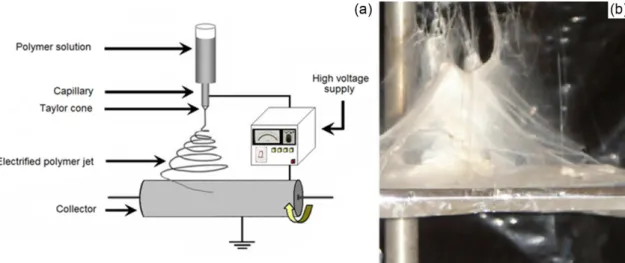
The electrospinning technique is based on electrostatic forces. Generally, the polymeric solution is maintained by its surface tension in the form of a drop at the end of a capillary metal. High voltage is applied to create an electric ield between the capillary and the iber collector, usually a grounded metal plate or a grounded rotating device (Figure 1).
Nanostructured membranes of CTS/PEO with and without AgNPs were made by the electrospinning technique, according to the methodology described by Vulcani et al. (2012). The resulting blend of CTS/PEO was stirred magnetically for two hours before being electrospun. The preparations of the solutions, as well as the preparation of the blend solution and electrospinning,
were run at room temperature. The low rate of 1 mL h-1
was determined by the solution viscosity, and the distance between the needle tips to collector cylinder was 7.5 cm at an applied voltage of 20 kV (Vulcani et al., 2012).
Preparation of CA polymer solutions
The polymer solutions were prepared in the presence and absence of AgNPs. The CA polymer solution was prepared by mixing 15% cellulose acetate into DMAc/ Acetone (1:2). This solution was stirred for approximately 2 hours to ensure its complete homogenization. The samples prepared in the presence of AgNPs were made by adding 5% of an aqueous solution (v/w) containing 20 mg L-1 of AgNPs into the CA solution.
Electrospinning of CA solutions
was connected to the metallic needle of the syringe, while the ground electrode was used to ground the copper plate collector, which had dimensions of 30 x 40 cm. The feed stream was controlled by a KD Scientiic pump, Model 100 (Campinas, Brazil) connected to a syringe. The distance from the needle to the collector was 10 cm; the
applied voltage was 15 kV, and the low rate was 1 mL h-1.
Nanostructured membrane samples were collected in aluminum foil used to coat the copper plate during the experiments (Nista et al., 2012).
SEM analyses
SEM measurements were carried out using a ield-emission scanning electron microscope (Zeiss Supra 55 VP) and an SEM microscope (JEOL JSM 5900 LV). The samples were analyzed after being deposited on aluminum supports and coated with evaporated carbon. Images were obtained for each sample, with different magnitudes, which were analyzed using Image Tool® software to measure the average diameter of the 50 measurements registered by each sample.
Antibacterial activity
The antimicrobial activity of nanoibers based on CA and CTS/PEO biopolymers and impregnated with AgNPs was evaluated for their effectiveness against the Gram-positivebacteria Staphylococcus aureus (S. aureus)(ATCC 29213) and Propionibacterium acnes (P. acnes) (ATCC
6919), and the Gram-negativebacteria Escherichia coli
(E. coli) (ATCC 25992) and Pseudomonas aeruginosa (P. aeruginosa) (ATCC 17933). The microbiological analyses
were performed in 24-well culture plates. The polymeric nanofibers in the absence and presence of AgNPs were deposited in the wells of a culture plate containing Nutrient Broth medium and bacterial solution at a concentration of 105 CFU mL-1. A positive control was performed only with the culture medium and bacteria. The plates were incubated at 37 °C and rotated at a rate of 60 rpm for 24 hours. After this incubation period, plating was performed by depositing 20 mL of each well into Petri dishes containing Plate Count Agar (PCA) culture medium. A positive control (growth) formed by culture broth containing bacteria and a negative control (sterile sample) formed by culture broth in which no microorganisms were included for each tested bacterial stock (Ridoli et al., 2012). Each concentration was tested in triplicate. The plates were analyzed in order to observe the inhibitory efect of bacterial growth relative to the positive control.
RESULTS AND DISCUSSION
UV–Vis spectra and DLS analysis of AgNPs
Figure 2a shows the UV-Vis spectrum of AgNPs synthesized in aqueous medium, and Figure 2b shows the AgNPs incorporated in CA polymer solution.
FIGURE 2 -(a) UV-Vis spectrum of AgNPs synthesized in aqueous medium (b), UV-Vis spectrum of CA/AgNPs solution and )c) DLS spectrum of AgNPs.
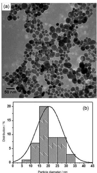
FIGURE 3 -(a) HR-TEM of the AgNPs synthesized in aqueous medium and (b) histogram showing the particle size distribution.
through the appearance of the band characteristic in 400 nm UV-Vis spectrums, as seen in Figure 2b.
Nanosized Ag Particles
The morphology and size of the nano-scale Ag particles were observed and confirmed by HR-TEM. Figure 3 shows an image of AgNPs prepared in aqueous medium.
Analysis of the images obtained by HR-TEM conirmed the spherical shape tendency of the AgNPs and revealed a monomodal particle size distribution with an average diameter of 20 nm, estimated by measuring the diameter of 100 randomly selected particles in enlarged TEM images. The histogram in Figure 3b shows the particle size distribution itted by one Gaussian curve. In addition, an average size for these AgNPs can be seen, corroborating the results observed for the DLS in Figure 2c.
Nanostructured membranes of CA and CTS/PEO blends in the absence and presence of AgNPs by electrospinning
and presence of AgNPs synthesized in an aqueous media and incorporated into biopolymers by in situ techniques described above. This method consists of adding the AgNPs colloidal suspension to each one of the polymeric solutions, as CA and CTS/PEO. Figure 4 shows the CTS/ PEO images obtained by SEM and Figure 5, by FEG, of CTS/PEO and CA, respectively, with AgNPs.
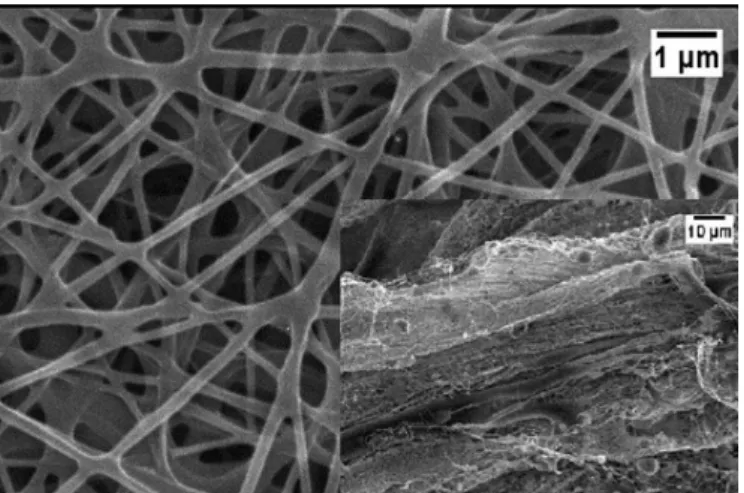
Nanoiber membranes were well formed, without beading, as shown in Figure 5 for both electrospun biopolymers. The average diameters of the CA and CTS/ PEO nanofibers electrospun with AgNPs were both in the nanometer range. As seen, the diameter of the ibers obtained in electrospinning experiments increased a little due to the amount of AgNPs added, but this did not interfere with the nanofiber membrane formation process. It was observed that the conductivities of the CA solutions increased in the presence of AgNPs, jumping from 8 to 12.5 µS cm-1, without and with AgNPs, respectively. Consequently, the potential applied during the electrospinning process needed to be decreased by 5 kV in order to form good nanoiber membranes.
Figure 6 shows FEG images of nanoibers formed in the presence of AgNPs. With the aid of high-resolution FEG and higher magniication, AgNPs can be visualized as many white spots dispersed inside nanoibers of CA and CTS/PEO formed in the presence of AgNPs, shown in Figure 6b and Figure 6c, respectively. This phenomenon is not observed for those samples (nanoibers) made without Ag NPs (Figure 6a).
The observed morphological aspects of the AgNPs (preferably spherical, see Figure 3) and the particle sizes as measured by DLS (Figure 2b) and conirmed by TEM (see Figures 6c and 3) corroborate this interpretation. Further consideration of the micrographs obtained of the
CA (Figure 6a) and the CTS/PEO nanoibers by FEG in the absence of AgNPs, makes it clear that these white spots are not observed in these samples (nanofibers without AgNPs, Figure 6a). Therefore, in comparing images made simultaneously on the same equipment and after the same nanoiber surface treatment, but only difering in whether they were made with or without AgNPs, it is possible that the white dots observed in the nanoibers that formed in the presence of Ag are indeed on the scale of NPs.
Antibacterial activity
The antibacterial activity of nanostructured biopolymer membranes made of CA and the CTS/ PEO nanocomposites with AgNPs and prepared using the in situ method, were tested against Gram-positive FIGURE 4 - Micrographs of SEM of CTS/PEO nanoibers in
the presence of AgNPs.
FIGURE 5 - FEG micrographs of a) bare CTS/PEO CA
nanoibers and b) CTS/PEO CA nanoibers in the presence of
FIGURE 6 - Micrographs of FEG of CA nanoibers made in the a) absence of AgNPs and the b) presence of AgNPs, and c) CTS/ PEO electrospun membranes made with AgNPs. The inset at the top right corner shows AgNPs powder.
S. aureus (ATCC 29213) and P. acnes (ATCC 6919)
microorganisms, as well as the Gram-negative E. coli
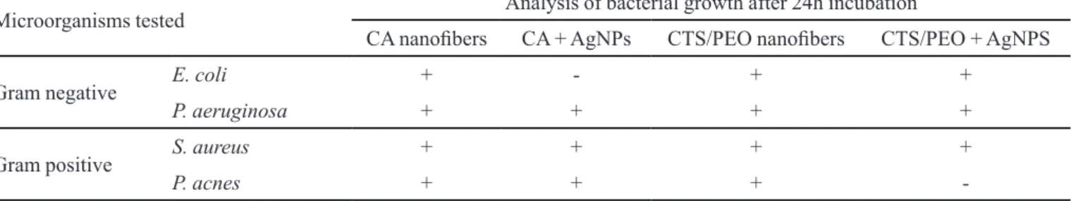
(ATCC 25992) and P. aeruginosa (ATCC 17933). Table I illustrates the results obtained.
The microbiological analyses were performed on solid medium (plate method), so that the CA and CTS/ PEO nanofibers can be observed as in Figure 7. In the case of the CA nanofibers, it was observed that in the presence of AgNPs (black dotted line) there was growth inhibition only for E. coli, a Gram-negative bacterium (Figure 7a). The same methodology was performed with nanostructured electrospun membranes of CTS/PEO. In this case, bacterial growth inhibition was observed for P. acnes, a Gram-positive bacterium (Figure 7d, white dotted line). These results indicate that the nanofibers in the presence of Ag NPs are leaching onto some microorganism.
The results observed in the incubated Petri dishes corroborate the visual observations of the culture plates in relation to turbidity, which indicates bacterial
FIGURE 7 - Image of solid medium (plate method) after 24 hours of bacterial growth of (a)E. coli, (b)P. aeruginosa,(c)S. aureus, (d)P. acnes in CA (black) and CTS/PEO (white) nanoibers in the absence (solid line) and presence of AgNPs (dotted line).
growth. These results showed that both microbiological tests (carried out in triplicate) were reproducible and comparable.
the surface charge of the NPs, the functional groups present on the surface, and many others. Pal et al. (2007) showed that the triangular shape of AgNPs exhibited higher biocide action on the bacterium E. coli when compared to spherical or cylindrical NPs, and even to
AgNO3, making it clear that diferent geometrical shapes
of NPs have diferent reactivities and potencies.
A relevant factor in the evaluation of biocide action on different materials is their behavior against the bactericidal dose-dependent effect on the strains. Due to the complexity involved in determining the real bioavailability of Ag ions, values of minimum inhibitory concentration (MIC) to approximately 100 strains of S. aureus were reported, with a range between 8-80 mg L-1
when using AgNO3 (Chopra, 2007). Similarly, MIC
values for Ag studied in approximately 100 strains of P. aeruginosa showed a range of 8 to 70 mg L-1. These
studies showed that some strains are resistant to Ag, and, in fact, the resistant mechanism of bacterial strains was isolated more than 70 years ago, and their Ag resistance mechanism has been investigated (Pal, Tak, Song, 2007; Yudkin, 1937).
A very important aspect of Ag action on bacterial cells, which must be considered when discussing the inhibitory effect of Ag tested on different types of microorganisms, is the classiication of the bacterium. Bacteria are commonly classified into Gram positive and Gram negative, depending on their cell walls. Gram-positive bacteria have an extra thick peptidoglycan layer on the outer surface, while Gram-negative bacteria have an outer membrane behind which a thin peptidoglycan layer exists. This drastic diference in the nature of the cell boundaries is a great challenge for having a general antibacterial material against the bacteria (Gangadharan
et al., 2010).
Therefore, the biopolymeric nanoibers prepared by incorporating AgNPs can provide antiseptic properties required of new materials that could be used to great beneit in medical devices.
CONCLUSION
Microbial experiments suggested that CA electrospun nanofibers containing AgNPs showed a much better
antimicrobial efect on the bacterium E. coli, a common
pathogen affecting humans and other animals that is an important bacterium associated with hospital infections. In the case of the electrospun nanoiber membrane made of CTS/PEO impregnated with AgNPs, the growth inhibition efect was observed for Gram-positive bacteria, such as P. acnes, indicating that these membranes present bactericidal action through both contact and leaching action. The results observed in the incubated Petri plates corroborate the visual observation performed on culture plates in relation to turbidity. Thus, it is supposed that the electrospun CTS/PEO and CA nanofibers, both biopolymers and both made in the presence of AgNPs, with the demonstrated and strong antimicrobial activity could be used in various biomedical ields such as wound dressings, mesh grafts in burns, and tissue scafolds. An assessment of the antimicrobial activity of the inal products and other issues related to the understanding of this new material is under way in the laboratories of the authors.
ACKNOWLEDGEMENTS
The authors thank National Council for Scientiic and Technological Development (CNPq), Coordination for the Improvement of Higher Education Personnel (CAPES) and São Paulo Research Foundation (FAPESP) for the financial support and scholarship. The authors would like to thank the Brazilian Nanotechnology National Laboratory/Center for Research in Energy and Materials (LNNano/CNPEM) for technical support during electron microscopy work. We also acknowlwedge Brazilian Network on Nanotoxicology (MCTI/CNPq) and NanoBioss (MCTI) for support. We would also like to thank Nature Publishing Group for the certiication of this manuscript review into an English Certiied Edition. TABLE I - Analysis of bacterial growth after a 24 h incubation period for microorganisms tested in both plate medium and test tubes
Microorganisms tested Analysis of bacterial growth after 24h incubation
CA nanoibers CA + AgNPs CTS/PEO nanoibers CTS/PEO + AgNPS
Gram negative E. coli + - + +
P. aeruginosa + + + +
Gram positive S. aureus + + + +
P. acnes + + +
REFERENCES
ABDELGAWADA, A.M.; HUDSONA, S.M.; ROJAS, O.J.
Antimicrobial wound dressing nanofiber mats from multicomponente (chitosan/silver-NPs/polyvinyl alcohol) systems. Carbohydr. Polym., v.100, p.166-178, 2014.
ABOU-ZEIDA, N.Y.; WALYA, A.I.; KANDILEB, N.G.;
RUSHDYC, A.A.; EL-SHEIKHA, M.A.; IBRAHIM, H.M. Preparation, characterization and antibacterial properties of cyanoethylchitosan/cellulose acetate polymer blended
ilms. Carbohydr. Polym., v.84, n.1, p.223-230, 2011.
ALEXEEV, V.L.; KELBERG, E.A.; EVMENENKO, G.A.;
BRONNIKOV, S.V. Improvement of the mechanical
properties of chitosan ilms by the addition of poly(ethylene
oxide). Polym. Eng. Sci., v.40, n.5, p.1211-1215, 2000.
BIZARRIA, M.T.; DAVILA, M.A.; MEI, L.H.I.
Non-woven nanoiber chitosan/PEO membranes obtained by
electrospinning. Braz. J. Chem. Eng., v.31, n.1, p.57-68, 2014.
CHALOUPKA, K.; MALAM, Y.; SEIFALIAN, A.M. Nanosilver as a new generation of nanoproduct in biomedical applications. Trends Biotechnol., v.28, n.11, p.580-588, 2010.
CHEN, X.; PARKER, S.G.; ZOU, G.; SU, W.; ZHANG, Q. β-Cyclodextrin functionalized silver nanoparticles for the
naked eye detection of aromatic isomers. ACS Nano., v.4, n.11, p.6387-6394, 2010.
CHOPRA I. The increasing use of silver-based products as antimicrobial agents: a useful development or a cause for concern. J. Antimicrob. Chemother., v.59, n.4, p.587-590, 2007.
DALLAS, P.; SHARMA, V.K.; ZBORIL, R. Silver polymeric nanocomposites as advanced antimicrobial agents:
Classiication, synthetic paths, applications, and
perspectives. Adv. Colloid Interface Sci., v.166, n.1-2, p.119-135, 2011.
DUTRA, R.L.; SEGALA, K.; DE OLIVEIRA, E.M.N.; DE
SOUZA, E.P.; ROSSI, L.M.; MATOS, J.R.; NODA, L.K.; PAULA, M.M.S.; FRANCO, C.V. Preparation and characterization of the novels terpolymers of poly-{trans-[RuCl2(vpy)4]-divinylbenzene} and styrene-divinylbenzene-vinylpiridine impregnated with silver nanoparticles. Polym. Bull., v.60, p.809-819, 2008.
ELLISON, J.; WYKOFF, G.; PAUL, A.; MOHSENI, R.; VASILIEV, A. Eficient dispersion of coated silver
nanoparticles in the polymer matrix. Colloids Surf., A: Physicochem. Eng. Aspects, v.447, p.67-70, 2014.
FERJANI, E.; LAJIM, R.H.; DERATANIB, A.; ROUDESLIB,
M.S. Bulk and surface modiication of cellulose diacetate
based RO/NF membranes by polymethylhydrosiloxane. Preparation and characterization. Desalination, v.146, n.1-3, p.325-330, 2002.
G A N G A D H A R A N , D . ; H A R S H VA R D A N , K . ; GNANASEKAR, G.; DIXIT, D.; POPAT, K.M.; ANAND,
P.S. Polymeric microspheres containing silver nanoparticles as a bactericidal agent for water disinfection. Water Res., v.44, n.18, p.5481-5487, 2010.
GERANIO, L.; HEUBERGER, M.; NOWACK, B. The behavior
of silver nanotextiles during washing. Environ. Sci. Technol., v.43, p.8113-8118, 2009.
GUO, L.; YUAN, W.; LU, Z.; LI, C.M. Polymer/nanosilver
composite coatings for antibacterial applications. Colloids and Surf. A: Physicochem. Eng. Aspects, v.439, p.69-83, 2013.
HAN, B.; ZHANG, D.; SHAO, Z.; KONG, L.; SHAOYI,
Lv. Preparation and characterization of cellulose acetate/ carboxymethyl cellulose acetate blend ultrafiltration membranes. Desalination, v.311, p.80-89, 2013.
JANA, N. R.; GEARHEARTA, L.; MURPHY, C. J. Wet chemical synthesis of silver nanorods and nanowiresof controllable aspect ratio. Chem. Commun., p.617-618, 2001.
KANMANI, P.; RHIM, J. Physicochemical properties of gelatin/
silver nanoparticle antimicrobial composite ilms. Food
Chem., v.148, p.162-169, 2014.
KHAN, N. Applications of electrospun nanofibers in the
biomedical ield: a review. Stud. Undergrad. Res. Guelph, v.5, n.2, p.63-73, 2012.
KITTLER, S.; GREULICH, C.; DIENDORF, J.; KOELLER,
KLEINUBING, S.A.; SERAPHIM, D.C.; VIEIRA, M.G.A.;
CANEVESI, R.L.S.; SILVA, E.A.; CÉSAR, C.L.;
MEI, L.H.I. Gastro-resistant controlled release of OTC
encapsulated in alginate/chitosan matrix coated with
acryl-EZE MP in luidized bed. J. Appl. Polym. Sci., v.131, n.12, p.1-9, 2014.
KVITEK, L.; PANACEK, A.; SOUKOPOVA, J.; KOLAR, M.; VECEROVA, R.; PRUCEK, R.; HOLECOVA, M.; ZBORIL, R.J. Effect of surfactants and polymers on stability and antibacterial activity of silver nanoparticles (NPs). J. Phys. Chem. C, v.112, n.15, p.5825-5834, 2008.
LIU, C.J.; BURGHAUS, U.; BESENBACHER, F.; WANG,
Z.L. Preparation and characterization of nanomaterials for sustainable energy production. ACS Nano, v.4, n.10, p.5517-5526, 2010.
M A D H U M AT H I , K . ; S U D H E E S H , K U M A R , P. T. ; ABHILASH, S.; SREEJA, V.; TAMURA, H.; MANZOOR, K.; NAIR, S.V.; JAYAKUMAR, R. Development of novel
chitin/nanosilver composite scafolds for wound dressing
applications. J. Mater. Sci: Mater. Med., v.21, n.2, p.807-813, 2010.
MURPHY, C. J.; JANA, N. R. Controlling the Aspect Ratio of Inorganic Nanorods and Nanowires. Adv. Mater., v.14, n.1, p.80-82, 2002.
NISTA, S.V.G.; BETTINI, J.; MEI, L.H.I. Coaxial nanoibers
of chitosan-alginate-PEO polycomplex obtained by electrospinning. Carbohydr. Polym., v.127, p.222-228, 2015.
NISTA, S.V.G.; AKIRA, M.D.; MARTINEZ, E.F; SILVA,
A.S.F., MEI, L.H.I. Nanostructured membranes based on cellulose acetate obtained by electrospinning. Part II.
Controlled release proile and microbiological behavior. J. Appl. Polym. Sci., v.130, n.4, p.2772-2779, 2013.
NISTA, S.V.G.; PERES, L.; D’ÁVILA, M.A.; SCHMIDT, F.L.;
MEI, L.H.I. Nanostructured membranes based on cellulose acetate obtained by electrospinning, part 1: Study of the best solvents and conditions by design of experiments. J. Appl. Polym. Sci., v.126, n.S1, p.E70-E78, 2012.
O K A M O T O , M . ; J O H N , B . S y n t h e t i c b i o p o l y m e r nanocomposites for tissue engineering Scaffolds. Prog. Polym. Sci., v.38, n.10-11, p.1487-1503, 2013.
PADIL, V.V.T.; NGUYEN, N.H.A.; ŠEVCR, A.; HERNÍK, M.
Fabrication, Characterization, and Antibacterial Properties
of Electrospun Membrane Composed of Gum Karaya,
Polyvinyl Alcohol, and Silver Nanoparticles. J. Nanomater., v.2015, ID 750726, 2015.
PAL, S.; TAK, Y.K.; SONG, J.M. Does the antibacterial
activity of silver nanoparticles depend on the shape of the
nanoparticle? A study of the Gram-negative bacterium
Escherichia coli. Appl. Environ. Microbiol., v.73, n.6, p.1712-1720, 2007.
PANACEK, A.; KVITEK, L.; PRUCEK, R.; KOLAR, M.; VECEROVA, R.; PIZUROVA, N.; SHARMA, V.K.; NEVECNA, T.; ZBORIL, R. Silver colloid nanoparticles: synthesis, characterization, and their antibacterial activity.
J. Phys. Chem. B, v.110, n.33, p.16248-16253, 2006.
PRABHU, S.; POULOSE, E.K. Silver nanoparticles: mechanism of antimicrobial action, synthesis, medical applications, and
toxicity efects. Int. Nano. Lett., v.2, p.3-10, 2012.
PREMA, P.; RAJU, R. Fabrication and characterization of silver nanoparticle and its potential antibacterial activity.
Biotechnol. Bioproc. Eng., v.14, p.842-847, 2009.
RAI, M.; YADAV, A.; GADE, A. Silver nanoparticles as a new
generation of antimicrobials. Biotechnol. Adv, v.27, p.76-83, 2009.
RHIM, J.W.; WANG, L.F.; HONG, S.I. Preparation and
characterization of agar/silver nanoparticles composite
ilms with antimicrobial activity. Food Hydrocol., v.33, n.2, p.327-335, 2013.
RIDOLFI, D.M.; MARCATO, P.D.; JUSTO, G.Z.; CORDI, L.; MACHADO, D.; DURÁN, N. Chitosan-solid lipid
nanoparticles as carriers for topical delivery of tretinoin.
Colloids Surf. B Bioint., v.93, p.36-40, 2012.
SEVERIN, N.; KIRSTEIN, S.; SOKOLOV, I.M.; RABE, J.P. Rapid trench channeling of graphenes with catalytic silver nanoparticles. Nano Lett., v.9, n.1, p.457-461, 2009.
S I G N O R I , A . M . ; S A N TO S , K . O . ; E I S I N G , R . ; ALBUQUERQUE, B.L.; GIACOMELLI, F.C.; DOMINGOS, J.B. Formation of catalytic silver
SOLOMON, S.D.; BAHADORY, M.; JEYARAJASINGAM, A.V.; RUTKOWSKY, S.; BORITZ, C. MULFINGER, L.
Synthesis and study of silver nanoparticles. J. Chem. Educ., v.84, n.2, p.322-325, 2007.
SOYEKWO, F.; ZHANG, Q.G.; DENG, C.; GONG; ZHU,
A.M.; LIU, Q.L. Highly permeable cellulose acetate
nanoibrous composite membranes by freeze-extraction. J. Membr. Sci, v.454, p.339–345, 2014.
TRICOLI, A.; PRATSINIS, S.E. Dispersed nanoelectrode devices. Nat. Nanotechnol., v.5, p.54-60, 2009.
VULCANI, V.A.S.; BIZARRIA, M.T.M.; D’ÁVILA, M.A.;
MEI, L.H.I.; BERNALD, C.; PERUSSI, J.R. Cytotoxicity tests for nanostructured chitosan/PEO membranes using the
agar difusion method. Mat. Res., v.15, p.213-217, 2012.
YUDKIN J. Efect of silver ions on enzymes of Bacterium coli.
Enzymologia, v.2, p.161-70, 1937.
ZENG, Q.; JIANG, X.; YU, A.; LU, G. Growth mechanisms
of silver nanoparticles: a molecular dynamics study.
Nanotechnology, v.18, n.3, p.035708, 2007.
ZIVANOVIC, S.; LI, J.; DAVIDSON, P.M.; KIT, K. Physical, mechanical and antibacterial properties of chitosan/PEO blend ilms. Biomacromolecules, v.8, n.5, p.1505-10, 2007.
Received for publication on 25th November 2014