*Correspondence: R. H. C. Queiroz. Departamento de Análises Clínicas, Toxicológicas e Bromatológicas, Faculdade de Ciências Farmacêuticas de Ribeirão Preto - USP. Av. do Café, s/n, 14040-903 - Ribeirão Preto - SP, Brazil. E-mail: [email protected].
A
vol. 48, n. 1, jan./mar., 2012
L-arginine, a nitric oxide precursor, reduces dapsone-induced
methemoglobinemia in rats
Natália Valadares de Moraes, Mateus Machado Bergamaschi, Maria de Lourdes Pires Bianchi,
Juliana Bordinassi Bragheto, Wilson Roberto Malfará, Regina Helena Costa Queiroz
Department of Clinical, Toxicological and Food Sciences Analysis, School of Pharmaceutical Sciences of Ribeirão Preto, University of São Paulo
Dapsone use is frequently associated to hematological side effects such as methemoglobinemia and hemolytic anemia, which are related to N-hydroxylation mediated by the P450 enzyme system. The aim
of the present study was to evaluate the inluence of L-arginine supplementation, a precursor for the
synthesis of nitric oxide, as single or multiple dose regimens on dapsone-induced methemoglobinemia.
Male Wistar rats were treated with L-arginine at 5, 15, 30, 60 and 180 mg/kg doses (p.o., gavage) in single or multiple dose regimens 2 hours prior to dapsone administration (40 mg/kg, i.p.). The effect of the nitric oxide synthase inhibitor L-NAME was investigated by treatment with multiple doses of 30 mg/kg (p.o., gavage) 2 hours before dapsone administration. Blood samples were collected 2 hours after
dapsone administration. Erythrocytic methemoglobin levels were assayed by spectrophotometry. The
results showed that multiple dose supplementations with 5 and 15 mg/kg L-arginine reduced
dapsone-induced methemoglobin levels. This effect is mediated by nitric oxide formation, since the reduction in
methemoglobin levels by L-arginine is blocked by simultaneous administration with L-NAME, a nitric
oxide synthase inhibitor.
Uniterms: Dapsone. Methemoglobinemia. L-arginine. Nitric oxide. L-NAME.
O uso da dapsona é frequentemente associado a efeitos adversos hematológicos, como a metemoglobinemia e anemia hemolítica, ambos relacionados com a N-hidroxilação mediada pelo sistema P450. O objetivo
do estudo foi avaliar a inluência da suplementação de L-arginina, um precursor da síntese de óxido
nítrico, administrado em regime de dose única ou múltipla na metemoglobinemia induzida pela dapsona.
Ratos machos Wistar foram tratados com L-arginina (po, gavagem) em dose única ou múltipla de 5, 15, 30, 60 e 180 mg/kg 2 horas antes da administração de dapsona (40 mg/kg, ip). O efeito do L-NAME, um inibidor de óxido nítrico sintase (NOS), foi avaliado através do tratamento com doses múltiplas de 30 mg/kg. Amostras de sangue foram coletadas duas horas após a administração de dapsona. A
concentração de metemoglobina eritrocitária foi analisada por espectrofotometria. Os resultados
mostraram que a suplementação em dose múltipla de 5 e 15 mg/kg de L-arginina reduziu os níveis de
metemoglobina induzida pela dapsona. Este efeito é mediado pela formação de óxido nítrico, uma vez
que a redução nos níveis de metemoglobina pela L-arginina é bloqueada pela administração simultânea de L-NAME, um inibidor da óxido nítrico sintase.
Unitermos: Dapsona. Metemoglobinemia. L-arginina. Óxido nítrico. L-NAME.
INTRODUCTION
Dapsone (4,4’-diaminodiphenylsulfone, DDS) is
syndrome (Powell et al., 1967; Mills et al., 1988; Castro 1998; Tobin-D’Angelo et al., 2004; Nyunt, Plowe 2007).
The major metabolic pathway of DDS is acetylation, producing monoacetyldapsone (MADDS). DDS is also metabolized by N-hydroxylation mediated by cytochrome P450 isozymes CYP2C19, CYP2C9, CYP3A4 and CYP2E1 in man, and isozymes CYP2C6/11 and CYP3A1 in rats, producing DDS hydroxylamine (DDS-NOH) (Fleming et al., 1992; Vage, Svensson, 1994; Mitra et al., 1995; Gill et al., 1995; Ganesan et al., 2010). Glucuronidation of DDS and DDS-NOH is catalyzed by the enzyme UDP-glucuronosyltransferase (UGT) allowing its excretion in urine and bile (Coleman et al., 1996; Tingle et al., 1997).
The co-oxidation of DDS-NOH and hemoglobin produces nitroso derivatives and methemoglobin, thus causing methemoglobinemia and hemolysis (Tingle et al., 1990; Coleman, 1995), which are the major dose-dependent side effects of DDS chronic users (Kaluarachchi et al., 2001). MADDS-NOH has been shown to be a more potent methemoglobin (metHb) former than DDS-NOH in human erythrocytes in vitro (Coleman, Holden, 2004), whilst both metabolites present the same potency in rats and humans (Vage et al., 1994).
The reduction of xenobiotic-induced metHb forma-tion and the mechanisms underlying this effect have been widely investigated in the last few years. The various attempts to reduce methemoglobinemia have included: preventing CYP-mediated oxidative metabolism of xeno-biotics to hydroxylamines (Coleman et al., 1990; Malfará et al., 2002); biochemical attenuation of metHb formation with antioxidants (Prussick et al., 1992; Wright et al., 1996; Dötsch et al., 1998; Wright et al., 1998; Dötsch et al., 2000; Tanen et al., 2000; Matteuci et al., 2003; De Moraes et al., 2008; Jo et al., 2008); and reduction of metHb to hemoglobin by stimulating NADH diaphorase or NADPH diaphorase (Dötsch et al., 2000).
L-arginine (ARG), a semi-essential amino acid, is the nitrogenous precursor for the synthesis of nitric oxide (NO) by a NADPH-dependent NO synthase (NOS) and regulates vital metabolic pathways. NO is suficiently non -polar to cross membranes without a carrier and is known to modulate vasorelaxation and exhibit antioxidant properties due to superoxide scavenger and heme oxygenase inductor activities (Wood et al., 2008).
In spite of all the evidence pointing to the importance of ARG in vital pathways, the role of ARG supplementa-tion in DDS-induced methemoglobinemia has yet to be described in the literature. The aim of the present study was to evaluate the role of ARG in single and multiple dose regimens in DDS-induced methemoglobinemia. We also evaluated whether the effect of ARG on DDS-induced
methemoglobinemia can be modulated by pretreatment with N-nitro-L-arginine methyl ester (L-NAME), a non-speciic NOS inhibitor.
MATERIAL AND METHODS
Dapsone was supplied by FURP (Fundação para o Remédio Popular; Guarulhos, Brazil) and L-(+)-arginine was supplied by Acros organics (Morris Plains, NJ, USA). L-NAME was purchased from Sigma-Aldrich (St. Louis, MO, USA). KCN was supplied by Merck (Darmstadt, Germany) and K3Fe(CN)6 was supplied by Merck (Rio de Janeiro, Brazil). Water was puriied with the Milli-Q Plus system (Millipore, Bedford, MA, USA).
Experimental study
The experimental study was approved by the Eth-ics Committee for the Use of Animals of Ribeirão Preto Campus, University of São Paulo, Brazil, in accordance with the US National Institutes of Health Guide for the Care and Use of Laboratory Animals (Protocol number 06.1.461.53.6). Male Wistar rats (200 ± 20 g) were kept for 48 hours before the experiment in a room under controlled temperature (21-23 °C) and humidity (40-60%) and on a 12 h light:12 h dark cycle. The animals had free access to chow and water throughout the experiment. The animals (n = 8 per group) were treated in single or multiple dose regimens. ARG was administered orally (p.o. gavage, 200 μL), dissolved in sterile physiologic saline, whereas DDS was dissolved in dimethylsulphoxide (DMSO) and administered intraperitoneally (i.p., 200 μL). L-NAME at 30 mg/kg was administered using a multiple dose regimen in the same solution as ARG.
Single dose regimen
The control group received the vehicle of ARG (sterile physiological saline) p.o. by gavage two hours before the administration of the vehicle used to dissolve DDS (DMSO) i.p. The DDS group received 40 mg/kg DDS (i.p.) 2 hours after the administration of saline p.o. The groups DDS + 5 mg/kg ARG, DDS + 15 mg/kg ARG, DDS + 30 mg/kg ARG, DDS + 60 mg/kg ARG, DDS + 180 mg/kg ARG received ARG at 5, 15, 30, 60 and 180 mg/kg doses, respectively, 2 hours before the administration of 40 mg/kg DDS.
Multiple dose regimen
received saline for ive days p.o. and 40 mg/kg DDS on the ifth day, 2 hours after saline administration. Groups DDS + 5 mg/kg ARG, DDS + 15 mg/kg ARG, DDS + 30 mg/kg ARG, DDS + 60 mg/kg ARG, DDS + 180 mg/kg ARG received ARG at 5, 15, 30, 60 and 180 mg/kg doses, respectively, for ive days. On the ifth day, 2 hours after ARG administration, the animals received 40 mg/kg DDS. Heparinized blood samples were collected two hours after DDS or DMSO administration, in both dose regimens (Liquemine 5000 IU, Roche, Rio de Janeiro, Brazil). Met -hemoglobin levels were determined immediately.
Methemoglobin assay
Methemoglobin levels relative to hemoglobin levels were determined according to the method described by Evelyn and Malloy (1938) (modified by Harrison and Jollow, 1986). Briely, an aliquot (200 μL) of heparinized blood was added to 10 mL of 0.02 M phosphate buffer pH 7.8 with 0.05% triton X-100 and then shaken in a mixer for 30 seconds. The hemolysate was then fractionated into four tubes. Tube 1 (A1) remained with hemolyzed blood. An aliquot (50 μL) of 20% K3Fe(CN)6 was added to tubes 3 (A3) and 4 (A4). An aliquot (50 μL) of 10% KCN was then added to tubes 2 (A2) and 4. The absorbance of each tube was measured at 635 nm. Methemoglobin levels relative to hemoglobin levels were then calculated by the following equation:
The initial blood measurement (A1) is referent to MetHb and possible interferences. When blood is added to KCN (A2), MetHb is converted to cyanomethemo -globin (CNMetHb) and then possible interferences are eliminated because CNMetHb does not absorb at 635 nm. When blood is added to K3Fe(CN)6 all hemoglobin is converted to MetHb, and this measurement refers to total MetHb (A3). Finally, blood is added to K3Fe(CN)6 and KCN, with all hemoglobin converted to MetHb and then to CNMetHb (A4).
Statistical analysis
GraphPad InStat® software (version 3.01) was used for the calculation of means ± standard deviation. ANOVA and the Tukey-Kramer post test for multiple comparisons (p<0.05) were used to compare groups.
RESULTS
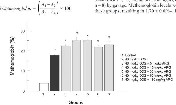
As there is no reference value for methemoglobin levels in rats, some preliminary studies were conducted in order to evaluate whether ARG or the vehicles could pro-duce methemoglobinemia. A single dose regimen control group was evaluated by administrating sterile physiologi-cal saline (p.o., 200 μL) and DMSO (i.p., 200 μL). The administration of these vehicles resulted in 3.77 ± 0.43% methemoglobin formation. In a pilot study, rats were treated with 5, 15, 30, 60 and 180 mg/kg of ARG (p.o., n = 8) by gavage. Methemoglobin levels were assayed in these groups, resulting in 1.70 ± 0.09%, 1.81 ± 0.08%,
FIGURE 1 - Effect of single dose ARG on methemoglobin levels (%). Animals were treated with ARG at 5, 15, 30, 60 and
1.59 ± 0.05%, 1.96 ± 0.08% and 2.03 ± 0.37% of met -hemoglobin, respectively (data expressed as means ± standard deviation). These results showed that ARG alone did not produce methemoglobin.
Administration of 40 mg/kg DDS (i.p.) resulted in methemoglobin levels of 17.18 ± 1.71% (Figure 1). The dose of 40 mg/kg (i.p.) of DDS was known to produce methemoglobinemia in rats based on previous studies by our group (Malfara et al., 2002; De Moraes et al., 2008; Bergamaschi et al., 2011). When ARG was administered in a single dose two hours prior to DDS (40 mg/kg) it failed to reduce DDS-induced methemoglobinemia (Figure 1).
ARG was also administered at doses 5, 15, 30, 60 and 180 mg/kg (po), in multiple dose regimens. Methe -moglobin levels of groups treated with ARG only, in a multiple drug regimen, did not produce signiicant levels of methemoglobin (1.88 ± 0.68%; 2.00 ± 0.13%; 2.56 ± 0.44%; 2.43 ± 0.43% and 1.75 ± 0.76%, respectively), as observed for a single dose regimen. Animals treated with 5 or 15 mg/kg ARG for 5 days prior to 40 mg/kg DDS administration showed a reduction in DDS-induced methemoglobinemia, with methemoglobin levels similar to the control group. However, higher doses of ARG (30, 60 and 180 mg/kg) in the multiple dose regimens did not inhibit methemoglobin formation (Figure 2).
The effect of L-NAME, a NOS inhibitor, was evalu -ated in order to understand the mechanisms rel-ated to the reduction of DDS-induced methemoglobinemia by ARG. L-NAME inhibited the reduction in DDS-induced methe
-moglobinemia promoted by ARG, leading to MetHb levels comparable to DDS administration alone (Figure 2).
DISCUSSION
The concentration of methemoglobin in erythro-cytes is regulated by three systems: nicotinamide adenine dinucleotide (NADH), nicotinamide adenine dinucleotide phosphate (NADPH) and glutathione systems. Methemo -globin is converted to hemo-globin by the NADH system when suficient NADH-methemoglobin reductase is avail -able; it contributes to 95% of methemoglobin reduction to hemoglobin. The NADPH system reduces methemoglobin to hemoglobin through the enzyme NADPH-methemo-globin reductase and contributes to 5% of methemoNADPH-methemo-globin reduction. Finally, the conversion of reduced glutathione to glutathione inluences methemoglobin levels by reduc -ing oxidiz-ing agents (Evelo et al., 1998; Ward, McCarthy, 1998; Umbreit, 2007).
The standard treatment for methemoglobinemia includes infusion with methylene blue, whose action depends on the availability of NADPH within the eryth-rocytes. This therapy requires glucose-6-phosphate dehy -drogenase (G6PD) optimal activity to produce suficient amounts of NADPH. In G6PD-deicient subjects, methy -lene blue therapy has been associated with hemolysis and methemoglobinemia (Rehman, 2001). Several other sub -stances have been investigated as alternatives to methylene blue therapy. These have included ascorbic acid (Dötsch
FIGURE 2 - Effect of multiple dose L-arginine (ARG) on methemoglobin level (%). Animals were treated with saline or ARG at 5,
et al., 1998), cimetidine (Coleman et al., 1990; Malfará et al., 2002), riboflavin (Dötsch et al., 2000), α-lipoic acid (Coleman, Taylor, 2003), sodium thiosulfate (Mat -teucci et al., 2003), ethyl pyruvate (Jo et al., 2008) and N-acetylcysteine (Wright et al., 1996; Wright et al., 1998; Dötsch et al., 2000; Tanen et al., 2000; De Moraes et al., 2008). N-acetylcysteine, a precursor of glutathione, used in combination with DDS in rats, has shown increased methemoglobin levels in these animals compared to rats treated with DDS alone. Some authors have suggested that glutathione can regenerate DDS-NOH from dapsone nitroso derivatives thus resulting in higher methemoglobin levels (De Moraes et al., 2008).
Considering the potency of ARG and NO as antioxi-dant agents, it was proposed that the co-administration of ARG and DDS might reduce the methemoglobin levels associated to DDS use. The cationic amino acid ARG is the precursor for NO biosynthesis mediated by NO syn-thase. Three isoforms of NO synthase (NOS) occur in a number of tissues: neuronal NOS (nNOS); inducible NOS (iNOS) located in glia cells, and endothelial NOS (eNOS) located in endothelial cells (Palmer et al., 1987; Thomas et al., 2008). The iNOS can form much larger amounts of NO compared with other isoforms. In many cells and pathological conditions the supply of extracellular ARG is rate-limiting for NO production (Brunini et al., 2007; Thomas et al., 2008).
In the present study, L-NAME administration suppressed the reduction in DDS-induced methemoglo-binemia mediated by 5 mg/kg and 15 mg/kg ARG in multiple dose regimens. Considering that L-NAME is a non-speciic NOS inhibitor, our data suggest that the ARG effect on methemoglobin is mediated by NO. NO is considered a potent antioxidant agent in vitro and in vivo. Its antioxidant activity has been proven by suppressing iron-induced generation of hydroxyl radicals (OH) via the Fenton reaction, interrupting lipid peroxidation chain reac-tion, increasing the glutathione antioxidant potency and inhibiting cysteine proteases (Chiueh, 1999). On the other hand, increased methemoglobin levels are a known toxic effect of inhaled NO therapy, commonly used for hypoxic neonates. NO can combine with hemoglobin to produce nitrosylhemoglobin and thus form methemoglobin by oxidation (Weinberger et al., 2001; Hamon et al., 2010). Based on our observations, we can hypothesize that lower doses of ARG were beneicial to decrease methemoglobin levels because of the antioxidant properties of NO. How-ever, higher doses of ARG do not decrease metHb levels because the antioxidant properties of NO are combined to its methemoglobinizant effect.
Excess ARG supplementation is also related with the
production of NG,NG-dimethyl-L-arginine (ADMA) which is a NOS inhibitor (Masuda et al. 2002). This metabolite can convert NO to a superoxide generator (Thomas et al., 2008). This may explain why NO is beneicial to DDS-induced methemoglobinemia at low ARG concentrations yet deleterious when excess ARG supplementation is administered to rats in multiple dose regimens.
NO also produces inhibitory effects in cytochrome P450 mediated drug metabolism. It is known that NO forms complexes with the catalytic center of P450 en-zymes which results in a decrease in enzymatic activities of rat microsomes (Khatsenko et al. 1993). The inhibi -tory effects of NO are rapid, concentration-dependent and mainly in CYP 2C11 > 2B1/2 > 2E1 = 3A2 > 1A1/2 (Vuppugalla, Mehvar 2004a, 2004b). If NO had inhibited DDS oxidative metabolism, animals treated with higher ARG concentration would present lower metHb levels. However, our results showed that the reduction in MetHb levels by ARG is observed only when animals are treated with multiple doses of 5 and 15 mg/kg ARG. Thus, CYP inhibition by NO does not seem to explain the reduction in MetHb levels.
In conclusion, ARG reduces DDS-induced methe-moglobinemia in rats when low doses (5 and 15 mg/kg) are administered as a multiple dose regimen. The effect can be blocked by the simultaneous administration of L-NAME (a NOS inhibitor). Thus, we can conclude that ARG supplementation can be an effective reducing agent for chronic treatment of DDS-induced methemoglobin-emia and that its effect is mediated by NO.
ACKNOWLEDGMENTS
This work was supported by CAPES (Coordenação de Aperfeiçoamento de Pessoal de Nível Superior). The authors gratefully thank FURP (Fundação para o Remé -dio Popular) for providing dapsone and Prof. Dr. Lusiane Maria Bendhackfor providing L-NAME.
REFERENCES
BERGAMASCHI, M.M.; ALCANTARA, G.K.; VALERIO,
D . A . ; Q U E I R O Z , R . H . C u r c u m i n c o u l d p r e v e n t methemoglobinemia induced by dapsone in rats. Food Chem. Toxicol., v.49, n.7, p.1638-1641, 2011.
BRUNINI, T.M.; MOSS, M.B.; SIQUEIRA, M.A.; SANTOS,
S.F.; LUGON J.R.; MENDES-RIBEIRO, A.C. Nitric
CASTRO, M. Treatment and prophylaxis of Pneumocystis carinii pneumonia. Semin. Respir. Infect., v.13, n.4,
p.296-303, 1998.
CHIUEH, C.C. Neuroprotective properties of nitric oxide. Ann. N.Y. Acad. Sci., v.890, p.301-311, 1999.
COLEMAN, M.D.; HOLDEN, L.J. The methaemoglobin
forming and GSH depleting effects of dapsone and monoacetyl dapsone hydroxylamines in human diabetic and non-diabetic erythrocytes in vitro. Environ. Toxicol. Pharmacol., v.17, n.1, p.55-59, 2004.
COLEMAN, M.D.; PAHAL, K.K.; GARDINER, J.M. The
effect of acetylation and deacetylation on the disposition of dapsone and monoacetyl dapsone hydroxylamines in human erythrocytes in-vitro. J. Pharm. Pharmacol., v.48,
n.4, p.401-406, 1996.
COLEMAN, M.D.; SCOTT, A.K.; BRECKENRIDGE, A.M.; PARK, B.K. The use of cimetidine as a selective inhibitor of
dapsone N-hydroxylation in man. Br. J. Clin. Pharmacol.,
v.30, n.5, p.761-767, 1990.
COLEMAN, M.D.; TAYLOR, C.T. Effects of dihydrolipoic acid (DHLA), α-lipoic acid, N-acetyl cysteine and ascorbate on
xenobiotic-mediated methaemoglobin formation in human erythrocytes in vitro. Environ. Toxicol. Pharmacol., v.14,
n.3, p.121-127, 2003.
COLEMAN, M.D. Dapsone toxicity: some current perspectives.
Gen. Pharmacol., v.26, n.7, p.1461-1467, 1995.
DE MORAES, N.V.; MELLO, M.H.; SOUZA, A.M.;
SAMPAIO, S.V.; QUEIROZ, R.H.C. Potentiation of dapsone induced methemoglobinemia by N-acetylcysteine in rats. Rev. Bras. Cienc. Farm., v.44, n.1, p.97-104, 2008.
DÖTSCH, J.; DEMIRAKÇA, S.; CRYER, A.; HÄNZE, J.; KÜHL, P.G.; RASCHER, W. Reduction of NO-induced
methemoglobinemia requires extremely high doses of ascorbic acid in vitro. Intensive Care Med., v.24, n.6,
p.612-615, 1998.
DÖTSCH, J.; DEMIRAKÇA, S.; KRATZ, M.; REPP, R.; KNERR, I.; RASCHER, W. Comparison of methylene blue,
ribolavin and N-acetylcysteine for the reduction of nitric
oxide-induced methemoglobinemia. Crit. Care Med., v.28,
n.4, p.958-961, 2000.
EVELO, C.T.; SPOOREN, A.A.; BISSCHOPS, R.A.; BAARS, L.G.; NEIS, J.M. Two mechanisms for toxic effects of
hydroxylamines in human erythrocytes: involvement of
free radicals and risk of potentiation. Blood Cells Mol. Dis.,
v.24, n.3, p.280-295, 1998.
EVELYN, K.A.; MALLOY, H.T. Microdetermination of
oxyhemoglobin, methemoglobin and sulfhemoglobin in a single sample of blood. J. Bio. Chem., v.126, p.655-662,
1938.
FLEMING, C.M.; BRANCH, R.A.; WILKINSON,
G.R.; GUENGERICH, F.P. Human liver microsomal N-hydroxylation of dapsone by cytochrome P-4503A4. Mol. Pharmacol., v.41, n.5, p.975-980, 1992.
GANESAN, S.; SAHU, R.; WALKER, L.A.; TEKWANI, B.L.
Cytochrome P450-dependent toxicity of dapsone in human erythrocytes. J. Appl. Toxicol., v.30, n.3, p.271-275, 2010.
GILL, H.J.; TINGLE, M.D.; PARK, B.K. N-hydroxylation
of dapsone by multiple enzymes of cytochrome P450: implications for inhibition of haematoxicity. Br. J. Clin. Pharmacol., v.40, n.6, p.531-538, 1995.
HAMON, I.; GAUTHIER-MOULINIER, H.; GRELET-DESSIOUX, E.; STORME, L.; FRESSON, J.; HASCOET, J.M. Methaemoglobinemia risk factors with inhaled nitric
oxide therapy in newborn infants. Acta Paediatr., v.99, n.10,
p.1467-1473, 2010.
HARRISON JR., J.H.; JOLLOW, D.J. Role of aniline
metabolites in aniline-induced hemolytic anemia. J. Pharmacol. Exp. Ther., v.238, n.3, p.1045-1054, 1986.
JO, Y.H.; KWON, W.Y.; LEE, J.H.; KIM, K.; SHIN, S.D.; KANG, Y.J.; SUH, G.J. The effect of ethyl pyruvate on
dapsone-induced methemoglobinemia in rats. Clin. Toxicol. (Phila)., v.46, n.9, p.811-814, 2008.
KALUARACHCHI, S.I.; FERNANDOPULLE, B.M.;
GUNAWARDANE, B.P. Hepatic and haematological adverse reactions associated with the use of multidrug
therapy in leprosy – a ive year retrospective study. Indian J. Lepr., v.73, n.2, p.121-129, 2001.
KATOCH, V.M. Advances in the diagnosis and treatment of
KHATSENKO, O.G.; GROSS, S.S.; RIFKIND, A.B.; VANE, J.R. Nitric oxide is a mediator of the decrease in cytochrome
P450-dependent metabolism caused by immunostimulants. Proc. Natl. Acad. Sci. USA, v.90, n.23, p.11147-11151, 1993.
MALFARÁ, W.R.; PEREIRA, C.P.; SANTOS, A.C.; QUEIROZ, R.H. Effects of H(2)-receptor antagonists on
dapsone-induced methaemoglobinaemia in rats. Pharmacol. Res.,
v.45, n.4, p.269-273, 2002.
MASUDA, H.; TSUJII, T.; OKUNO, T.; KIHARA, K.; GOTO,
M.; AZUMA, H. Accumulated endogenous NOS inhibitors, decreased NOS activity, and impaired cavernosal relaxation with ischemia. Am. J. Physiol. Regul. Integr. Comp. Physiol., v.282, n.6, p.R1730-1738, 2002.
M AT T E U C C I , M . J . ; R E E D , W. J . ; TA N E N , D . A .
Sodium thiosulfate fails to reduce nitrite-induced methemoglobinemia in vitro. Acad. Emerg. Med., v.10, n.4, p.299-302, 2003.
MILLS, J.; LEOUNG, G.; MEDINA, I.; HOPEWELL,
P.C.; HUGHES, W.T.; WOFSY, C. Dapsone treatment of Pneumocystis carinii pneumonia in the acquired immunodeficiency syndrome. Antimicrob. Agents Chemother., v.32, n.7, p.1057-1060, 1988.
MITRA, A.K.; THUMMEL, K.E.; KALHORN, T.F.; KHARASCH, E.D.; UNADKAT, J.D.; SLATTERY, J.T.
Metabolism of dapsone to its hydroxylamine by CYP2E1 in vitro and in vivo. Clin. Pharmacol. Ther., v.58, n.5,
p.556-566, 1995.
NYUNT, M.M.; PLOWE, C.V. Pharmacologic advances in
the global control and treatment of malaria: combination therapy and resistance. Clin. Pharmacol. Ther., v.82, n.5,
p.601-605, 2007.
PALMER, R.M.; FERRIGE, A.G.; MONCADA, S. Nitric oxide
release accounts for the biological activity of endothelium-derived relaxing factor. Nature, v.327, p.524-526, 1987.
POWELL, R.D.; DEGOWIN, R.L.; EPPES, R.B.; MCNAMARA, J.V.; CARSON, P.E. The antimalarial
and hemolytic properties of 4,4-diaminodiphenyl sulfone
(DDS). Int. J. Lepr. Other Mycobact. Dis., v.35, n.4,
p.590-604, 1967.
PRUSSICK, R.; ALI, M.A.; ROSENTHAL, D.; GUYATT,
G. The protective effect of vitamin E on the hemolysis associated with dapsone treatment in patients with dermatitis herpetiformis. Arch. Dermatol., v.128, n.2, p.210-213, 1992.
REHMAN, H.U. Methemoglobinemia. West J. Med., v.175, n.3,
p.193-196, 2001.
TANEN, D.A.; LO VECCHIO, F.; CURRY, S.C. Failure of
intravenous N-acetylcysteine to reduce methemoglobin produced by sodium nitrite in human volunteers: a randomized controlled trial. Ann. Emerg. Med., v.35, n.4,
p.369-373, 2000.
THOMAS, D.D.; RIDNOUR, L.A.; ISENBERG, J.S.; FLORES-SANTANA, W.; SWITZER, C.H.; DONZELLI, S.; HUSSAIN, P.; VECOLI, C.; PAOLOCCI, N.; AMBS, S.; COLTON, C.A.; HARRIS, C.C.; ROBERTS, D.D.; WINK,
D.A. The chemical biology of nitric oxide: implications in cellular signaling. Free Radic. Biol. Med., v.45, n.1,
p.18-31, 2008.
TINGLE, M.D.; COLEMAN, M.D.; PARK, B.K. An
investigation of the role of metabolism in dapsone-induced methaemoglobinemia using a two compartment in vitro test system. Br. J. Clin. Pharmacol., v.30, n.6, p.829-838, 1990.
T I N G L E , M . D . ; M A H M U D , R . ; M A G G S , J . L . ; PIRMOHAMED, M.; PARK, B.K. Comparison of the
metabolism and toxicity of dapsone in rat, mouse and man. J. Pharmacol. Exp. Ther., v.283, n.2, p.817-823, 1997.
TOBIN-D’ANGELO, M.J.; HOTEIT, M.A.; BROWN, K.V.; RAY, S.M.; KING M.D. Dapsone-induced hypersensitivity pneumonitis mimicking Pneumocystis carinii pneumonia
in a patient with AIDS. Am. J. Med. Sci., v.327, n.3,
p.163-165, 2004.
UMBREIT, J. Methemoglobin-it’s not just blue: a concise
review. Am. J. Hematol., v.82, n.2, p.134-144, 2007.
VAGE, C.; SAAB, N.; WOSTER, P.M.; SVENSSON, C.K.
VA G E , C . ; S V E N S S O N , C . K . E v i d e n c e t h a t t h e
biotransformation of dapsone and monoacetyldapsone to their respective hydroxylamine metabolites in rat liver
microsomes is mediated by cytochrome P450 2C6/2C11
and 3A1. Drug Metab. Dipos., v.22, n.4, p.572-577, 1994.
VUPPUGALLA, R.; MEHVAR, R. Hepatic disposition and
effects of nitric oxide donors: rapid and concentration-dependent reduction in the cytochrome P450-mediated drug metabolism in isolated perfused rat livers. J. Pharmacol. Exp. Ther., v.310, n.2, p.718-727, 2004.
VUPPUGALLA, R.; MEHVAR, R. Short-term inhibitory
effects of nitric oxide on cytochrome P450-mediated drug
metabolism: time dependency and reversibility proiles in
isolated perfused rat livers. Drug Metab. Dispos., v.32, n.12,
p.1446-1454, 2004.
WALKER, S.L.; LOCKWOOD, D.N. Leprosy. Clin. Dermatol.,
v.25, n.2, p.165-172, 2007.
WARD, K.E.; MCCARTHY, M.W. Dapsone-induced
methemoglobinemia. Ann. Pharmacother., v.32, n.5,
p.549-553, 1998.
WEINBERGER, B.; LASKIN, D.L.; HECK, D.E.; LASKIN, J.D. The toxicology of inhaled nitric oxide. Toxicol. Sci.,
v.59, n.1, p.5-16, 2001.
WOOD, K.C.; HSU, L.L.; GLADWIN, M.T. Sickle cell disease
vasculopathy: a state of nitric oxide resistance. Free Radic. Biol. Med., v.44, n.8, p.1506-1528, 2008.
WRIGHT, R.O.; MAGNANI, B.; SHANNON, M.W.; WOOLF,
A.D. N-acetylcysteine reduces methemoglobin in vitro. Ann. Emerg. Med., v.28, n.5, p.499-503, 1996.
WRIGHT, R.O.; WOOLF, A.D.; SHANNON, M.W.;
MAGNANI, B. N-acetylcysteine reduces methemoglobin
in an in-vitro model of glucose-6-phosphate dehydrogenase deiciency. Acad. Emerg. Med., v.5, n.3, p.225-229, 1998.