*Correspondence: M. C. B. Lira-Nogueira. Centro Acadêmico de Vitória - CAV, Universidade Federal de Pernambuco - UFPE. Rua do Alto do Reserva-tório s/n, Bela Vista, 55608-680 - Vitória de Santo Antão, PE - Brasil. E-mail: [email protected]
†in memoriam
A
vol. 51, n. 1, jan./mar., 2015 http://dx.doi.org/10.1590/S1984-82502015000100018
Validation of a UV-spectrophotometric analytical method for
determination of LPSF/AC04 from inclusion complex and liposomes
Rafaela Siqueira Ferraz
1, Elisângela Afonso Moura Mendonça
2, Jéssica Priscila Avelino Silva
1,
Isabella Macário Ferro Cavalcanti
1,3, Mariane Cajubá Britto Lira-Nogueira
1,3*, Suely Lins
Galdino
4,†, Ivan Rocha Pitta
4, Maria do Carmo Alves Lima
4, Nereide Stela Santos-Magalhães
11Universidade Federal de Pernambuco, UFPE, Laboratório de Imunopatologia Keizo-Asami, LIKA, Recife, PE, Brazil, 2Universidade Estadual da Paraíba, UEPB, Laboratório de Síntese e Vetorização de Moléculas, João Pessoa, PB, Brazil,
3Centro Acadêmico de Vitória, CAV, Recife, PE, Brazil, 4Universidade Federal de Pernambuco, UFPE, Laboratório de
Planejamento e Síntese de Fármacos, Departamento de Antibióticos, Recife, PE, Brazil
The aim of this study was to develop and validate a UV spectrophotometric method for determination of LPSF/AC04 from inclusion complex and encapsulated into liposomes. The validation parameters were determined according to the International Conference on Harmonisation (ICH) and National Health Surveillance Agency (ANVISA) guidelines. LPSF/AC04 was determined at 250 nm in methanol by a UV spectrophotometric method, exhibiting linearity in the range from 0.3 to 2 µg.mL−1 (Absorbance=0.18068 x [LPSF/AC04 µg.mL-1] + 0.00348), (r2=0.9995). The limits of detection and quantiication were 0.047µg.mL−1 and 0.143µg.mL−1, respectively. The method was accurate, precise, reproducible and robust
since all the samples analyzed had coeficient of variation of less than 5% and no statistically signiicant
difference between theoretical and practical concentrations was detected. Thus, a rapid, simple, low cost and sensitive spectrophotometric method was developed and validated for determining the content of inclusion complex and liposomes containing LPSF/AC04.
Uniterms: UV Spectrophotometry/quantitative analysis/method validation. Acridine derivatives/ determination. Inclusion complex. Liposomes.
O objetivo deste estudo foi desenvolver e validar um método espectrofotométrico para determinação do LPSF/AC04 em complexo de inclusão e encapsulado em lipossomas. Os parâmetros de validação foram determinados de acordo com o International Conference on Harmonisation (ICH) e Agência Nacional de Vigilância Sanitária (ANVISA). OLPSF/AC04 foi determinado a 250 nm em metanol pelo método
espectrofotométrico UV, que apresenta linearidade na faixa de 0,3 a 2 µg/mL (Absorbância = 0,18068 x
[LPSF/AC04 µg/mL] + 0,00348), (r2 = 0,9995). Os limites de detecção e quantiicação foi 0,047 µg/mL
e 0,143 µg/mL, respectivamente. O método foi exato, preciso, reprodutível e robusto e todas as amostras
analisadas apresentaram coeiciente de variação menor que 5% e não houve diferença estatisticamente signiicante entre a concentração teórica e a prática. Assim, um método espectrofotométrico rápido,
simples, sensível e de baixo custo foi desenvolvido e validado para determinar o conteúdo do LPSF/ AC04 em complexos de inclusão e encapsulados em lipossomas.
INTRODUCTION
LPSF/AC04
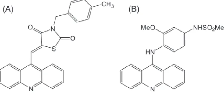
(5Z)-[5-acridine-9-yl-methylene-3-(4-methyl-benzyl)-thiazolidine-2,4-dione] (Figure 1A)
is an acridinylidene thiazolidinedione, a monoacridinine
structural analogue of amsacrine (Figure 1B) (Denny,
2002; Mourão et al., 2005). A variety of different acridine derivatives has been synthesized and promising results obtained in some cases, prompting the development of new acridine-based drugs that present a wide spectrum of biological activities, such as antibacterial, antimalarial, antitrypanosomial (Bonse et al., 1999), antileishmanial
and antiviral (Goodell et al., 2006) actions, and most notably antitumor activity (Goodell et al., 2008).
The rational design of drugs is one of the most useful approaches for the introduction of new drugs in therapy and
widely used by researchers in the medicinal chemistry ield.
In this context, LPSF/AC04 is an acridine-based derivative, part of a series of new anticancer agents synthesized for the purpose of developing more effective and less toxic anticancer drugs. This molecule, with potential antitumor activity, was synthesized at the Laboratory of Rational Design and Synthesis of Drugs (LPSF) of the Federal University of Pernambuco, Brazil. LPSF/AC04 has shown antitumor activity with tumor inhibition of more
than 85% in a murine sarcoma 180 model after 8 days of treatment with 100 mg/kg i.p/day (De Lima, Lins, Pitta,
2007). In addition, a recent study has reported preliminary
pharmacokinetics of LPSF/AC04 with a half-life of
66 h and accumulation in different tissues (Pigatto et
al., 2011). However, heterocyclic acridine derivatives
such as LPSF/AC04 are characterized by low solubility in aqueous solutions, limiting both clinical trials and its therapeutic use.
In order to minimize this problem, incorporation of the drug into controlled release systems is required. Liposomes are widely used in pharmaceutical applications,
such as for drug delivery vehicles of several therapeutic agents, given their versatility and clinical efficacy (Torchilin, 2006). In addition, cyclodextrins are used to improve the solubility of poorly hydrophilic drugs. Cyclodextrins (CyDs) have a relatively nonpolar cylindrical cavity that can bind, and thereby solubilize, a wide range of hydrophobic molecules (Loftsson, Hreinsdóttir, Másson, 2005; Loftsson, Duchêne, 2007). The main purpose of inclusion complex-loaded liposomes is to combine the advantages of cyclodextrins as drug solubility enhancement agents with those of liposomes as drug-targeting agents. Some authors have cited the use of cyclodextrins in the formation of inclusion complexes with hydrophobic drugs, including acridine derivatives, to enhance their solubility (Schuette et al., 1991; Correia et al., 2002; Loftsson, Hreinsdóttir, Másson, 2005; Loftsson, Duchêne, 2007; Mishur et al., 2011; Al Omari et al., 2011).
The quality control of pure drugs and dosage forms are carried out using official and validated methods. Despite the clear advantages of using the HPLC chromatographic technique, the method has several limitations such as the high cost of instrumentation and operation, relatively long analysis times and the need for experience in handling the equipment and in processing samples (Siqueira-Moura et al., 2008). Spectrophotometry is a highly convenient analytical technique that is widely used in laboratories for quality control given its simplicity, low cost and wide availability (Darwish et al., 2009).
The main objective of this work was to develop
and validate a simple, precise, accurate and economical analytical method for quantifying LPSF/AC04 by UV-spectrophotometry. Subsequently, the resultant method can be applied to determine pure drug and dosage forms, such as liposomes and inclusion complexes containing LPSF/AC04.
MATERIAL AND METHODS
Apparatus
A UV spectrophotometer (Ultrospec 3000 Pro, Amersham Pharmacia Biotech, Sweden) equipped
with 10 mm quartz cells was used for all the absorption
measurements. A magnetic stirrer (Variomag, Germany), Vibra Cell sonicator (Branson, USA), lyophilizer EZ-DRY (FTS Systems, USA) and thermostatically-controlled
water bath (Bioblock Scientiic, France) were employed.
Material and reagents
LPSF/AC04, obtained by the synthetic route
N S N O
O
CH3
N HN
MeO NHSO2Me
(A) (B)
FIGURE 1 - Chemical structures of
(Pitta et al., 2007), was kindly provided by the
Laboratory of Medicinal Chemistry of the Federal University of Pernambuco, Brazil, CAS:440367-56-6. Cholesterol (CHOL), trehalose, stearylamine (SA) and
2-hydroxypropyl-β-cyclodextrin (HP-β-CyD) were
purchased from Sigma-Aldrich (St. Louis, USA); Soybean
phosphatidylcholine (SPC, S100®) was obtained from
Lipoid GmbH (Ludwigshafen, Germany). Methanol
and chloroform (Merck, Darmstadt, Germany) were of
analytical grade.
LPSF/AC04 standard solutions
LPSF/AC04 has a molecular weight of 410 and melting point of 199 °C, being characterized as a
yellow-greenish amorphous powder. This molecule was characterized by spectroscopy (IR, 1H-NMR, Ms), C25H18N2O2S synthetic route has 96% yield: TLC
benzene:ethyl acetate (9:1) Rf:0.45. IR cm-1(KBr): υ 1746,
1697, 1630, 1378, 1339, 1149, 758. 1H NMR (δ ppm,
CDCl3): 2.37 (s, CH3); 4.92 (s, CH2); 7.21 (d, 2H benzyl,
J=7.8 Hz), 7.42 (d, 2H benzyl, J=8.1 Hz), 7.57-7.62 (m, 2H
acridine), 7.8-7.85 (m,2H acridine), 7.96 (d, 2H acridine,
J=7.8 Hz), 8.29 (d, 2H Benzyl J=8.1 Hz), 7.57-7.62 (m,
2H acridine), 7.96 (d, 2H acridine, J=7.8 Hz), 8.29 (d, 2H
acridine, J=8.7 Hz), 8.69 (s, 1H, CH). Ms, m/z (%):410 (M 98.1), 411 (29.3), 305 (24.2), 235 (52.9), 190 (9), 105 (100), 77 (8.3).
T h e s t a n d a r d s o l u t i o n s o f L P S F / A C 0 4 a t concentrations ranging from 0.3 to 2.0 µg.mL−1 were
prepared in methanol from a stock solution (100 µg.mL−1).
Standard solutions were prepared in triplicate to validate the analytical method.
Analytical method validation
The validation of the UV spectrophotometric analytical method was carried out based on parameters
including linearity, limits of detection and quantiication, speciicity, precision, accuracy and robustness (ANVISA, 2003; ICH, 1995A; ICH, 1996B). All assays were performed at 25 °C except for the robustness assay, where samples were also stored at 4 °C and 37 °C before analysis.
Initially, UV spectra of LPSF/AC04 at different
concentrations (0.3, 0.5, 1, 1.5 and 2 µg.mL-1) were
obtained to determine the wavelength of greatest absorptivity.
Specificity
The specificity of the method was evaluated by
comparing the UV spectra of blank samples (HP-β-CD
and unloaded liposomes) against LPSF/AC04 standard solution. The analysis of LPSF/AC04-loaded liposomes
and LPSF/AC04:HP-β-CD inclusion complex scanning was also performed from 225 to 340 nm and checked for
changes in absorbance at the respective wavelengths.
Linearity
The linearity of the proposed method was veriied by
preparing three different standard solutions of LPSF/AC04
(0.3, 0.5, 1.0, 1.5 and 2.0 µg.mL-1), analyzed in triplicate, to plot nine derived analytical curves. The linearity of the analytical curve was evaluated by linear regression analysis using the least squares method and analysis of variance (ANOVA).
Limits of detection and quantification
Limits of detection (LOD) and quantiication (LOQ) were estimated according to ICH guidelines (ICH, 1995A; ICH, 1995B). Limit of detection was calculated by: LOD = 3.3(σ/S), and limit of quantiication was calculated
by: LOQ = 10(σ/S), where σ is the standard deviation of the response of the blank and S is the slope of the analytical curve.
Accuracy
The accuracy of the method was determined by the
recovery of a known amount of LPSF/AC04 added to samples of unloaded liposomes and HP-β-CD. Briely,
to determine the accuracy of the proposed method, different levels of drug concentrations were used: lower concentration (0.5 µg.mL-1), intermediate concentration
(1.0 µg.mL-1) and higher concentration (1.5 µg.mL-1).
A known aliquot of LPSF/AC04 stock solution was transferred to a 10mL volumetric lask containing unloaded liposomes or an accurately weighed amount of HP-β-CD equivalent to the quantity in the LPSF/AC04:HP-β-CD inclusion complex (Section 2.6.1) and illed to volume with methanol. A 1:10 dilution in methanol of the sample
was then performed.
The samples were prepared in triplicate and analyzed by the proposed method. The relative standard deviation and LPSF/AC04 recovery percentage were employed to
evaluate the accuracy of the method by applying equation 1.
The paired t-test at a 95% level of significance was
performed to compare the mean absorbance of the samples.
theoretical drug concentration Recovery (%) =
Precision
Inter-day, intra-day and inter-analyst variations were studied to determine repeatability and intermediate precision of the proposed analytical method. Intermediate precision was determined by analyzing three different
levels of LPSF/AC04 concentrations at 0.5, 1.0, 1.5 µg.mL-1. Different solutions were prepared in triplicate by two different analysts at two different times during one day and analyzed for intra-day variations. The same procedure was followed for two different days to study inter-day and inter-analyst variations (ANVISA, 2003).
The percentage relative standard deviation (%RSD) of the
predicted concentrations from the regression equation was
taken as the measure of precision. The paired t-test at a
95% level of signiicance was performed to compare the
mean absorbance of the samples.
Robustness
The robustness of the method was evaluated by changing the solvent suppliers and the temperature of the
LPSF/AC04 samples (1.5 µg.mL1) at 4, 25 and 37 °C.
The samples were previously prepared and transferred
to sealed tubes and refrigerated at 4 °C or maintained at different temperatures (25 and 37 °C) for 24 h prior to
analysis. Assays were performed three times under the
same conditions (ANVISA, 2003; Ulu, Elmali, 2010).
Application of the method: determination of LPSF/AC04 from inclusion complex and liposomes
Preparation of LPSF/AC04:HP-b-CD inclusion complex
LPSF/AC-04:HP-β-CD complex was prepared using freeze-drying. First, stoichiometric amounts of HP-β-CD and LPSF/AC04 were dissolved in puriied water and the mixture stirred for 72 h at 25 °C, then frozen in liquid
nitrogen and inally lyophilized at 4x10⁻⁶ Bars for 72 h.
Preparation of LPSF/AC04 and LPSF/AC04:HP-b -CD-loaded liposomes
LPSF/AC04 and LPSF/AC04:HP-β-CD-loaded liposomes were prepared using the thin lipid ilm method
(Mendonça et al., 2012). Drug entrapment eficiency was determined using ultrailtration by the ultracentrifugation
technique (Ultrafree® units, Millipore, USA). Samples
of LPSF/AC04 and LPSF/AC04:HP-β-CD-loaded liposomes (400 µL) were placed into the iltration unit
and submitted to ultracentrifugation at 8,792g for 1 h. The
samples were analyzed at 250 nm for determination of LPSF/AC04 content using a LPSF/AC04 analytical curve
with concentrations ranging from 0.3 to 2.0 µg.mL-1. All samples were prepared in triplicate. Data are expressed as percentage of total content of LPSF/AC04 entrapped in liposomes.
RESULTS AND DISCUSSION
Analytical method validation
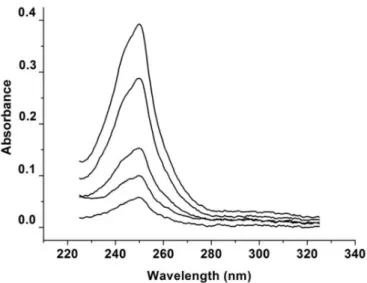
The absorption spectra of the LPSF/AC04 in methanol were recorded at concentrations ranging from
0.3 to 2.0 μg.mL-1 (Figure 3). LPSF/AC04 was found
to exhibit a maximum absorption peak (λmax) at 250 nm
with a molar absorptivity (ε) of 7.60×10⁴ L.mol−1.cm−1. In addition, no interference from the solvent in LPSF/AC04
shoulder peaks near λmaxwas veriied. Methanol can thus be
considered a suitable solvent for validating the proposed method, since it showed no interference in the analysis, thus supporting the reproducibility of the results.
Specificity
The method described was specific for the determination of LPSF/AC04 in inclusion complex and
liposomes. A well-defined peak of LPSF/AC04 was
observed at 250 nm. In addition, the absorption spectrum
of blank samples (HP-β-CD without LPSF/AC04) and unloaded liposomes showed no peak at the specific
wavelength of LPSF/AC04 (Figure 3). The absorption
peak of LPSF/AC04 at 250 nm was unchanged in the
presence of the constituents of the liposomal formulation
and HP-β-CD, thereby demonstrating the speciicity of
the method.
FIGURE 2 - Scanning UV spectra of LPSF/AC04 at
TABLE I - Experimental results of validation parameters
Concentration (µg. mL -1)
Mean absorbance (± SD)a
0.3 0.056 ± 0.002
0.5 0.095 ± 0.004
1.0 0.186 ± 0.007
1.5 0.271 ± 0.004
2.0 0.365 ± 0.007
aValues represent the mean of three analytical curves.
FIGURE 3 - Scanning UV spectra of: (A) LPSF/AC04 (1 μg.mL-1) (a); LPSF/AC04:HP-β-CD inclusion complexes (b) and HP-β-CD
(c), (B) LPSF/AC04 (1 μg.mL-1) (a); unloaded liposomes (b) and LPSF/AC04-loaded liposomes 2 μg.mL-1) (c) diluted in methanol.
Linearity
Absorbance at λmax= 250 nm and concentrations of LPSF/AC04 ranging from 0.3 to 2.0 µg.mL-1 presented a linear relationship (Table I), and the regression analysis data are summarized in Table II. The analytical curves
were itted by least squares treatment to give the following mean regression equation: Absorbance = 0.18068 x [LPSF/
AC04, µg.mL−1] + 0.00348 (r2 = 0.9995, n=9) (Figure 4).
Limits of detection and quantification
The limit of detection (LOD) and the limit of
quantiication (LOQ) of LPSF/AC04 were 0.047 µg.mL-1
and 0.143 µg.mL-1, respectively. These results showed
that the method was sensitive even at low concentrations of LPSF/AC04.
Accuracy
The accuracy of the procedure was determined by
recovery tests of known quantities of LPSF/AC04 (0.5, FIGURE 4 – Analytical curve or Plot linearity.
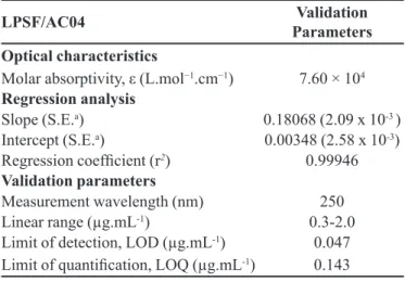
TABLE II - Optical characteristics of LPSF/AC04, statistical data of regression equations and validation parameters
LPSF/AC04 Validation
Parameters Optical characteristics
Molar absorptivity, ε (L.mol−1.cm−1) 7.60 × 10⁴
Regression analysis
Slope (S.E.a) 0.18068 (2.09 x 10⁻³ )
Intercept (S.E.a) 0.00348 (2.58 x 10⁻³)
Regression coeficient (r2) 0.99946
Validation parameters
Measurement wavelength (nm) 250
Linear range (µg.mL-1) 0.3-2.0
Limit of detection, LOD (µg.mL-1) 0.047
1.0 and 1.5 µg.mL-1) spiked in unloaded liposomes and
HP-β-CD. LPSF/AC04 recoveries ranged from 101.06 ± 2.09% to 100.71 ± 1.35% for HP-β-CD (Table III) and 99.36 ± 0.67% to 101.51 ± 1.91% (Table IV) for liposome.
The mean LPSF/AC04 recovery values were close to
100%, and their low standard deviation values evidence
the high accuracy of the analytical method. These results reveal that small changes in the drug concentration in the solutions can be accurately determined by the proposed analytical method.
Precision
Repeatability (RSD%) ranged from 4.20 to 4.34 (%)
for three levels of LPSF/AC04 concentrations (Table V). The results of repeatability (intra-day precision) of the method indicated the precision under the same operating conditions over a short period of time. Inter-day precision expresses within-laboratory variations on different days by different analysts (ANVISA, 2003). In the precision
study, RSD% values were less than 5% in all cases and
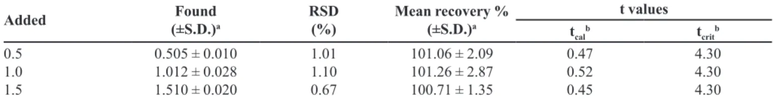
TABLE III - Recovery of LPSF/AC04 in HP-β-CD matrix to evaluate the accuracy of the UV method
Added Found
(±S.D.)a
RSD (%)
Mean recovery % (±S.D.)a t values tcal b t crit b 0.5 1.0 1.5
0.505 ± 0.010 1.012 ± 0.028 1.510 ± 0.020
1.01 1.10 0.67
101.06 ± 2.09 101.26 ± 2.87 100.71 ± 1.35
0.47 0.52 0.45 4.30 4.30 4.30
aValues represent the mean of nine measurements. bt
cal is the calculated value and tcrit is the theoretical value based on the paired
t-test at the level of signiicance of p = 0.05.
TABLE IV - Recovery of LPSF/AC04 in liposomes to assess the accuracy of the proposed method
Added Found (±S.D.)a RSD (%) Mean recovery% (±S.D.)a t values
tcalb t
crit b
0.5 1.0 1.5
0.502 ± 0.006 1.015 ± 0.022 1.490 ± 0.010
1.02 0.76 0.49
100.56 ±1.33 101.51 ±1.91
99.36 ± 0.67
0.53 0.40 0.24 4.30 4.30 4.30
aValues represent the mean of nine measurements. bt
cal is the calculated value and tcrit is the theoretical value based on the paired
t-test at the level of signiicance of p = 0.05.
TABLE V - Precision of proposed analytical method
Precision
[LPSF/AC04] Added (µg.mL-1)
[LPSF/AC04] Found ± S.D.
(µg.mL-1)
R.S.D.
(%) tcal
a t
tab b
Inter-day
Same analyst (days 1 and 2)
0.5 0.494 ± 0.02 4.36 0.57 2.57
1.0 1.011 ± 0.02 2.50 0.31 2.57
1.5 1.496 ± 0.02 1.52 0.73 2.57
Inter-day
Analysts 1 and 2 (days 1 and 2)
0.5 0.498 ± 0.02 4.01 0.83 2.57
1.0 1.008 ± 0.05 4.78 0.68 2.57
1.5 1.529 ± 0.07 4.46 0.33 2.57
Intra-day
Different tests (same analyst; day 1)
0.5 0.494 ± 0.02 4.20 0.57 2.57
1.0 1.019 ± 0.04 4.34 0.32 2.57
1.5 1.534 ± 0.06 4.20 0.25 2.57
at
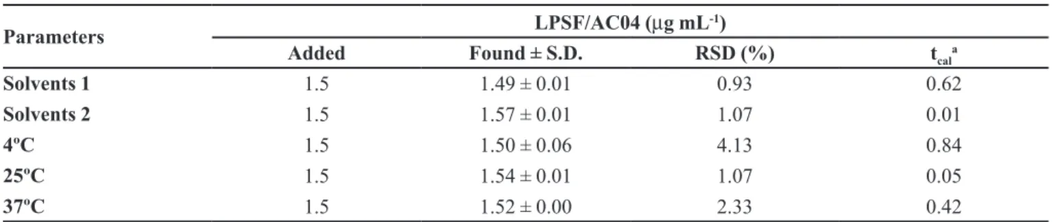
TABLE VI - Robustness of the UV method using different solvent suppliers and temperatures of LPSF/AC04 samples
Parameters LPSF/AC04 (µg mL
-1)
Added Found ± S.D. RSD (%) tcal
a
Solvents 1 1.5 1.49 ± 0.01 0.93 0.62
Solvents 2 1.5 1.57 ± 0.01 1.07 0.01
4ºC 1.5 1.50 ± 0.06 4.13 0.84
25ºC 1.5 1.54 ± 0.01 1.07 0.05
37ºC 1.5 1.52 ± 0.00 2.33 0.42
at
cal is the calculated value. The theoretical value tcrit = 4.30 is based on the paired t-test at the level of signiicance of p = 0.05 (n=3).
were well within the acceptable range, indicating that the method has good repeatability and inter-day precision (ANVISA, 2003).
Robustness
The analytical method proved to be robust (Table
VI), since no statistically signiicant differences (Student´s t-test) were found when samples were subjected to temperature variations and diluted in solvents from different manufacturers. The satisfactory recovery of
LPSF/AC-04 from liposomes (>99%) stored at different
temperatures confirmed that the molar absorptivity of LPSF/AC04 is not dependent on temperature.
Application of the method: determination of LPSF/AC04 in inclusion complex and liposomes
The encapsulation efficiencies of LPSF/AC04
and LPSF/AC04:HP-β-CD in liposomes were 99.02 ± 1.56% and 93.57 ± 0.37%, respectively. In the literature, the quantiication of hydrophobic molecules in
inclusion complexes and liposomes have been performed successfully using the proposed spectrophotometry method (Siqueira-Moura et al., 2008; Cavalcanti et al.,
2012; Lapenda et al., 2012).
The high encapsulation eficiency of LPSF/AC04 and LPSF/AC04:HP-β-CD from liposomes indicated the effectiveness of the proposed method in the quantiication
of LPSF/AC04.
CONCLUSION
The proposed spectrophotometric analytical method for determination of LPSF/AC04 proved simple, rapid, accurate, precise and low-cost. The method was applied to quantify the LPSF/AC04 in inclusion complex as well in liposomes and can therefore be used for routine analysis.
ACKNOWLEDGMENTS
The authors wish to thank the Conselho Nacional de Desenvolvimento Cientíico e Tecnológico (CNPq) for grant # 474071/2007-3. The irst author is also grateful to
the Coordenação de Aperfeiçoamento de Pessoal de Nível Superior (CAPES) for a scholarship grant.
REFERENCES
AGÊNCIA NACIONAL DE VIGILÂNCIA SANITÁRIA. ANVISA. R E, nº 899 de 29 de maio de 2003. Guia para validação de métodos analíticos e bioanalíticos. Diário Oicial da União Poder Executivo, Brasilia, 02 jun. 2003. Available at: <http://portal.anvisa.gov.br>. Acessed on: 05 Sept. 2013.
AL OMARI, A. A.; AL OMARI, M .M.; BADWAN, A. A.; AL-SOU’OD, K. A. Effect of cyclodextrins on the solubility and stability of candesartan cilexetil in solution and solid state.
J. Pharm. Biomed., v.54, p.503-509, 2011.
B O N S E , S . ; S A N T E L L I - R O U V I E R , C . ; B A R B E J . ; KRAUTH-SIEGEL, R. L. Inhibition of Trypanosomacruzi
trypanothione reductase by acridines: kinetic studies and structure−activity relationships. J. Med. Chem., v.42, p.5448-5454, 1999.
CORREIA, I.; BEZZENINE, N.; RONZANI, N.; PLATZER, N.; BELOEIL, J-C.; DOAN, B-T. Study of inclusion complexes of acridine with β-and (2,6-dio-methyl)-β-cyclodextrin by use of solubility diagrams and NMR spectroscopy. J. Phys. Org. Chem., v.15, p.647-659, 2002.
DARWISH, I. A.; ABDINE, H. H.; AMER, S. M.; AL-RAYES, L. I. Spectrophotometric study for the reaction between fluvoxamine and 1,2-naphthoquinone-4-sulphonate: Kinetic, mechanism and use for determination of luvoxamine in its dosage forms. Spectrochim. Acta A,
v.72, p.897-902, 2009.
DE LIMA, M.C.A.; LINS, G.S.; PITTA, I.R. Acridine derivatives with antitumoral activity. Patent number WO/2007/109871A2.PCT/BR2007/000074.2007.04.10, 23 Mar. 2007, 04 Out. 2007. Federal University of Pernambuco, UFPE, Brasil.
DENNY, W. A. Acridine derivatives as chemotherapeutic agents.
Curr. Med. Chem., v.9, p.1655-1665, 2002.
GIRAULT, S.; GRELLIER, P.; BERECIBAR, A.; MAES,L.; MOURAY, E.; LEMIERE, P.; DEBREU, M. A.; DAVIOUD-CHARVET E.; SERGHERAERT, C. Antimalarial, antitrypanosomal, and antileishmanial activities and cytotoxicity of Bis(9-amino-6-chloro-2-methoxyacridines): Inluence of the linker. J. Med. Chem., v.43, p.2646-54, 2000.
GOODELL, J. R.; MADHOK, A. A.; HIASA, H.; FERGUSON, D. M.. Synthesis and evaluation of acridine-andacridone-based anti-herpes agents with topoisomerase activity.
Bioorg. Med. Chem., v.14, p.5467-5480, 2006. .
GOODELL, J. R.; OUGOLKOV, A. V.; HIASA, H.; KAUR, H.; REMMEL, R.; BILLADEU, D. D.;FERGUNSON, D. M. Acridine-based agents with topoisomerase II activity inhibit pancreatic cancer cell proliferation and induce apoptosis. J. Med. Chem., v.51, p.179-182, 2008.
INTERNATIONAL CONFERENCE ON HARMONISATION. ICH. Validation of analytical procedures: deinitions and terminology. Q2A (CPMP/ICH/381/95). 1995A. Available at: <http://www.ich.org/products/guidelines/quality/article/ quality-guidelines.html>. Accessed on: 28 Jan. 2013.
INTERNATIONAL CONFERENCE ON HARMONISATION. ICH. Validation of analytical procedures: methodology. Q2B (CPMP/ICH/281/95), 1996B. Available at: ttp://www.ich. org/products/guidelines/quality/article/qualityguidelines. html>. Accessed on: 28 Jan. 2013.
LAPENDA, T. L. S.; MORAIS, W. A.; LIRA, M. C. B.; MACIEL, M. A. M.; SANTOS-MAGALHÃES, N. S. Validation of a UV spectrophotometric method for determining trans-dehydrocrotonin in inclusion complexes with hydroxypropyl-β-cyclodextrin. Lat. Am. J. Pharm.,
v.31, p.1-7, 2012.
LOFTSSON, T.; HREINSDÓTTIR, D.; MÁSSON, M. Evaluation of cyclodextrin solubilization of drugs. Int. J. Pharm., v.302, p.18-28, 2005.
LOFTSSON, T.; DUCHÊNE, D. Cyclodextrins and their pharmaceutical applications. Int. J. Pharm., v.329, p.1-11,
2007.
MENDONÇA, E. A. M.; LIRA, M.C.B.; RABELLO, M. M.; CAVALCANTI, I. M. F.; GALDINO, S. L.; PITTA, I. R.; LIMA, M. C. A.; PITTA, M. G. R.; MARCELO, M. Z.; SANTOS-MAGALHÃES, N. S. Enhanced Antiproliferative Activity of the New Anticancer Candidate LPSF/AC04 in Cyclodextrin Inclusion Complexes Encapsulated into Liposomes. AAPS Pharm. Sci. Tech., v.13, p.1355-1366,
2012.
MISHUR, R. J.; GRIFFIN, M. E.; BATTLE, C. H.; SHAN, B.; JAYAWICKRAMRAJAH, J. Molecular recognition and enhancement of aqueous solubility and bioactivity of CD437 by β-cyclodextrin. Bioorg. Med. Chem. Lett., v.21,
p.857-860, 2011.
MOURÃO, R. H.; SILVA, T. G.; SOARES, A. L.; VIEIRA, E. S.; SANTOS, J. N.; LIMA, M. C. A.; LIMA, V. L. M.; GALDINO, S. L.; BARBE, J.; PITTA, I. R. Synthesis and biological activity of novel acridinylidene and benzylidene thiazolidinediones. Eur. J. Med. Chem., v.40, p.1129-1133,
2005.
SCHUETTE, J. M.; DOU, T. N.; DE LA PEÑA, A. M.; GREENE, K. L.; WILLIAMSON, C. K.; WARNER, I. M. Characterization of the β-cyclodextrin/acridine complex. J. Phys. Chem., v.95, p.4897-4902, 1991.
SIQUEIRA-MOURA, M. P.; LIRA, M. C. B.; SANTOS-MAGALHÃES, N. S. Validação de método analítico espectrofotométrico UV para determinação de ácido úsnico em lipossomas. Braz. J. Pharm. Sci., v.44, p.621-628, 2008.
TORCHILIN, V. P. Multifunctional nanocarriers. Adv. Drug. Deliver. Rev., v.58, p.1532-1555, 2006.
ULU, S. T.; ELMALI, F. T. Spectrophotometric method for the determination, validation, spectroscopic and thermal analysis of diphenhydramine in pharmaceutical preparation.
Spectrochim. Acta, v.77, p.324-329, 2010.
Received for publication on 04th October 2013