Causes, effects and connectivity changes
in MS-related cognitive decline
Carolina de Medeiros Rimkus1,2,Martijn D. Steenwijk1,3, Frederik Barkhof1
ABSTRACT. Cognitive decline is a frequent but undervalued aspect of multiple sclerosis (MS). Currently, it remains unclear what the strongest determinants of cognitive dysfunction are, with grey matter damage most directly related to cognitive impairment. Multi-parametric studies seem to indicate that individual factors of MS-pathology are highly interdependent causes of grey matter atrophy and permanent brain damage. They are associated with intermediate functional effects (e.g. in functional MRI) representing a balance between disconnection and (mal) adaptive connectivity changes. Therefore, a more comprehensive MRI approach is warranted, aiming to link structural changes with functional brain organization. To better understand the disconnection syndromes and cognitive decline in MS, this paper reviews the associations between MRI metrics and cognitive performance, by discussing the interactions between multiple facets of MS pathology as determinants of brain damage and how they affect network efficiency.
Key words: multiple sclerosis, cognition, brain mapping, functional neuroimaging, diffusion tensor imaging.
CAUSAS, EFEITOS E ALTERAÇÕES DE CONECTIVIDADE NO DECLÍNIO RELACIONADO À ESCLEROSE MÚLTIPLA
RESUMO. Declínio cognitivo é uma situação frequente mas ainda pouco compreendida na esclerose múltipla (EM). Atualmente, não são totalmente conhecidos os principais determinantes da disfunção cognitiva na doença, tendo sido apontadas fortes associações entre danos à substância cinzenta e declínio cognitivo. Estudos multiparamétricos mostram que os diferentes fatores patológicos da EM participam como causas interdependentes de atrofia da substância cinzenta e dano cerebral permanente. Eles são associados a efeitos funcionais intermediários (detectados por RM funcional) representando um equilíbrio entre desconexão cerebral e alterações (mal) adaptativas. Portanto, uma abordagem de imagem mais abrangente é necessária, com o objetivo de encontrar associações entre alterações estruturais e a organização funcional cerebral. Para melhor compreender o declínio cognitivo na EM, esse artigo propões uma revisão dos principais métodos de imagem por RM e suas correlações com função cognitiva, discutindo as múltiplas faces patológicas da EM e seu impacto na eficiência das redes neurais.
Palavras-chave: esclerose múltipla, cognição, mapeamento encefálico, neuroimagem funcional, imagem por tensor de difusão.
INTRODUCTION
M
ultiple sclerosis (MS) is a common inlammatory and demyelinating dis-ease of the central nervous system (CNS). Although about 40 to 70% of MS patients develop signiicant cognitive decline,1,2 it wasnot until the past few decades that MS-related cognitive deicit is being systematically stud-ied. Overt dementia in MS is rare, but the
cog-nitive impairment can be substantially debili-tating, impacting on patients daily activities.3
Although MS has been traditionally classi-ied as a white matter (WM) disease, involve-ment of grey matter (GM) by demyelination and neurodegeneration has become evident in all stages of the disease.4-6
Neurodegen-erative GM processes, culminating in axonal and neuronal injury, are being recognized as
This study was conducted at the Department of Radiology, Laboratory of Medical Investigation (LIM-44), Faculty of Medicine of the University of São Paulo, São Paulo SP, Brazil
1,2Department of Radiology, Laboratory of Medical Investigation (LIM-44), Faculty of Medicine of the University of São Paulo, São Paulo SP, Brazil and Department of Radiology and Nuclear Medicine, Neuroscience Campus Amsterdam, VU University Medical Center, Amsterdam, The Netherlands. 1,3Department of Radiology and Nuclear Medicine, Neuroscience Campus Amsterdam, VU University Medical Center, Amsterdam, The Netherlands and Department of Physics and Medical technology, Neuroscience campus Amsterdam, VU University Medical Center, Amsterdam, The Netherlands.
Carolina M Rimkus. Department of Radiology / Laboratory of Medical Investigation (LIM-44) / Faculty of Medicine of the University of São Paulo – 05403-010 Av. Dr. Enéas de Carvalho Aguiar, 269 – São Paulo SP – Brazil. E-mail: [email protected]
Disclosure: The authors report no conflits of interest.
Received December 24, 2015. Accepted in final form February 26, 2016.
more direct causes of clinical disability and cognitive impairment.4,7 However, in vivo characterization of GM pathology remains challenging. Detection of focal GM lesions, especially in the cortical GM, requires advanced magnetic resonance imaging (MRI) techniques, such as high-resolution 3D-T1 and 3D-FLAIR,8 double
inver-sion recovery (DIR)9 (Figure 1) and (ultra) high-ield/
ultra high-ield scanners.10 Furthermore, it has been
suggested that other factors, such as difuse pathology in normal appearing brain tissues (NABT) are signiicant determinants of GM degeneration and atrophy,11,12 partly
due to axonal transection in MS lesions13 are signiicant
determinants of GM degeneration and atrophy.
It has been suggested that WM lesions and inlamma-tory processes have stronger inluence as determinants of GM damage in early stages of MS,14-16 while
neuro-degenerative aspects and difuse pathology in normal appearing WM (NAWM) and GM (NAGM)12,14,17 are
more important determinants of brain damage in long-standing disease. However, a recent longitudinal study has shown that inlammatory changes and T2 lesion volumes remained predictive of disability even after 20 years in patients who presented with clinical isolated syndromes (CIS).18 Other studies have also demonstrated
that diferent patterns of lesion distribution correlate to regional GM atrophy13 and rate of disease progression19
in primary progressive (PPMS) and secondary progres-sive (SPMS) MS.
MRI is the main modality used to assess patho-logical changes in MS and it has been used to quantify WM lesions,15,20,21 GM lesions,8,10,22 difuse
abnormali-ties11,12,15,23,24 and atrophy24-26 (Figure 1). All these factors
are present from early stages of MS,16,27-29 associate with
higher disability scores and cognitive decline,11,15,16,30 and
are more pronounced in long-standing disease.14,31 A few
multi-parametric studies assessed several MRI measure-ments concurrently and have shown that MRI measures of MS pathology are highly interdependent and most of them will eventually correlate with disease severity.12,14,32
he neurodegenerative process in MS is likely to pre-cipitate changes in brain function. Conventional MRI data (i.e. lesion volume, lesion location and atrophy) has been compared to neuropsychological tests scores to relate MS pathology to cognitive performance.20,21,25,33
However, this type of investigation provides indirect associations between structural damage and brain func-tion. In contrast to conventional MRI, functional MRI (fMRI) is providing more direct visualization of brain activity. Recent studies have observed diferent syn-chronization patterns of neuronal activity that can be described as large-scale networks.34-36 hese networks
appear to be closely related to complex brain functions and can be used to study the organization of cognitive functions.34 Neuronal activity in individual components
of functional networks is presumably integrated through WM pathways, building up structural modules of brain networks.37 Understanding the structural basis of
func-tional connectivity requires comprehensive maps of structural connections of human brain. Recent advances in difusion tensor imaging (DTI) and tractography are providing insights about WM microstructure, allowing noninvasive mapping of inter-cortical and deep GM anatomic connections.37,38 Recent assessments of brain
networks in MS patients show that changes in cognitive performance are accompanied by variable modiications in functional39,40 and structural38,41 connectivity. hese
emerging imaging processing techniques are promising tools to surmount the limitations of conventional MRI in providing information about the integration of brain regions and systems.
his paper reviews the associations between MRI metrics and cognitive performance in MS, by discussing the interactions between multiple facets of MS pathol-ogy as determinants of brain damage. We also present an overview of new imaging analysis techniques studying structural and functional brain networks towards access-ing the interplay between structural damage, functional changes and cognitive outcome.
COGNITIVE DYSFUNCTION IN MS
Cognitive dysfunction in MS is heterogeneous and usually afects multiple cognitive domains. he more frequently afected domains are memory, attention, information processing speed and executive func-tions.25,32 Essential verbal skills (word naming and
comprehension) remain relatively preserved, and it is rare to observe aphasia.1 A recent study showed that
the spectrum of afected domains is relatively constant throughout the course of the disease: in long-standing MS patients the same cognitive domains were afected as in early MS, although the changes were more pronounced.32
Risk factors of cognitive symptoms in MS are not completely understood, but studies have suggested that sex, age and genetic predisposition may play a role. For instance, several studies reported that cognitive symp-toms are more severe and occur more often in male patients than in female patients.25,42 Moreover, in adult
patients with MS, cognitive symptoms seem to be worse with older age of onset.43 Lastly, the apolipoprotein E4
Figure 1. MRI metrics associated to brain damage and MS-related cognitive impairment. Examples of the multiple facets of MS pathology, white matter [A, B and C), grey matter lesions [D and E], diffuse MS pathology [F] and cortical grey matter atrophy [G and H] are demonstrated in the picture. White matter lesions are assorted in MS patients, with different distributions, according to the cognitive status. A cognitively preserved RRMS patient shows a lower lesion load, with predominance of centrally distributed WM lesions [A]; a SPMS patient [B] with mild cognitive impairment presents a higher lesion load, with confluent lesions, some of them reaching juxta-cortical locations; the more cognitively impaired subject [C], although this patient presents a lower overall lesion load than patient “b”, the image shows an increased rate of (juxta)cortical lesions. Double inversion recovery (DIR) images [D and E] are sensible to detect cortical lesions. A cognitively impaired MS patient [E] has a significant higher number of cortical lesions (white arrows) than a cognitively preserved patient [D]. Fractional anisotropy differences between cognitively impaired and cognitively preserved MS patients [F - reproduced
from Hulst et al.77, with permission of Wolters Kluwer). The brain areas in yellow display reduced fractional anisotropy in cognitively impaired patients
disease progression and cognitive decline in MS, whereas the more common susceptibility genes for MS, such as human leukocyte antigen class II (HLA-II), do not specii-cally relate to cognition.44
Neuropsychiatric factors are an important aspect to consider when assessing cognitive performance in patients with MS: studies have shown that cognitive fatigue might be partially responsible for decreases in performance of tasks that require sustained mental efort.45 Depression is also common in MS, being present
in up to 60% of the patients, and may impair the perfor-mance in working memory, processing speed, learning, abstract reasoning and executive functioning.1
Not surprisingly, progressive MS phenotypes pres-ent worse cognitive performance than early relapsing remitting MS (RRMS) or CIS patients.1 Some studies32,46
found poorer cognitive performance in both PPMS and SPMS compared to RRMS, though SPMS patients had a worse performance than PPMS when tasks required higher-order working memory processes.46
Although cognitive deicits can be found in early stages of the disease, they are usually a feature of advanced disease. Natural history studies suggest that the cognitive decline tends to progress with increased disease duration and, once it appears in MS patients, it is unlikely to remit.47,48 Hence, it seems that the
cogni-tive deterioration in MS is a neglected aspect of disease progression.
MRI CORRELATES OF COGNITIVE DYSFUNCTION
White matter (WM) lesions. Depicting WM lesions
dissemi-nated in the CNS is indispensible for the diagnosis and early clinical management of MS.49 Although some
studies have reported associations between lesion volumes and cognitive impairment in speciic cognitive domains, such as information and processing speed, verbal memory,16,50 sustained attention and executive
functions,16 overall WM lesion load has shown only
poor correlations with average cognition50 and
progres-sion of cognitive impairment.32,51 hese poor
correla-tions in part relect the lack of pathological speciicity of conventional MRI techniques52 and underestimation
of the amount of demyelination and degeneration in NABT.11,17
It has been suggested that lesion location might be a stronger determinant of neurological dysfunction in MS,20,21,53 rather than overall lesion burden. Some
stud-ies have found signiicant correlations between peri-ventricular lesion burden and physical disability.53,54
However, presence of lesions in more peripheral loca-tions has shown stronger associaloca-tions to cognitive
decline (Figure 1C); a higher number and volume of juxta-cortical lesions has been observed in cognitively impaired patients,21,33,55,56 demonstrating signiicant
cor-relations with average cognition,21,33 executive function
and memory.55 Furthermore, speciic correlations with
regional lesion load have been reported, e.g. for verbal memory with lesions in temporal20 and parietal lobes;57
sustained attention, information and processing speed and executive function with lesions in frontal20,55,57,58 and
temporal lobes;20 abstract reasoning with frontal lobe
lesions57 and visuospatial memory with parietal20,57 and
temporal20 lesions. It thus seems that periventricular and
deep WM lesions might play a more important role in causing physical disability,54 while juxta-cortical lesions
and lesions afecting associative pathways have greater impact on neuropsychological functioning.20,21
Most studies evaluating associations between lesion load and cognition were performed in patients with rel-ative short disease duration. Baseline T2-hyperintense and T1-hypointense lesion loads are signiicant short-term predictors of disability and cognitive decline in patients with clinical isolated syndromes (CIS)15 and
relatively short-term follow-up of RRMS16,59 and PPMS19.
Increases in lesion volumes show greater impact on clin-ical outcome in the irst 5 to 10 years of MS,18,19 with
decreasing importance in the subsequent years suggest-ing that mechanisms of brain damage may change over time, and other factors such as GM lesions, difuse MS-pathology and brain atrophy may be more important determinants of cognitive dysfunction in later stages.
Gray matter (GM) lesions. Renewed interest in MS GM
pathology has revealed that the prevalence of GM lesions is high4,60 and can afect cortical GM60 and also
deep GM structures, such as thalamus, basal ganglia61
and hippocampus.62 Furthermore, it has been shown
that abnormalities in and around the cortex are related to both physical and cognitive deicits.10,21 However, it
is also known that more routinely used MRI sequences to investigate MS lesions, such as FLAIR and dual-echo T2-weighted images, fail to detect a large number of GM lesions.60
Double inversion-recovery (DIR) is a relatively new MRI sequence that selectively nulls the signals from WM and cerebrospinal luid (CSF), leaving only GM and lesions visible62,63 (Figure 1D and 1E). When applied to
a MS population it resulted in a 5-fold increase in detec-tion rate of cortical lesions compared to standard MRI pulse-sequences.60. DIR also improved lesion detection in
other GM regions, such as hippocampus62 and cerebellar
pres-ent higher cortical lesion number and volume than their cognitively unimpaired counterparts.65 A recent study
has demonstrated that the GM lesions detected by DIR frequently afect cortical regions involved in informa-tion processing,66 being mainly distributed in frontal and
temporal lobes, with prominent involvement of motor and anterior cingulate cortices. Moreover, GM lesions in speciic associative cortices and hippocampal lesions are strongly associated with impaired visuospatial memory and information processing speed.62,67
Although DIR has considerably improved GM lesion detection, a post-mortem evaluation uncovered that about 80% of cortical lesions remain undetected at 1.5T.68 Especially purely intra-cortical and subpial
lesions are still frequently missed. Several new strategies are being pursued to increase the detection of cortical lesions in MS. hese include the combination of DIR with other MRI sequences, such as phase-sensitive inversion recovery (PSIR),69 magnetization-prepared rapid
acquisi-tion gradient echo (MPRAGE),10 or the use of ultra-high
ield scanners.10 A recent study found an improved rate of
cortical lesion detection using MPRAGE using a 7T scan-ner, observing stronger correlations between the number of cortical lesions and cognitive tests and EDSS scores.10
Additionally, the use of ultra-high ield scanners facili-tates the characterization of cortical lesion subtypes, especially improving in vivo visualization of the most prevalent subtype - subpial lesions.70 he latter study
showed that, within the cortical lesion subtypes, mixed grey-white matter lesions and subpial lesions have the strongest correlations with cognitive and physical dis-ability status in MS.
Despite the fact that multiple studies have shown the clinical relevance of cortical lesions, there are still several limitations to the assessment of cortical lesions in routine clinical practice. First, speciic MRI sequences to detect cortical lesions, such as DIR and PSIR, are not available on most clinical scanners. he availability of ultra-high ield scanners for clinical practice is even more limited. Second, DIR imaging is prone to artifacts62
and still has low sensitivity to detect subpial lesions.68
hird, the inter-observer agreement for the assessment of cortical lesions on DIR sequences is sub-optimal71
and assessors need to be trained to read these images. Finally, the majority of cortical thinning occurs inde-pendently of cortical lesions, suggesting that there are mechanisms contributing to GM degeneration beyond focal demyelination.72
Diffuse MS pathology. Quantitative MRI techniques, such
as magnetization transfer imaging (MTI) and
difu-sion tensor imaging (DTI), enable quantiication of the extent and severity of structural changes occurring in NABT outside focal lesions.52 MTI quantiies the
macro-molecular content in brain tissues through the transfer of magnetization between immobile (protein-bound) and free (water) protons.52,73 Loss of myelin and axonal
membranes in MS lesions or NABT reduces the pool of protons bound to macromolecules and increases the mobile proton pool, decreasing the magnetization transfer ratio (MTR).73 he most commonly used DTI
indexes to assess the microstructural characteristics of brain tissues include fractional anisotropy (FA) and mean difusivity (MD).12,74 Extra-cellular edema, myelin
and axonal membrane disruption deviate the water difusion from a highly oriented (i.e. anisotropic) to a more isotropic condition, decreasing FA and increasing MD.12
Several studies have shown that correlations between cognitive deterioration in MS and markers of difuse MS pathology are stronger than correlations observed between cognitive decline and WM lesion load.16,32,42,75,76
Cognitively impaired MS patients present lower aver-age MTR and peak location in (NABT) histograms11 and
greater extent and severity of DTI parameters abnormal-ities.32,42,77 A longitudinal study showed that changes in
NABT MTR are stronger predictors of cognitive outcome in MS patients than increases in lesion volumes.16
Fur-thermore, a recent study was able to distinguish cogni-tively preserved from cognicogni-tively impaired MS patients, based on characteristics of microstructural WM damage measured with DTI.77 MS patients showed reduced FA
in several brain areas, being signiicantly worse in cog-nitively impaired subjects (Figure 1F). In addition, some areas critical for cognition, such as the thalamus, unci-nate fasciculus and cerebellum were only afected in the cognitively impaired group. Aligned to these observa-tions, another recent multi-parametric study found that NAWM FA and deep GM atrophy were the strongest pre-dictors of cognitive dysfunction in long-standing MS.32
Evidence suggests that MTR and DTI can be used to probe neurodegeneration (i.e. indirect signs of neuronal loss) and might have prognostic value. First, MTR23 and
DTI12,14 abnormalities worsen along the course of MS,
being more pronounced in progressive subtypes. Sec-ond, DTI changes in NABT are closely related to GM atrophy.14,78 Finally, changes in MTR NAWM progress
in SPMS partially independent from the formation of new MS lesions79 and were considered to be resistant to
irst-line immunomodulatory drugs, such as interferon beta-1.80
inves-tigate NAWM and NAGM abnormalities (i.e. MTR, FA and MD) are pathologically unspeciic, and might cor-respond to (combinations of) demyelination, inlamma-tory processes or neurodegeneration.79 Furthermore,
the observed abnormalities are quantitatively small and there are no current standardized parameters to be applied on an individual level.79
Brain atrophy. Brain atrophy is considered to be an
end-stage phenomenon, strongly correlated to perma-nent brain damage / neurodegeneration. Patterns of GM atrophy are being identiied from early stages of MS onward81 and show diferential efects on
neuro-psychological functioning. Deep GM atrophy is more pronounced than cortical atrophy in the initial stages of the disease and in patients with clinical isolated syndromes (CIS).5,82 Mesial temporal (i.e. hippocampal
and amygdala)83,84 and thalamic25,78,85 atrophy are the
most frequent deep GM structures associated with cognitive impairment in MS. halamic atrophy seems to afect multiple cognitive domains, such as executive functions, information and processing speed, attention and psychomotor processing speed,25 while the
hippo-campal damage is more speciically associated with memory impairment.82 halamus and caudate atrophy
also play a role in memory impairment, but with diferent manifestations than found for mesial temporal structures. While thalamus and caudate atrophy were the primary predictors of impaired learning and new memory acquisition (i.e. memory encoding), mesial temporal atrophy showed stronger correlations with recognition of recently learned information (i.e. memory retrieval).86
Global cortical thinning is mild in early stages of MS such as CIS and increases with disease severity27,87 and in
conjunction with cognitive decline65 (Figure 1G). here
are some patterns of regional cortical atrophy in MS that correlate with disability,88 speciic clinical symptoms27,89
and cognitive decline.90 Calabrese et al.27 found
signii-cant cortical atrophy in frontal, precentral and occipital areas in very early stages of MS and the degree and dis-tribution of cortical thinning was associated with symp-toms at disease onset. In another study, atrophy in the striatum, posterior parietal cortex and middle frontal gyrus correlated with fatigue.89 Charil et al.88 observed
correlations between EDSS and cortical thickness in the anterior cingulate gyri, insula and frontal and temporal associative cortices. One study showed that GM volumes in bilateral prefrontal cortex, precentral gyrus, superior parietal lobe, left precuneus and right cerebellum corre-late with Paced Auditory Serial Addition Task (PASAT),91
a neuropsychological test used to assess information pro-cessing speed and sustained attention. A recent study detected non-random patterns of cortical atrophy in MS, demonstrating that the spatial distribution of cor-tical atrophy is closely related to the dominant clinical proile.90 Interestingly, the patterns that showed most
pronounced cortical atrophy overlap with regions associ-ated to the limbic system and the default mode network (DMN), and atrophy in the posterior cingulate cortex and temporal pole was associated to cognitive dysfunction.
Despite these eforts, it is still not well understood which pathological factors determine GM atrophy in MS. Previous studies showed associations between GM atro-phy with WM,87,88 GM72 lesions and NAWM damage.14,29
Most of these studies were restricted to a single MRI modality or brain region, and investigated patients with a relatively short disease duration, which makes it hard to discern the importance of individual measurements. A recent multi-modality study showed that all these factors play independent roles as determinants of GM atrophy.14
Lesion load and difuse WM pathology were important explanatory variables for cortical thickness and deep GM atrophy in RRMS. On the other hand, WM atrophy and changes in NAWM (on DTI) were stronger predictors of GM atrophy in progressive types of MS. Although the pathological mechanisms in MS might change over time, it seems that GM degeneration occurs at least partly sec-ondary to WM damage. he complex substrate of brain damage and cognitive dysfunction in MS emphasizes the need for more comprehensive approaches, investigat-ing the interplay of diferent imaginvestigat-ing markers, towards assessing the links between structural damage and func-tional changes.
BRAIN CONNECTIONS AND NETWORKS:
THE MISSING LINKS BETWEEN STRUCTURAL
DAMAGE AND FUNCTIONAL OUTCOME
Traditional models of brain function employ modular paradigms, in which brain areas are postulated to act as independent processors for speciic neural functions.34
Accumulating evidence suggests that segregation of brain areas in highly specialized modules is overly simplistic and presents several limitations in explaining higher cognitive functions. Newer paradigms in cogni-tive neuroscience show evidence of complex cross-modal interactions where conjoint functions of brain areas work together in large-scale networks.34 A more
(fMRI)35,40,92 (Figure 2), while structural WM pathways
are being accessed by DTI tractography38,93-95 (Figure 3).
Most DTI studies show signs of microscopic damage/ disconnection in several WM pathways involved in cogni-tion. he iber bundles more frequently associated with
neuropsychological performance are the cingulum,93,94
uncinate fasciculus (UF),93,95 corpus callosum,30 superior
and medial cerebellar peduncles.93 he damage in these
iber bundles is associated with interlobar, interhemi-spheric and long-distance brain disconnection. In
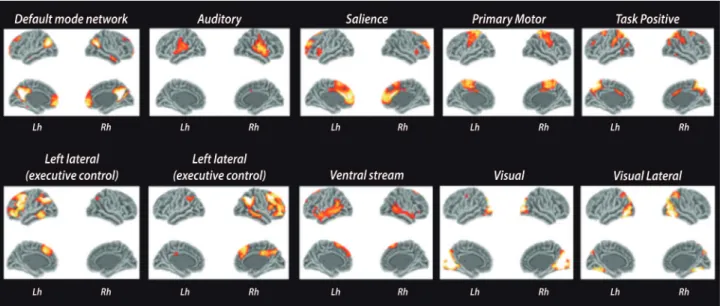
addi-Figure 2. Resting-state fMRI networks. The ten main components of resting-state networks. Images were obtained after processing functional images of 30 healthy controls using FSL toolbox.
tion to the disconnection of long-distance WM pathways, two studies observed signs of decreased local eiciency in prefrontal and occipital associative areas.38,94
Some of the disrupted WM connections are anatomi-cally related to the default mode network (DMN).38,94 he
DMN is one of the most exuberant resting-state func-tional networks and appears to be relevant in maintain-ing normal cognition and higher executive functions. It concatenates parietal, occipital and prefrontal corti-cal functions,96 with important participation of
precu-neus and cingulate cortex. Some fMRI studies showed decreased connectivity in the DMN35,36,96,97 in MS, which
are related to cognitive impairment. Other studies found increased functional activity/connectivity in components of the DMN92,98 and diferent cognitive networks, such
as attention,97 hippocampal98 executive and auditory.36
Increased functional activity was most often reported in cognitively preserved MS patients. However, stud-ies reported positive,97,99 negative36 or both positive and
negative98 correlations between increased connectivity
and cognitive performance or disability level.
hese results are diicult to interpret. Increased con-nectivity in cognitively preserved patients could be a sign of beneicial functional reorganization or compensatory mechanisms to sustain normal function, delaying the cognitive decline. Alternatively, it could be a maladap-tive response secondary to the disruption of inhibitory WM connections. In this context, one would expect positive correlations between signs of WM disconnec-tion and increased funcdisconnec-tional connectivity. Louapre et al.97 observed signiicant correlations between increased
functional activity in the attention network and DTI abnormalities in anterior and posterior cingulum. A positive correlation between WM microstructural dam-age and increased functional activity was also observed in hippocampal pathways.41 On the other hand, a recent
multimodality study100 showed results in the opposite
direction, reporting difuse WM microstructural damage being associated with decreased functional connectivity in 5 resting-state networks, with both positive and nega-tive correlations between WM damage and functional activity.
Diferent studies found similar results in struc-tural38,94 and functional40,92 networks in thalamic and
cin-gulate connectivity. hey showed that, in mildly impaired
patients, both functional and structural connectivity are increased. However, there are no studies that systemati-cally investigated global structural and functional con-nectivity in the same patients. Also, most of the studies are cross-sectional, thus it cannot be excluded that dif-ferent results between studies are secondary to cognitive reserve and inter-subject diferences.
CONCLUSION
Cognitive decline is an important source of disability in MS patients and an undervalued aspect of disease progression. Even though cognitive impairment is partly related to macroscopic lesion load, focal damage might play an indirect role as a cause of further tissue damage. Relective of permanent damage and tissue loss, atrophy markers show stronger correlations with cognitive tests, but regional damage in target struc-tures provides limited information about their speciic relevance for a given cognitive domain and the integra-tion between brain areas and systems. A more holistic investigation of brain networks seems to better capture the mechanisms of brain damage and (mal)adapta-tion encountered in cognitive dysfunc(mal)adapta-tion. However, multimodal imaging techniques and network analyses are still in their infancy and, although it is possible to visualize the associative areas more frequently afected in cognitive impaired MS subjects, we still lack a more speciic comprehension of the interplay between func-tion and structure to better understand initial adapta-tion and subsequent collapse of brain networks.
Aknowledgments. To São Paulo Research Foundation
(FAPESP – 2014/23299-4) for the research grant to Carolina de Medeiros Rimkus. To Paulo Rodrigo Bazan, researcher in the Laboratory of Medical Investigation (LIM-44), Faculty of Medicine of the University of São Paulo, for the resting-state images (Figure 2). To Hanneke Hulst and Menno M Schoonheim for contrib-uting with the TBSS image in Figure 1.
Author contribution: Carolina de Medeiros Rimkus:
design, conceptualization and drafting the study. Martijn D Steenwijk: review the manuscript for intellec-tual content. Frederik Barkhof: review the manuscript for intellectual content.
REFERENCES
1. Chiaravalloti ND, DeLuca J. Cognitive impairment in multiple sclerosis. Lancet Neurol 2008;7:1139-1151.
2. Schoonheim MM, Meijer KA, Geurts JJ. Network collapse and cognitive impairment in multiple sclerosis. Front Neurol 2015;6:82.
3. Guimaraes J, Sa MJ. Cognitive dysfunction in multiple sclerosis. Front Neurol 2012;3:74.
5. Audoin B, Zaaraoui W, Reuter F, et al. Atrophy mainly affects the limbic system and the deep grey matter at the first stage of multiple sclerosis. J Neurol Neurosurg Psychiatry 2010;81:690-695.
6. Eshaghi A, Bodini B, Ridgway GR, et al. Temporal and spatial evolution of grey matter atrophy in primary progressive multiple sclerosis. Neuro-image 2014;86:257-264.
7. Dutta R, Trapp BD. Pathogenesis of axonal and neuronal damage in multiple sclerosis. Neurology 2007;68(22 Suppl 3):S22-31; discussion S43-S54.
8. de Graaf WL, Kilsdonk ID, Lopez-Soriano A, et al. Clinical applica-tion of multi-contrast 7-T MR imaging in multiple sclerosis: increased lesion detection compared to 3 T confined to grey matter. Eur Radiol 2013;23:528-540.
9. Calabrese M, Poretto V, Favaretto A, et al. Cortical lesion load asso-ciates with progression of disability in multiple sclerosis. Brain 2012; 135:2952-2961.
10. Harrison DM, Roy S, Oh J, et al. Association of Cortical Lesion Burden on 7-T Magnetic Resonance Imaging With Cognition and Disability in Multiple Sclerosis. JAMA Neurol 2015.
11. Filippi M, Tortorella C, Rovaris M, et al. Changes in the normal appearing brain tissue and cognitive impairment in multiple sclerosis. J Neurol Neurosurg Psychiatry 2000;68:157-161.
12. Vrenken H, Pouwels PJ, Geurts JJ, et al. Altered diffusion tensor in multiple sclerosis normal-appearing brain tissue: cortical diffusion changes seem related to clinical deterioration. J Magn Reson Imaging 2006;23:628-636.
13. Ceccarelli A, Rocca MA, Pagani E, et al. A voxel-based morphometry study of grey matter loss in MS patients with different clinical pheno-types. Neuroimage 2008;42:315-322.
14. Steenwijk MD, Daams M, Pouwels PJ, et al. What explains gray matter atrophy in long-standing multiple sclerosis? Radiology 2014;272: 832-842.
15. Summers M, Swanton J, Fernando K, et al. Cognitive impairment in multiple sclerosis can be predicted by imaging early in the disease. J Neurol Neurosurg Psychiatry 2008;79:955-958.
16. Deloire MS, Ruet A, Hamel D, Bonnet M, Dousset V, Brochet B. MRI predictors of cognitive outcome in early multiple sclerosis. Neurology 2011;76:1161-1167.
17. Kutzelnigg A, Lucchinetti CF, Stadelmann C, et al. Cortical demy-elination and diffuse white matter injury in multiple sclerosis. Brain 2005;128:2705-2712.
18. Fisniku LK, Brex PA, Altmann DR, et al. Disability and T2 MRI lesions: a 20-year follow-up of patients with relapse onset of multiple sclerosis. Brain 2008;131:808-817.
19. Bodini B, Battaglini M, De Stefano N, et al. T2 lesion location really matters: a 10 year follow-up study in primary progressive multiple scle-rosis. J Neurol Neurosurg Psychiatry 2011;82:72-77.
20. Lazeron RH, Boringa JB, Schouten M, et al. Brain atrophy and lesion load as explaining parameters for cognitive impairment in multiple scle-rosis. Mult Scler 2005;11:524-531.
21. Lazeron RH, Langdon DW, Filippi M, et al. Neuropsychological impair-ment in multiple sclerosis patients: the role of (juxta)cortical lesion on FLAIR. Mult Scler 2000;6:280-285.
22. Bagnato F, Butman JA, Gupta S, et al. In vivo detection of cortical plaques by MR imaging in patients with multiple sclerosis. AJNR Am J Neuroradiol 2006;27:2161-2167.
23. Samson RS, Cardoso MJ, Muhlert N, et al. Investigation of outer cortical magnetisation transfer ratio abnormalities in multiple sclerosis clinical subgroups. Mult Scler 2014;20:1322-1330.
24. Modica CM, Zivadinov R, Dwyer MG, Bergsland N, Weeks AR, Benedict RH. Iron and volume in the deep gray matter: association with cognitive impairment in multiple sclerosis. AJNR Am J Neuroradiol 2015;36:57-62. 25. Schoonheim MM, Popescu V, Rueda Lopes FC, et al. Subcortical
atrophy and cognition: sex effects in multiple sclerosis. Neurology 2012; 79:1754-1761.
26. Sanfilipo MP, Benedict RH, Weinstock-Guttman B, Bakshi R. Gray and white matter brain atrophy and neuropsychological impairment in multiple sclerosis. Neurology 2006;66:685-692.
27. Calabrese M, Atzori M, Bernardi V, et al. Cortical atrophy is relevant in multiple sclerosis at clinical onset. J Neurol 2007;254:1212-1220. 28. De Stefano N, Matthews PM, Filippi M, et al. Evidence of early cortical
atrophy in MS: relevance to white matter changes and disability. Neurology 2003;60:1157-1162.
29. Henry RG, Shieh M, Amirbekian B, Chung S, Okuda DT, Pelletier D. Connecting white matter injury and thalamic atrophy in clinically isolated syndromes. J Neurol Sci 2009;282(1-2):61-66.
30. Rimkus Cde M, Junqueira Tde F, Lyra KP, et al. Corpus callosum micro-structural changes correlate with cognitive dysfunction in early stages of relapsing-remitting multiple sclerosis: axial and radial diffusivities approach. Mult Scler Int 2011;2011:304875.
31. Steenwijk MD, Vrenken H, Jonkman LE, et al. High-resolution T1-relax-ation time mapping displays subtle, clinically relevant, gray matter damage in long-standing multiple sclerosis. Mult Scler 2015.
32. Daams M, Steenwijk MD, Schoonheim MM, et al. Multi-parametric struc-tural magnetic resonance imaging in relation to cognitive dysfunction in long-standing multiple sclerosis. Mult Scler 2015.
33. Rovaris M, Filippi M, Minicucci L, et al. Cortical/subcortical disease burden and cognitive impairment in patients with multiple sclerosis. AJNR Am J Neuroradiol 2000;21:402-408.
34. Bressler SL, Menon V. Large-scale brain networks in cognition: emerging methods and principles. Trends Cogn Sci 2010;14:277-290. 35. Cruz-Gomez AJ, Ventura-Campos N, Belenguer A, Avila C, Forn C. The
link between resting-state functional connectivity and cognition in MS patients. Mult Scler 2014;20:338-348.
36. Rocca MA, Valsasina P, Martinelli V, et al. Large-scale neuronal network dysfunction in relapsing-remitting multiple sclerosis. Neurology 2012;79:1449-1457.
37. Hagmann P, Cammoun L, Gigandet X, et al. Mapping the structural core of human cerebral cortex. PLoS Biol 2008;6:e159.
38. Shu N, Liu Y, Li K, et al. Diffusion tensor tractography reveals disrupted topological efficiency in white matter structural networks in multiple scle-rosis. Cereb Cortex 2011;21:2565-2577.
39. Hawellek DJ, Hipp JF, Lewis CM, Corbetta M, Engel AK. Increased functional connectivity indicates the severity of cognitive impairment in multiple sclerosis. Proc Natl Acad Sci U S A 2011;108:19066-19071. 40. Tewarie P, Schoonheim MM, Schouten DI, et al. Functional brain
networks: linking thalamic atrophy to clinical disability in multiple scle-rosis, a multimodal fMRI and MEG study. Hum Brain Mapp 2015; 36:603-618.
41. Zhou F, Zhuang Y, Wang L, et al. Disconnection of the hippocampus and amygdala associated with lesion load in relapsing-remitting multiple sclerosis: a structural and functional connectivity study. Neuropsychiatr Dis Treat 2015;11:1749-1765.
42. Schoonheim MM, Vigeveno RM, Rueda Lopes FC, et al. Sex-specific extent and severity of white matter damage in multiple sclerosis: implica-tions for cognitive decline. Hum Brain Mapp 2014;35:2348-2358. 43. Amato MP, Ponziani G, Siracusa G, Sorbi S. Cognitive dysfunction in
early-onset multiple sclerosis: a reappraisal after 10 years. Arch Neurol 2001;58:1602-1606.
44. Benedict RH, Zivadinov R. Risk factors for and management of cogni-tive dysfunction in multiple sclerosis. Nat Rev Neurol 2011;7:332-342. 45. DeLuca J, Genova HM, Hillary FG, Wylie G. Neural correlates of
cogni-tive fatigue in multiple sclerosis using functional MRI. J Neurol Sci 2008; 270(1-2):28-39.
46. Huijbregts SC, Kalkers NF, de Sonneville LM, de Groot V, Reuling IE, Polman CH. Differences in cognitive impairment of relapsing remitting, secondary, and primary progressive MS. Neurology 2004;63:335-339. 47. Achiron A, Polliack M, Rao SM, et al. Cognitive patterns and progression
in multiple sclerosis: construction and validation of percentile curves. J Neurol Neurosurg Psychiatry 2005;76:744-749.
48. Bagert B, Camplair P, Bourdette D. Cognitive dysfunction in multiple sclerosis: natural history, pathophysiology and management. CNS Drugs 2002;16:445-455.
49. Montalban X, Tintore M, Swanton J, et al. MRI criteria for MS in patients with clinically isolated syndromes. Neurology 2010;74:427-434. 50. Fulton JC, Grossman RI, Udupa J, et al. MR lesion load and cognitive
function in patients with relapsing-remitting multiple sclerosis. AJNR Am J Neuroradiol 1999;20:1951-195.
51. Zivadinov R, Sepcic J, Nasuelli D, et al. A longitudinal study of brain atrophy and cognitive disturbances in the early phase of relapsing-remit-ting multiple sclerosis. J Neurol Neurosurg Psychiatry 2001;70:773-780. 52. Filippi M, Rocca MA, Barkhof F, et al. Association between pathological and MRI findings in multiple sclerosis. Lancet Neurol 2012;11:349-360. 53. Vellinga MM, Geurts JJ, Rostrup E, et al. Clinical correlations of brain
54. Charil A, Zijdenbos AP, Taylor J, et al. Statistical mapping analysis of lesion location and neurological disability in multiple sclerosis: application to 452 patient data sets. Neuroimage 2003;19:532-544.
55. Miki Y, Grossman RI, Udupa JK, et al. Isolated U-fiber involvement in MS: preliminary observations. Neurology 1998;50:1301-136.
56. Comi G, Filippi M, Martinelli V, et al. Brain MRI correlates of cognitive impairment in primary and secondary progressive multiple sclerosis. J Neurol Sci 1995;132:222-227.
57. Swirsky-Sacchetti T, Mitchell DR, Seward J, et al. Neuropsychological and structural brain lesions in multiple sclerosis: a regional analysis. Neurology 1992;42:1291-1295.
58. Foong J, Rozewicz L, Quaghebeur G, et al. Executive function in multiple sclerosis. The role of frontal lobe pathology. Brain 1997;120 (Pt 1):15-26. 59. Patti F, De Stefano M, Lavorgna L, et al. Lesion load may predict long-term cognitive dysfunction in multiple sclerosis patients. PLoS One. 2015;10:e0120754.
60. Geurts JJ, Bo L, Pouwels PJ, Castelijns JA, Polman CH, Barkhof F. Cortical lesions in multiple sclerosis: combined postmortem MR imaging and histopathology. AJNR Am J Neuroradiol 2005;26:572-577. 61. Gilmore CP, Donaldson I, Bo L, Owens T, Lowe J, Evangelou N. Regional
variations in the extent and pattern of grey matter demyelination in multiple sclerosis: a comparison between the cerebral cortex, cerebellar cortex, deep grey matter nuclei and the spinal cord. J Neurol Neurosurg Psychiatry 2009;80:182-187.
62. Roosendaal SD, Moraal B, Vrenken H, et al. In vivo MR imaging of hippocampal lesions in multiple sclerosis. J Magn Reson Imaging 2008;27:726-731.
63. Geurts JJ, Pouwels PJ, Uitdehaag BM, Polman CH, Barkhof F, Castelijns JA. Intracortical lesions in multiple sclerosis: improved detection with 3D double inversion-recovery MR imaging. Radiology 2005;236:254-260. 64. Damasceno A, Damasceno BP, Cendes F. The clinical impact of
cere-bellar grey matter pathology in multiple sclerosis. PLoS One 2014; 9:e96193.
65. Calabrese M, Agosta F, Rinaldi F, et al. Cortical lesions and atrophy asso-ciated with cognitive impairment in relapsing-remitting multiple sclerosis. Arch Neurol 2009;66:1144-1150.
66. Calabrese M, Battaglini M, Giorgio A, et al. Imaging distribution and frequency of cortical lesions in patients with multiple sclerosis. Neurology 2010;75:1234-1240.
67. Roosendaal SD, Moraal B, Pouwels PJ, et al. Accumulation of cortical lesions in MS: relation with cognitive impairment. Mult Scler 2009;15:708-714.
68. Seewann A, Kooi EJ, Roosendaal SD, et al. Postmortem verification of MS cortical lesion detection with 3D DIR. Neurology 2012;78:302-308. 69. Nelson F, Poonawalla AH, Hou P, Huang F, Wolinsky JS, Narayana PA.
Improved identification of intracortical lesions in multiple sclerosis with phase-sensitive inversion recovery in combination with fast double inver-sion recovery MR imaging. AJNR Am J Neuroradiol 2007;28:1645-1649. 70. Nielsen AS, Kinkel RP, Madigan N, Tinelli E, Benner T, Mainero C. Contri-bution of cortical lesion subtypes at 7T MRI to physical and cognitive performance in MS. Neurology 2013;81:641-649.
71. Geurts JJ, Roosendaal SD, Calabrese M, et al. Consensus recommen-dations for MS cortical lesion scoring using double inversion recovery MRI. Neurology 2011;76:418-424.
72. Klaver R, De Vries HE, Schenk GJ, Geurts JJ. Grey matter damage in multiple sclerosis: a pathology perspective. Prion 2013;7:66-75. 73. Berry I, Barker GJ, Barkhof F, et al. A multicenter measurement of
magnetization transfer ratio in normal white matter. J Magn Reson Imaging 1999;9:441-446.
74. Schmierer K, Wheeler-Kingshott CA, Boulby PA, et al. Diffusion tensor imaging of post mortem multiple sclerosis brain. Neuroimage 2007; 35:467-477.
75. Rovaris M, Iannucci G, Falautano M, et al. Cognitive dysfunction in patients with mildly disabling relapsing-remitting multiple sclerosis: an exploratory study with diffusion tensor MR imaging. J Neurol Sci 2002;195:103-109.
76. Lin X, Tench CR, Morgan PS, Constantinescu CS. Use of combined conventional and quantitative MRI to quantify pathology related to cogni-tive impairment in multiple sclerosis. J Neurol Neurosurg Psychiatry 2008;79:437-441.
77. Hulst HE, Steenwijk MD, Versteeg A, et al. Cognitive impairment in MS: impact of white matter integrity, gray matter volume, and lesions. Neurology 2013;80:1025-1032.
78. Benedict RH, Hulst HE, Bergsland N, et al. Clinical significance of atrophy and white matter mean diffusivity within the thalamus of multiple sclerosis patients. Mult Scler 2013;19:1478-1484.
79. Miller DH, Thompson AJ, Filippi M. Magnetic resonance studies of abnormalities in the normal appearing white matter and grey matter in multiple sclerosis. J Neurol 2003;250:1407-1419.
80. Inglese M, van Waesberghe JH, Rovaris M, et al. The effect of interferon beta-1b on quantities derived from MT MRI in secondary progressive MS. Neurology 2003;60:853-860.
81. Sailer M, Fischl B, Salat D, et al. Focal thinning of the cerebral cortex in multiple sclerosis. Brain 2003;126:1734-1744.
82. Henry RG, Shieh M, Okuda DT, Evangelista A, Gorno-Tempini ML, Pelle-tier D. Regional grey matter atrophy in clinically isolated syndromes at presentation. J Neurol Neurosurg Psychiatry 2008;79:1236-1244. 83. Roosendaal SD, Hulst HE, Vrenken H, et al. Structural and functional
hippocampal changes in multiple sclerosis patients with intact memory function. Radiology 2010;255:595-604.
84. Sicotte NL, Kern KC, Giesser BS, et al. Regional hippocampal atrophy in multiple sclerosis. Brain 2008;131:1134-1141.
85. Houtchens MK, Benedict RH, Killiany R, et al. Thalamic atrophy and cognition in multiple sclerosis. Neurology 2007;69:1213-1223. 86. Benedict RH, Ramasamy D, Munschauer F, Weinstock-Guttman B,
Ziva-dinov R. Memory impairment in multiple sclerosis: correlation with deep grey matter and mesial temporal atrophy. J Neurol Neurosurg Psychiatry 2009;80:201-206.
87. Fisher E, Lee JC, Nakamura K, Rudick RA. Gray matter atrophy in multiple sclerosis: a longitudinal study. Ann Neurol 2008;64:255-265. 88. Charil A, Dagher A, Lerch JP, Zijdenbos AP, Worsley KJ, Evans AC. Focal
cortical atrophy in multiple sclerosis: relation to lesion load and disability. Neuroimage 2007;34:509-517.
89. Calabrese M, Rinaldi F, Grossi P, et al. Basal ganglia and frontal/parietal cortical atrophy is associated with fatigue in relapsing-remitting multiple sclerosis. Mult Scler 2010;16:1220-1208.
90. Steenwijk MD, Geurts JJ, Daams M, et al. Cortical atrophy patterns in multiple sclerosis are non-random and clinically relevant. Brain 2016;139: 115-126.
91. Morgen K, Sammer G, Courtney SM, et al. Evidence for a direct asso-ciation between cortical atrophy and cognitive impairment in relapsing-remitting MS. Neuroimage 2006;30:891-898.
92. Schoonheim M, Geurts J, Wiebenga O, et al. Changes in functional network centrality underlie cognitive dysfunction and physical disability in multiple sclerosis. Mult Scler 2013;20:1058-1065.
93. Mesaros S, Rocca MA, Kacar K, et al. Diffusion tensor MRI tractog-raphy and cognitive impairment in multiple sclerosis. Neurology 2012; 78:969-975.
94. Li Y, Jewells V, Kim M, et al. Diffusion tensor imaging based network analysis detects alterations of neuroconnectivity in patients with clinically early relapsing-remitting multiple sclerosis. Hum Brain Mapp 2013;34: 3376-3391.
95. Kern KC, Gold SM, Lee B, et al. Thalamic-hippocampal-prefrontal disruption in relapsing-remitting multiple sclerosis. Neuroimage Clin 2015;8:440-447.
96. Rocca MA, Valsasina P, Absinta M, et al. Default-mode network dysfunction and cognitive impairment in progressive MS. Neurology 2010;74:1252-1259.
97. Louapre C, Perlbarg V, Garcia-Lorenzo D, , et al. Brain networks discon-nection in early multiple sclerosis cognitive deficits: an anatomofunctional study. Hum Brain Mapp 2014;35:4706-4717.
98. Hulst HE, Schoonheim MM, Van Geest Q, Uitdehaag BM, Barkhof F, Geurts JJ. Memory impairment in multiple sclerosis: Relevance of hippo-campal activation and hippohippo-campal connectivity. Mult Scler. 2015. 99. Loitfelder M, Filippi M, Rocca M, et al. Abnormalities of resting state
functional connectivity are related to sustained attention deficits in MS. PLoS One 2012;7:e42862.
![Figure 1. MRI metrics associated to brain damage and MS-related cognitive impairment. Examples of the multiple facets of MS pathology, white matter [A, B and C), grey matter lesions [D and E], diffuse MS pathology [F] and cortical grey matter atrophy [G a](https://thumb-eu.123doks.com/thumbv2/123dok_br/15189388.527213/3.892.147.708.102.855/associated-cognitive-impairment-examples-multiple-pathology-pathology-cortical.webp)