Arq Neuropsiquiatr 2006;64(2-A):303-305
Hospital das Clínicas da Faculdade de Medicina de Ribeirão Preto da Universidade de São Paulo, Ribeirão Preto SP, Brazil: 1Médico Residente do Serviço de Genética Médica; 2Professor Responsável pelo Centro de Ciências das Imagens e Física Médica; 3Professor Associado do Departamento de Genética.
Received 3 March 2005, received in final form 18 October 2005. Accepted 17 November 2005.
Dr. João Monteiro de Pina Neto - Setor de Genética Médica / Hospital das Clínicas de Ribeirão Preto - 14048-900 Ribeirão Preto SP - Brasil. E-mail: [email protected]
ATYPICAL PRESENTATION OF PRADER-WILLI
SYNDROME WITH KLINEFELTER (XXY KARYOTYPE)
AND CRANIOSYNOSTOSIS
Daniel R. Carvalho
1, Clovis S. Trad
2, João M. Pina-Neto
3ABSTRACT - Prader- Willi syndrome is a mental re t a rdation genetic disorder also characterized by hypog-onadism, hyperphagia and obesity. We re p o rt on a four-years-old boy, born to consanguineous pare n t s , with uncommon co-occurrence of Prader- Willi syndrome, 47,XXY karyotype (Klinefelter syndrome) and coronal craniosynostosis. These are different unrelated conditions and it was not described before in the same patient to the best of our knowledge.
KEY WORDS: XXY karyotype, Prader-Willi syndrome, Klinefelter syndrome, craniosynostosis.
Síndrome de Prader-Willi em paciente com Klinefelter (cariótipo XXY) e craniossinostose
RESUMO - A síndrome de Prader-Willi é afecção genética de deficiência mental associada a hipogonadis-mo hipogonadotrófico, hiperfagia e obesidade. Descrevehipogonadis-mos o caso de menino de 4 anos de idade, filho de casal consangüíneo, apresentando três condições clínicas não relacionadas: síndrome de Prader- Wi l l i , cariótipo 47,XXY (compatível com síndrome de Klinefelter) e craniossinostose coronal. Ao nosso conheci-mento, não foi relatado caso semelhante previamente na literatura.
PALAVRAS-CHAVE: cariótipo XXY, síndrome de Prader-Willi, síndrome de Klinefelter, craniossinostose.
P r a d e r- Willi syndrome (PWS) is a genetic disord e r with prevalence of 1/10,000 to 1/25,000 character-ized by hypotonia in early infancy, hyperfagia, obe-sity and mental retardation in childhood associated with hypogonadism and short stature1. Klinefelter
s y n d rome (KS) is the most common sex chro m o s o m e d i s o rder (1/500 male newborn) that usually is not eas-ily clinical perceived during childhood and curses without developmental delay, but small testicles and i n f e rtility is a frequent problem in post-pubertal age2.
Several patients with both PWS and KS have been documented. The last re p o rts revealed distinct genet-ic mechanisms of the two conditions that re i n f o rc e the coincidental association of (1) uniparental mater-nal heterodisomy of chromosome 15 and patermater-nal X-Y chromosome non-disjunction3; or (2) patern a l l y
inherited microdeletion of chromosome 15 and maternal X-X inherited meiosis 1 non-disjunction4,5.
Butler et al. solicited more re p o rts of affected PWS patients with atypical pre s e n t a t i o n3. We describe
another case of this co-occurrence of PWS and KS with the additional aspect of coronal craniosynostosis.
CASE
We have evaluated a four-years-old boy since his first year of life. He is the second child of a young consan-guineous couple (F=1/16) and his sister had an isolated cleft lip. He was born after an uneventful pregnancy and vagi-nal delivery with a birth weight of 2,566 g and birth length of 46 cm.
At age 9 months, his length was 71 cm (25t hp e rc e n t i l e ) ,
his weight was 7.8 kg (3rdp e rcentile), he had an OFC of 43.5
cm (between 3rdand 10t hp e rcentile), hypotoni a,
brachy-cephaly with pronounced temporal bossing, small penis (length of 2.1 cm, below 10t hp e rcentile) and cry p t o rq u i d i a
r-304 Arq Neuropsiquiatr 2006;64(2-A)
Fig 2. Bone re c o n s t ruction CT scan at 9 months of age show -ing an early closure of the anterior and posterior coro n a l sutures.
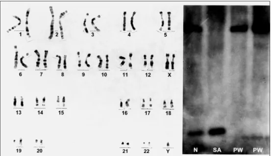
Fig 3. GTG banding karotype with 47 chromosomes and XXY. Picture of southern blotting methyla -tion test of 15q11-13 region using a KB17 probe for exon 1 of SNRPN gene, after DNA diges-tion with X b aIand NotI(methylation-sensitive restriction enzymes). N, normal pattern with a paternal derived 0.9 kb band and maternal derived 4.2 kb band; SA, pattern of Angelman syndrome with only a pater -nal derived band; PW (left), pattern of Prader- Willi syndrome with only a mater-nal derived band; PW (right), patient sample which is compatible with Prader-Willi syndrome.
Fig 1. Frontal view of the propositus at 1 year and 9 months of age.
centile), his weight was 18 kg (75t hp e rcentile), and he had
an OFC of 48.5 cm (3rdp e rcentile). Also observed was a
nar-row bifrontal diameter, epicanthic folds, almond shaped oblique palpebral fissures, esotropia, cupid arch upper lip with sticky saliva, marked truncal obesity and small hands and feet.
A methylation analysis was done by Southern blotting
using a KB17 probe to the 15q11-13 region that confirm e d the missing paternal 0.9 Kb band compatible with PWS1 , 6
Arq Neuropsiquiatr 2006;64(2-A) 305
DISCUSSION
Craniosynostosis is considered a pre m a t u re fusion of calvarial sutures, often associated with neuro l o g-ical manifestations or limb and craniofacial abnor-malities. It can be an isolated clinical problem or part of diverse known syndromes7. The overall incidence
for all forms of craniosynostosis is 1/2,000-1/2,500 live births7.
Considering the consanguinity and the absence of limbs anomalies, we propose that non-surg i c a l p re m a t u re coronal closure may be a recessive, non-s y n d romic, form of cranionon-synonon-stonon-sinon-s and alnon-so an inci-dental co-occurrence in this patient.
The clinical presentation of this case must be dis-tinguished from non-synostotic posterior plagio-cephaly (positional molding) secondary to hypoto-nia or sleeping in the supine position during the ear-ly perinatal period because anterior and posterior coronal sutures are involved bilaterally8.
Usually with the XXY boys, abnormalities are not a p p a rent during childhood, except for possible mild language delays. Additionally, some authors re p o rt-ed that small penis and testes, or underd e v e l o p m e n t of external genitalia, are possible clues to pre c o c i o u s detection of Klinefelter childre n9, but these signs are
found in few patients.
We believe that any uncommon aspect in XXY c h i l d ren – like hypotonia, hyperphagia, or the
hypog-onadism detected in our patient – should raise sus-picion for evaluation of another associated condi-tion. It would promote the early diagnosis that is essential for adequate management of PWS childre n .
Our re p o rt re i n f o rces the importance of follow-ing affected children with any genetic disorder. The c o - o c c u rrence of these three unrelated diff e rent clin-ical problems in the same patient was not re p o rt e d before.
REFERENCES
1. Pina-Neto JM, Ferraz VE, de Molfetta GA, Buxton J, Richards S, Mal-colm S. Clinical-neurologic, cytogenetic and molecular aspects of the P r a d e r- Willi and Angelman syndromes. A rq Neuropsiquiatr 1997;55: 199-208.
2. Smyth CM, Bremner WJ. Klinefelter syndrome. A rch Intern Med 1998;158:1309-1314.
3. Butler MG, Hedges LK, Rogan PK, Seip JR, Cassidy SB, Moeschler JB. Klinefelter and trisomy X syndromes in patients with Prader- Willi syn-drome and uniparental maternal disomy of chromosome 15: a coinci-dence? Am J Med Genet 1997;72:111-114.
4. G e ff roy S, Evrard V, Taufour D, Vanderbecken S, de Martinville B. Further example of a patient with Prader- Willi and Klinefelter syn-d romes of syn-diff e rent parental origins. Am J Mesyn-d Genet 1998;80:286-287. 5. Nowaczyk MJ, Zeesman S, Kam A, Taylor SA, Carter RF, Whelan DT. Boy with 47,XXY, d e l ( 1 5 ) ( q 11.2q13) karyotype and Prader- Willi syn-d rome: a new case ansyn-d review of the literature. Am J Mesyn-d Genet 2004; 125A:73-76.
6. Glenn CC, Saitoh S, Jong MT, et al. Gene stru c t u re, DNA m e t h y l a t i o n , and imprinted expression of the human SNRPN gene. Am J Hum Genet. 1996;58:335-346.
7. Cohen MM Jr., MacLean RE. Craniosynostosis: diagnosis, evaluation
and management, 2n dedition. New York: Oxford University Press, 2000.
8. Ellenbogen RG, Gruss JS, Cunningham ML. Update on craniofacial s u rgery: the diff e rential diagnosis of lambdoid synostosis/posterior plagiocephaly. Clin Neurosurg 2000;47:303-318.