A DUAL ACTION OF
α
-LIPOIC ACID IN THE BRAIN
An electrophysiological evaluation
Otoni Cardoso do Vale
1, Daniel Sá Roriz Fonteles
2,
Francisco Romero Cabral
3, Manassés Claudino Fonteles
4ABSTRACT - Oxidative stress causes metabolic and structural abnormalities during reperfusion. In an animal model of electrophysiological evaluation of cerebral ischemia-reperfusion, α-lipoic acid effect on the oxidative
stress was studied by mean absolute amplitude of EEG spectra evaluation. The left carotideal infusion of 3.03 mM α-lipoic acid in Wistar rats after cerebral ischemia and reperfusion caused initial reduction and partial
final recuperation of the various EEG spectral frequency mean absolute amplitudes (p<0.05). The left intracarotideal infusion of 6.06 mM α-lipoic acid significantly reverted the induced depression of mean absolute
amplitude of theta and delta spectra. Nevertheless there was an increasing pattern of ischemia demonstrated by mean absolute amplitude depression of almost all EEG spectra with 60.6 mM α-lipoic acid infusion. These
observations suggest that, depending on the administered concentration, α-lipoic acid may act in a dual way,
protecting from ischemia at lower concentrations and worsening this process at higher doses.
KEY WORDS: α-lipoic acid, anti-oxidant, pro-oxidant, cerebral ischemia, electroencephalogram (EEG), spectral
mean absolute amplitude.
Dupla ação do ácido Dupla ação do ácido Dupla ação do ácido
Dupla ação do ácido Dupla ação do ácido ααααα-lipoico no encéfalo: uma avaliação eletrofisiológica-lipoico no encéfalo: uma avaliação eletrofisiológica-lipoico no encéfalo: uma avaliação eletrofisiológica-lipoico no encéfalo: uma avaliação eletrofisiológica-lipoico no encéfalo: uma avaliação eletrofisiológica
RESUMO - Estresse oxidativo causa anormalidades metabólicas e estruturais durante reperfusão após isquemia. Em um modelo animal de avaliação eletrofisiológica de isquemia-reperfusão, o efeito da infusão do ácido α
-lipóico sobre o estresse oxidativo foi estudado por meio da avaliação da média das amplitudes absolutas dos espectros eletrencefalográficos. A infusão intracarotídea esquerda de ácido α-lipoic 3,03 mM em ratos Wistar
após isquemia-reperfusão cerebral causou significante redução inicial e recuperação parcial final da média das amplitudes absolutas dos vários espectros eletro - encefalográficos (p<0,05). A infusão intracarotídea esquerda de ácido α-lipoic 6,06 mM significantemente reverteu a depressão induzida da média das amplitudes
absolutas médias dos espectros teta e delta. Houve, contudo, maior depressão da média das amplitudes absolutas de quase todos os espctros com infusão de ácido α-lipóico 60,6 mM. Estas observações sugerem
que, dependendo da concentração administrada, o ácido α-lipoic tem duplo efeito, protegendo da isquemia
em baixas concentrações e piorando este processo em doses altas.
PALAVRAS-CHAVE: ácido α-lipóico, anti-oxidante, pró-oxidante, isquemia cerebral, eletrencefalograma (EEG),
amplitude absoluta média espectral.
Clinical Research Unit of the University Hospital Walter Cantidio, Department of Pharmacology and Clinical Medicine, Federal University of Ceará and Ceará State University Fortaleza CE, Brazil: 1Professor Adjunto de Neurologia e pesquisador, Departamento de Medicina Clínica, Faculdade de Medicina, Universidade Federal do Ceará; 2Psicólogo e bolsista; 3Farmacêutico-Bioquímico, bolsista; 4Professor titular de Farmacologia e Reitor da Universidade Estadual do Ceará, pesquisador. O presente estudo experimental foi financiado pelo CNPq. Received 4 December 2002, received in final form 26 March 2003. Accepted 10 April 2003.
Dr.Otoni Cardoso do Vale - Rua Vicente Linhares 614/700 - 60135-270 Fortaleza CE - Brasil. We have previously studied the
electroencepha-lographic effects on mean absolute amplitudes of electroencephalogram (EEG) waves in Wistar rats submitted to global cerebral ischemia due to common carotid arteries ligation, arterial hypoten-sion and reperfuhypoten-sion1. There are only few
experimen-tal reports in the literature concerning the value of the EEG as a marker of induced cerebral ischemia in animals2-4. According to Siesjö and Siesjö5 two
me-chanisms are responsible for secondary cerebral in-volvement after brain ischemia. The first mechanism involves cellular signal transduction modifications from glutamate and growth factor receptors stimu-lation with intracellular signal modustimu-lation, delete-rious calcium enzyme activation and genetic protein synthesis expression abnormalities6. The second one
production. Oxygen reactive species and free radicals could structurally compromise various macromole-cules by oxidation and reduction reactions causing, among others, DNA strand braking7, with products
formation such as 8-hydroxy-2’-deoxyguanosine8,
lipid peroxydation products9,10 and carbonyl group
formation11.
Otherwise, one of the most important aims in the treatment of cerebral ischemia, during the acute phase, is to minimize or even avoid oxygen reactive species structural lesions, and consequently broaden the therapeutic window in order to implement thrombolytic therapy. Although without beneficial clinical evidence, many experimental procedures sug-gest that pharmacological entities, blocking cytotoxic cascade events during ischemic process or acting as free radical or reactive oxygen species scavengers, could reduce infarct volume due to focal ischemia or late effects of global ischemia with reperfusion.
Thioctic acid, 1,2-dithiolane-3-pentanoic acid or α-lipoic acid and its reduced form, di-hydrolipoic acid, are natural components of biologic membranes, acting as mytochondrial lipoamid co-factor of α -ceto-acid de-hydrogenase12,13. Its participation in
oxy-reduction state from external sources has been focu-sed in various recent studies. It is easily absorbed and taken by cells, being finally reduced to di-hy-drolipoic acid with participation of the systems NADH and NADPH14. The mitochondria are the main site of
α-lipoic acid anti-oxidant action. Within the intracel-lular space the di-hydrolipoic is produced from α-lipoic acid and acts as anti-oxidant in the extracellular space15.
The α-lipoic acid also increases glutathione intra-cellular levels16,17, and as a result, it could be an
im-portant component of the therapeutic arsenal of cerebral vascular ischemic diseases. Other α-lipoic acid and di-hydrolipoic acid actions in ischemia and reperfusion have been described. Ion Fe+2 chelation
by di-hydrolipoic acid18, and nitric oxide19 and other
oxygen reactive species scavenging, as well as E vitamin and other anti-oxidant regeneration20 are
important α-lipoic acid roles. Using our previous idealized model1, we decided on studying the
electro-physiological effects of intracarotideal α-lipoic acid infusion.
METHOD
Male Wistar rats, weighing 300-500g at the time of surgery were used in the present study. The animals were fasted with free access to water during 24 hours before the experimental procedure. Three paired groups of ani-mals (n=6, each paired group) were used in the first part of this study. Intracarotid α-lipoic acid infusion (different
concentrations: 3.03 mM, 6.06 mM and 60.6 mM) started 30 minutes after both common carotid arteries occlusion. The animals of group 1 were infused with 3.03 mM a-lipoic acid, the animals of group 2 with 6.06 mM α-lipoic
acid. The animals of group 3 received 60.6 mM α-lipoic
acid infusion. In the second parf of this study, three other groups of six animals each were studied in order to deter-mine brain tissue reduced glutathione (GSH) contents by spectrophotometry: one made of rats with both common carotid arteries occlusion, one with both common carotid arteries occlusion plus 6.06 mM α-lipoic acid intracarotid
infusion and the last one with both common carotid arte-ries occlusion plus 60.6 mM α-lipoic acid intracarotid
infusion. The mean weight of the animals was 339.3 ± 50.6 g. The weights of the animals of all studied groups were subject to Kolmogorov-Smirnov statitistical evalua-tion and considered as a normal distibuevalua-tion. Under intra-peritoneal urethane anesthesia (1.5g/Kg), all the animals were submitted to surgical procedures that briefly consis-ted of cervical median incision with exposure of the tra-chea; identification and occlusion of the common carotid arteries; insertion and fixation of a polyethylene cannula (PE 50) into the left internal carotid artery in order to infuse
α-lipoic acid solution; insertion and fixation of a
po-lyethylene cannula into right internal carotid artery in order to record the intracarotideal blood pressure. The carotid blood pressure was monitored by using a Narco Bio-system physiograph. The right carotid cannula was connected to a P23 Statham transducer, being the carotideal blood pressure registered by a physiograph.
The animals were kept under continuous electrocardio-graphic and electroencephaloelectrocardio-graphic monitoring. This sys-tem consisted of a preamplifier, an amplifier with adjus-table time constants, an AD converter (12 bits), a 486 PC compatible microcomputer and Braintech® software to
study the spectral frequencies and the absolute amplitu-de average of the chosen events of the EEG. This software analyses the frequency components with precision of 0.35 Hz and uses epochs of 2.84 seconds. Subcutaneous electrodes were inserted in right frontal (F4), left frontal (F3), right parietal (P4), left parietal (P3), vertex (Cz) regions with reference to bilateral auricular (A1 and A2) surface electrodes. The ground electrode was subcutaneously inserted in the nose region. Surface electrodes kept on anterior paws were used for electrocardiographic moni-toring. The technical details of the system are similar to those presented in a recent review21. The
electroencepha-lographic and electrocardiographic activities of each ani-mal were monitored during about 30 minutes before and 30 minutes after the beginning of α-lipoic acid infusion
(26.37-32.70 Hz), theta (4.22-7.73 Hz) and delta (0.35-3.87 Hz) was done by choosing 2.84 s epochs (about 30 epochs from each animal group) of the saved EEG on hard disk. Epochs with artifacts were rejected. The rate of 3.03 mM α-lipoic
acid infusion was 41.91 µl/100g/min.With 3.03 mM α-lipoic
infusion, we considered initial, intermediate and final infusion periods and ANOVA was used to evaluate statistical significant differences (p<0.05) of the absolute amplitudes of the EEG main spectra. When the normality test failed, variance of repeated measures (Friedman) was considered. Two other paired groups were included in this study: 6.06 mM α-lipoic acid infusion (45.87 µl/100g/min; n=6)
and 60.6 mM α-lipoic acid infusion (44.84 µl/100g/min;
n=6); paired t-test was used to evaluate statistical significance. We also performed spectrophotometric analysis of GSH in brain tissue fragments after both common carotid arteries occlusion, and after both common carotid arteries occlusion plus carotid infusion of α-lipoic (6.06 mM and 60.6 mM). The infusion of
a-lipoic acid began 30 minutes after both common carotid arteries occlusion and it was done during 30 minutes.
Six cortical fragments from each rats were removed after craniectomy and kept on liquid nitrogen before their preparation for spectrophotometric analysis by modified Beutler et al.22 method. Briefly, brain tissue fragments were
weighted, homogenized and put into tubes with 6% tri-chloroacetic acid (TCA) solution (50 mg of brain tussue into 1 ml of 6 % TCA). After centrifugation (4000 rpm), 500 µl of each tube float solution were added to 500 µl of
6 % TCA; 1 ml of 6 % TCA acted as a blank tube. After
that, 4 ml of 4.3% NaHPO4 solution plus 500 µl of
5,5-dithio (2´-nitrobenzoic) acid (DTNB) (40 mg of DTNB/100 ml of 1 % sodium citrate) were added to each tube. Each tube was analized by spectrophotometry at 412 nm of wavelength and the absorbance was registred. From pre-vious determination of GSH standard curve, it was possible to know GSH tissue content in the brain fragments. The groups of the second part of this study were used to this analysis. The choice of those groups was based on the electrophysiological analysis of the first part of this study. All the animals were euthanazed with lethal dosis of urethane after the end of the experiment.
RESULTS
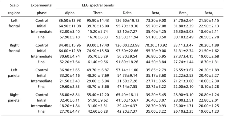
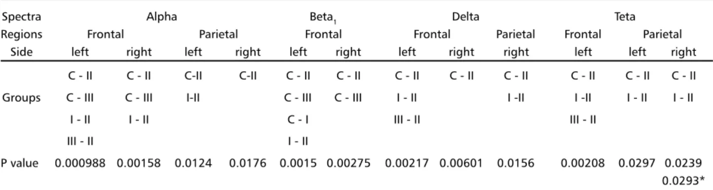
We observed that intracarotideal 3.03 mM α -lipoic acid infusion induced initial reduction (phases I and II) and final (phase III) partial reversal of mean absolute amplitude EEG waves (Table 1, Fig 1, Fig 2 - Panel A). Many mean absolute amplitude dif-ferences of EEG spectra between paired sub-groups reflecting initial reduction (control and initial or in-termediate α-lipoic infusion phase) and final reversal (intermediate and final α-lipoic infusion phase) were statistically significant (p<0,05). More details about such mean EEG spectra absolute amplitude differences are presented in Tables 1 and 2, and Fig 2 - Panel A. The least significant differences were observed with left and right parietal alpha, right parietal delta and left and parietal theta spectra.
Table 1. Mean absolute amplitudes (µV) ± standard errors of mean of EEG spectra from scalp frontal and parietal regions. Control means EEG activity after ischemia and reperfusion; Initial means initial phase of 3.03 mM α-lipoic acid infusion; Intermediate means intermediate
phase of 3.03 mM α-lipoic acid infusion; Final means final phase of 3.03 mM α-lipoic acid infusion. Initial reduction and final partial
reversal of mean absolute amplitude EEG waves were observed during 3.03 mM α-lipoic acid infusion. See more details in the text.
Scalp Experimental EEG spectral bands
regions phase Alpha Theta Delta Beta1 Beta2 Beta3
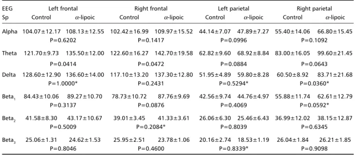
This pattern of EEG spectra mean absolute amplitu-de variation with 3.03 mM α-lipoic acid intracarotideal infusion was not observed when we used other α-lipoic acid concentrations. With 6.06 mM α-lipoic acid infusion, there was a significant mean absolute am-plitude increase (p<0.05) of right parietal delta, and left and right frontal theta spectra (Table 3, Fig 2B).
With 60.6 mM α-lipoic acid infusion, we observed a significant (p<0.05) mean absolute amplitude de-crease of all EEG studied spectra with exception of
right beta3 and left frontal delta spectra (Table 4, Fig 3). The greatest statistical differences were found with right frontal (p=0.0024) and left parietal beta2 (p=0.0064), right (p=0.0052) and left parietal beta1 (p=0.0054), right frontal (p=0.0083) and left pa-rietal alpha (p=0.0066), right frontal theta (p=0.0098), and right (p=0.0030) and left parietal delta spectra (p=0.0066).
There was a statistical significant increase of the brain tissue level of GSH evaluated by spectrophoto-Fig 1. Rat EEG and EKG before (A), during (B) intracarotideal 60.6 mM α-lipoic acid infusion and before (C),
during inicial (I), intermediate (II) and final (III) 3,03 mM α-lipoic acid infusion phases. F3 means left frontal; F4,
metric method after infusion of 6.06 mM α-lipoic acid by intracarotid route. So, we found (0.372±0.021 µg of GSH/ml)/mg of brain tissue in rats with both common carotid arteries occlusion; in rats with both common carotid arteries occlusion plus 6.06 mM α -lipoic acid infusion, we found (0.444 µg of GSH/ml)/ mg of brain tissue (p=0.024). With 60.6 mM α-lipoic acid infusion, the variations of GSH brain tissue contents were not significantly different (p>0.05).
DISCUSSION
In a previous observation, we demonstrated that computerized study of EEG spectral brain activity could
be a valuable electrophysiological tool for evaluating drug cytoprotective action1. We also demonstrated
that intracarotid GSH infusion could significantly revert to the previous wave amplitude the wave depression imposed by ischemia2 and this effect was atributed to
its physiological protection against oxidative stress. This fact has driven us to study α-lipoic acid intracarotideal infusion in Wistar rats with bilateral carotid arteries occlusion as previously described.
Previous studies have demonstrated α-lipoic acid cytoprotective action against reperfusion injury in animal models of global23,24 and focal25 cerebral
ischemia.
Fig 2. Panel A - EEG alpha spectrum mean absolute amplitudes: groups control (C), initial (I), intermediate (II) and final (III) 3.03 mM α-lipoic acid infusion phases. S.E.M. means standard error of mean; * means p<0.05. Panel B - EEG theta
spectrum mean absolute amplitudes; TF3 means left frontal theta; TF4, right frontal theta; TP3, left parietal theta, TP4, right parietal theta; C, control; AAL, 6.06 mM α-lipoic acid infusion; S.E.M., standard error of mean; *, p<0.05.
Table 2. Paired sub-groups with statistically significant EEG spectra mean absolute amplitudes differences (P<0.05) by ANOVA one way repeated measures (Student-Newman-Keuls method). Normality test failed when sub-groups C-II and I-II (parietal beta1), I-III, I-II (right frontal theta) and C-II and I-II (right parietal theta) were analysed. C means control; I means inicial fase, II, intermediate and III, final fase of intracarotideal 3.03 mM α-lipoic infusion, *means P value by ANOVA Friedman method (failed normality test). Se more details in the text.
Spectra Alpha Beta1 Delta Teta
Regions Frontal Parietal Frontal Frontal Parietal Frontal Parietal
Side left right left right left right left right right left left right C - II C - II C-II C-II C - II C - II C - II C - II C - II C - II C - II C - II Groups C - III C - III I-II C - III C - III I - II I -II I -II I - II I - II
I - II I - II C - I III - II III - II
III - II I - II
Our demonstrations that 3.03 mM α-lipoic acid infusion promotes initial reduction and final partial EEG spectra mean absolute amplitude recuperation, allowed us to speculate that, in this condition, this substance has an initial pro-oxidant and a late anti-oxidant action. As we can see in Tables 1 and 2 and in Fig 2 – Panel A, many amplitude reductions and some amplitude final augmentations (left frontal alpha, theta and delta) are statistically significant. Some anti-oxidants, in some circumstances, produce eventually more oxidative stress , acting as pro-oxidant. Di-hydro-lipoic acid (reduced form from α -lipoic acid) reduces Fe+++ to Fe++, activating Fenton
reaction with OH. and OH- production as a
pro-oxi-dant mechanism26. The di-hydro-lipoic acid also
ori-ginates sulfur free radicals, and so, compromises so-me proteins such as 1- antiproteinase, with previous
.OH radicals generation27. The exogenous α-lipoic
acid is rapidly reduced to di-hydrolipoic acid; in mi-tochondria the enzyme lipoate de-hydrogenase induces reduction of α-lipoic acid using NADH as co-factor28; in the cytosol the enzyme glutathione
reductase actively participates of this reduction using NADPH as co-factor29. We speculate that such α-lipoic
acid reductions could act as a possible cytotoxic me-chanism. Roy and colleagues30 suggested that lipoic
acid could reduce NADH levels, using it as a co-factor during its reduction stress; during oxidative stress, both α-lipoic acid and di-hydrolipoic acid act as free radicals scavengers. It is possible that, at least, in α
-lipoic acid infusion initial phases, the cerebral metabolic conditions of our experimental model behaved as it was in reduction stress. The final oxidative stress justifies the observed α-lipoic acid anti-oxidant activity.
This pattern of pro and anti-oxidant effect was not observed when we used 6.06 mM α-lipoic acid infusion. There was a significant mean absolute am-plitude increase of right parietal and left and right frontal theta EEG spectra. So, we suppose that this anti-oxidant effect is more prominent in generating theta activity structure (the hippocampus). We ex-Table 3. Spectral mean absolute amplitudes (µV) ± standard error of mean and P values of EEG spectra during 6.06 mM α-lipoic acid
intracarotideal infusion; * means P value from non-parametric test (normality test failed), AAL means α-lipoic acid; Sp means spectra.
There is a significant mean absolute amplitude wave increase (P<0.05) of right parietal delta and both right and left frontal theta spectra during 6.06 mM α-lipoic acid intracarotideal infusion. See more details in the text.
EEG Left frontal Right frontal Left parietal Right parietal
Sp Control α-lipoic Control α-lipoic Control α-lipoic Control α-lipoic
Alpha 104.07±12.17 108.13±12.55 102.42±16.99 109.97±15.52 44.14±7.07 47.89±7.27 55.40±14.06 66.80±15.45
P=0.6202 P=0.1417 P=0.0996 P=0.1092
Theta 121.70±9.73 135.50±12.00 122.60±16.27 142.70±19.58 62.82±9.60 68.92±8.84 83.00±16.05 99.60±21.45
P=0.0414 P=0.0472 P=0.0884 P=0.0643
Delta 128.60±12.90 136.60±14.00 117.10±13.20 137.30±12.80 51.95±4.89 59.80±8.28 60.50±8.92 83.71±21.68
P=1.0000* P=0.2431 P=0.5294* P=0.0360*
Beta1 84.43±10.06 89.27±10.70 78.73±10.72 87.76±9.69 42.56±9.74 44.76±4.97 55.88±11.74 62.61±12.79
P=0.3137 P=0.0876 P=0.4069 P=0.0592*
Beta2 41.58±8.30 43.17±10.67 39.01±3.45 41.33±3.61 26.06±6.30 25.46±6.43 36.99±12.02 38.15±12.87
P=0.5009 P=0.2084* P=0.8039 P=0.6345
Beta3 25.06±1.31 24.62±1.53 25.95±2.51 23.78±1.06 20.16±2.74 18.53±1.19 26.04±1.84 26.21±1.85
P=0.8046 P=0.4600 P=0.8339* P=0.9098
Fig 3. EEG alpha spectrum mean absolute amplitudes; AF3 means left frontal alpha; AF4, right frontal alpha; AP3, left parietal alpha, TP4, right parietal alpha; C, control; AAL, 60.6 mM α-lipoic acid
Table 4. Spectral mean absolute amplitudes (µV) ± standard error of mean and P values of EEG spectra during 60.6 mM α-lipoic acid
intracarotideal infusion; * means P value from non-parametric test (normality test failed), AAL means α-lipoic acid; Sp means spectra.
Significant mean absolute decrease of all EEG spectra (P<0.05) with exception of right parietal beta3 is observed with 60.6 α-lipoic acid
infusion. See more details in the text.
EEG Left frontal Right frontal Left parietal Right parietal
Sp Control α-lipoic Control α-lipoic Control α-lipoic Control α-lipoic
Alpha 67.20±12.37 29.50±9.26 69.20±8.63 32.00±6.99 147.20±16.12 60.50±6.96 116.8±11.88 52.70±7.27
P=0.0193 P=0.0083 P=0.0066 P=0.0064
Theta 77.90±17.90 40.20±14.60 84.30±11.10 43.30±12.30 195.70±22.00 77.70±13.00 146.10±15.80 71.50±13.10
P=0.0444 P=0.0098 P=0.0138 P=0.0176
Delta 77.50±27.30 48.10±17.30 84.00±17.38 53.40±16.43 196.50±17.30 92.80±15.40 146.50±17.20 81.90±14.40
P=0.0714 P=0.0205 P=0.0066 P=0.0030
Beta1 59.90±10.69 28.00±7.90 63.80±8.86 30.70±5.19 124.70±11.84 55.50±7.35 101.60±8.24 52.50±7.26
P=0.0208 P=0.0360* P=0.0054 P=0.0052
Beta2 30.00±5.18 16.00±3.47 30.50±2.99 18.20±2.71 55.40±5.08 29.60±3.15 47.80±4.27 30.10±3.62
P=0.0239 P=0.0024 P=0.0064 P=0.0102
Beta3 17.66±2.72 10.85±1.81 17.35±1.67 12.21±1.73 29.90±2.93 18.60±1.76 26.19±2.85 20.04±3.01
P=0.0360* P=0.0067 P=0.0220 P=0.0795
perimentally observed by spectrophotometric method a significant increase of GSH in Wistar rat cerebral parenchyma with both common carotid ar-teries occlusion after 6.06 mM α-lipoic acid intra-carotideal infusion (data not yet published). The anti-oxidant effect of 6.06 mM α-lipoic acid in our ex-periments could be due to GSH regeneration, after its reduction to di-hydrolipoic acid. Di-hydro-lipoic acid could actively participate of GSH regeneration mechanisms according to the chemical reaction: di-hydro-lipoic acid + oxidized glutatione (GSSG)® re-duced glutathione (2GSH) + α-lipoic acid20.
With our present experimental model, the intra-carotideal infusion of more concentrated (60.6 mM) α-lipoic acid induced significant mean absolute am-plitude reduction of all EEG spectra in almost all stu-died regions (Fig 3, Table 4). Therefore our data suggest that intracarotideal direct infusion of 60.6 mM α-lipoic acid in rats with both common carotid arteries occlusion has pro-oxidant effect. This could result of a counter regulatory effect on GSH produc-tion. Sen and colleagues31, studying the intracellular
thiol regulation by α-lipoic acid, verified that high concentrations of this acid produced cytotoxic lesions of stimulated mitogenic lymphocytes, in which thiol depletion and DNA fragmentation were detected. It was also verified that α-lipoic acid increases caspase-3 activation, having a role in cellular apoptosis32.
The-refore, apoptotic events may be occurring with the higher concentrations used herein.
In the present experimental model high concen-trations of α-lipoic acid was directly infused into the common carotid artery and rapidly gained access to cerebral parenchyma without systemic enzymatic re-duction. So, the α-lipoic acid could be reduced directly in brain cells by glutathione reductase NADPH-dependent action with concomitant decre-asing of GSH contents, and de-hydrogenase NADH-dependent action. Decreased contents of GSH could explain the pro-oxidant activity of higher concen-trations of α-lipoic acid in our experimental model.
In a recent review Moini et al.33 emphasized the
antioxidant and prooxidant activities of α-lipoic and dihidrolipoic acid. According to these researchers, These are many evidences that both α-lipoic and di-hidrolipoic acid exhibit direct free radical scavenging propreties and that there are only in vitro evidence of their prooxidant propreties. Our data suggest by first time the presence of this phenomena in an in vivo preparation induced by α-lipoic acid.
Acknowledgment Acknowledgment Acknowledgment Acknowledgment
Acknowledgment – The authors would like to thank Dr. Domingos Barreto da Silva and Maria Sílvia Helena Freire de França by their valuable technical assistance.
REFERENCES
1. Cardoso do Vale O, Fonteles DSR, Fonteles MC. Electrophysiological studies in a cerebral ischemic model of the anesthetized rat. J Brain Sci 1998;24:88-100.
3. Dora E, Tanaka K, Greenberg JH, Gonatas NH, Reivich M. Kinetics of microcirculatory NAD/NADH, and electrocorticographic changes in cat brain during ischemia and recirculation. Ann Neurol 1986;19:536-544. 4. Dezsi L, Greenberg JH, Sladky J, Araki N, Hamar J, Reivich M.
Prolonged effects of MK-801 in the cat during focal cerebral ischemia and recovery: survival, EEG and histopathology. J Neurol Sci 1994;121:110-120.
5. Siesjö BK, Siesjö P. Mechanisms of secondary brain injury. Eur J Anaesthesiol 1996;13:247-268.
6. Wieloch T, Hu B-R, Boris-Möller A, et al. Intracellular signal transduction in the postischemic brain. In Siesjö BK, Wieloch T (eds). Advances in neurology: cellular and molecular mechanisms of ischemic brain damage. Philadelphia: Lippincott-Raven, 1996:371-388. 7. Devasagayam TP, Steenken S, Obendorf MS, Schulz WA, Sies H.
Formation of 8-hydroxy(deoxy)guanosine and generation ofstrand breaks at guanine residues in DNA by singlet oxygen. Biochemistry 1991;30:6283-6289.
8. Shigenaga MK, Ames BN. Assays for 8-hydroxy-2'-deoxyguanosine: a biomarker of in vivo oxidative DNA damage. Free Radic BiolMed 1991;10:211-216.
9. Wilson JX. Antioxidant defense of the brain: a role for astrocytes. Can J PhysiolPharmacol 1997;75:1149-1163.
10. Hall ED Lipid peroxidation. In Siesjö BK, Wieloch T (eds). Advances in neurology: cellular and molecular mechanisms of ischemic brain damage. Philadelphia: Lippincott-Raven, 1996:247-258.
11. Smith CD, Carney JM, Starke-Reed PE, et al. Excess brain protein oxidation and enzyme dysfunction in normal aging and in Alzheimer disease. Proc Natl Acad Sci USA 1991;88:10540-10543.
12. Reed LJ, Hackert ML. Structure-function relationships in dihydrolipoamide acyltransferases. Biol Chem 1990;265:8971-8974. 13. Liu S, Baker JC, Andrews PC, Roche TE. Recombinant expression and
evaluation of the lipoyl domains of the dihydrolipoyl acetyltransferase component of the human pyruvate dehydrogenase complex. Arch Biochem Biophys 1995;316:926-940.
14. Haramaki N, Han D, Handelman GJ, Tritschler HJ, Packer L. Cytosolic and mitochondrial systems for NADH- and NADPH-dependent reduction of α-lipoic acid. Free Radic Biol Med 1997;22:535-542. 15. Packer L, Witt EH, Tritschler HJ. Alpha-lipoic acid as a biological
anti-oxidant. Free Radic Biol Med 1995;19:227-250.
16. Busse E, Zimmer G, Schopohl B, Kornhuber B. Influence of α-lipoic acid on intracellular glutathione in vitro and in vivo. Arzneim-Forsch/ Drug Res 1992;42:829-831.
17. Han D, Tritschler HJ, Packer L. Alpha-lipoic acid increases intracellular glutathione in a human T-lymphocyte Jurkat cell line. Biochem Biophys Res 1995;207:258-264.
18. Ou P, Nourooz ZJ, Tritschler HJ, Wolff S. Activation of aldose reductase in rat lens and metal-ion chelation by aldose reductase inhibitors and lipoic acid. Free Radic Res 1996;25:337-346.
19. Vriesman MF, Haenen GR, Westerveld GJ, Paquay JB, Voss HP, Bast A. A method of measuring nitric oxide radical scaveging activity, scaveging properties of sulfur-containing compounds Pharm World Sci 1997;19:283-286.
20. Biewenga GP, Haenen GR, Bast A. The pharmacology of the antioxidant lipoic acid. Gen Pharmacol 1997;29:315-331.
21. Kamp A, Pfutscheller G, Silva FL. Special techniques of recording and transmission. In Niedermeyer E, Silva FL (eds). Electroencephalo-graphy: basic principles, clinical applications, and related fields. Baltimore: Williams & Wilkins, 1999:761-775.
22. Beutler E, Duron O, Kelly BM. Improved method for the determination of blood glutathione. J Lab Clin Med 1963;61:882-888.
23. Cao X, Phillis JW. The free radical scavenger, alpha-lipoic acid, protects against ischemia-reperfusion injury in gerbils. Free Radic Res 1995;23:365-370.
24. Panigrahi M, Sadguna Y, Shivakumar BR, et al. α-lipoic acid protects against reperfusion injury following cerebral ischemia in rats. Brain Res 1996;717:184-188.
25. Wolz P, Krieglstein J. Neuroprotective effects of α-lipoic acid and its enantiomers demonstrated in rodent models of focal cerebral ischemia. Neuropharmacology, 1995;35:369-375.
26. Kawabata T, Tritschler HJ, Packer L. Reaction of (R,S)-dihydrolipoic acid and homologs with iron. Methods Enzymol 1995;251:325-332. 27. Scott BC, Aruoma OI, Evans PJ, et al. Lipoic and dihydrolipoic acids as
antioxidants. a critical evaluation. Free Radic Res 1994;20:119-133. 28. Biewenga GP, Dorstijn MA, Verhagen JV, Haenen GRMM, Bast A.
Reduction of lipoic acid by lipoamide dehydrogenase. Biochem Pharmacol 1996;51:233-238.
29. Handelmann GJ, Han D, Tritschler H, Packer L. α-lipoic acid reduction by mammalian cells to the dithiol form, and release into culture medium. Biochem Pharmacol 1994;47:1725-1730.
30. Roy S, Sen CK, Tritschler HJ, Packer L. Modulation of cellular reducing equivalent homeostasis by alpha-lipoic acid. Mechanisms and implications for diabetes and ischemic injury. Biochem Pharmacol 1997;53:393-399. 31. Sen CK, Roy S, Han D, Packer L. Regulation of cellular thiols in human
lymphocytes by alpha-lipoic acid: a flow cytometric analysis. Free Radic Biol Med 1997;22:1241-1257.
32. Sen CK, Sashwati R, Packer L. Fas mediated apoptosis of human Jurkat T-cells: intracellular events and potentiation by redox-active alpha-lipoic acid. Cell Death Differ 1999;6:481-491.