HS were studied retrospectively. All the examinations were performed on 1.5 T units (SIGNA, GE, Milwaukee, WI) and included high resolution coronal T2-weighted images (3 mm thickness, 0.6 mm gap). Results: The patient’s age ranged between 3.5 and 80 years (mean 34.1); 62 (53.9%) were female and 53 (46.1%) were male. There were HS on the left side in 53 (46.0%), on the right side in 51 (44.3%), and bilateral in 11 (9.7%). In 43 (37.3%) cases there were ipsilateral PHG volume loss and signal hyper intensity on T2-weighted imaging. In 29 (25.2%) cases there were ipsilateral fornix volume loss and in 10 (34.5%) of this there were also ipsilateral MB changes. In abnormal PHG, 23 (53.4%) were on the left side, 17 (39.5%) were on the right side, and 3 (7.1%) were bilateral. There were fornix changes in 15 (34.8%) cases and MB volume loss in 5 (11.6%) cases. Pertinent clinical data were obtained in only 18 (41.8%) of the PHG lesion cases and 11 (61.1%) of these patients had epileptic attacks for more than 20 years before the examination. Conclusion: PHG involvement must be investigated in patients with HS and we suggest that the term mesial temporal sclerosis should be used only if there are also changes at this anatomical site.
KEY WORDS: hippocampal sclerosis, epilepsy, MRI, parahippocampal gyrus.
Análise do giro para-hipocampal em pacientes com esclerose hipocampal: estudo de 115 casos Análise do giro para-hipocampal em pacientes com esclerose hipocampal: estudo de 115 casos Análise do giro para-hipocampal em pacientes com esclerose hipocampal: estudo de 115 casos Análise do giro para-hipocampal em pacientes com esclerose hipocampal: estudo de 115 casos Análise do giro para-hipocampal em pacientes com esclerose hipocampal: estudo de 115 casos
RESUMO - Objetivo: Analisar o envolvimento do giro para-hipocampal (GPH) em 115 pacientes com esclerose hipocampal (EH) à RM. Estudou-se a porcentagem dos casos com redução volumétrica do fórnix (F) e corpo mamilar (CM) ipsilaterais, lado acometido, sexo, idade e tempo de convulsão. Método: Estudo retrospectivo de 115 casos retirados do nosso arquivo. Foram realizados cortes coronais STIR (3mm de espessura com 0,6mm de espaçamento) em aparelhos GE, Signa Horizon, LX e CVI, 1,5T. Resultados: Nos 115 casos estudados, a idade dos pacientes variava entre 3,5 e 80 anos (média 34,1 anos); 62(53,9%) eram mulheres e 53(46,1%) eram homens; 53(46,0%) à esquerda, 51(44,3%) à direita e 11(9,7%) bilaterais; 43(37,3%) apresentavam GPH com dimensões reduzidas e hipersinal ipsilateral, 29(25,2%) fornix reduzido ipsilateralmente e destes, 10(34,5%) tinham CM alterado ipsilateral. Dos GPH alterados, 23(53,4%) à esquerda e 3(7,1%) bilaterais; 15(34,8%) redução de volume CAF e 5(11,6%) no CM. Sabíamos o tempo de crises de 18(41,8%) pacientes com alteração do GPH e destes, 11(61,1%) apresentavam crises há mais de 20 anos. Conclusão: Concluímos que o radiologista deve estar ciente do envolvimento do GPH nos pacientes com esclerose do lobo temporal e sugerimos que o termo esclerose mesial temporal deva somente ser utilizado na presença de alterações do mesmo.
PALAVRAS-CHAVE: esclerose hipocampal, epilepsia, ressonância magnética, giro para-hipocampal.
MEDIMAGEM, Hospital Beneficência Portuguesa, São Paulo SP, Brasil: 1Médico neurorradiologista da MedImagem; 2Médico estagiário do
setor de ressonância magnética da MedImagem; 3Chefe do Departamento de Imagens da MedImagem
Received 29 November 2002, received in final form 28 March 2003. Accepted 7 April 2003.
Dr. Nelson Fortes Ferreira - Rua Martiniano de Carvalho 611/123 - 01321-001 São Paulo SP - Brasil. E-mail: [email protected]
Hippocampal sclerosis (HS) is the most common abnormality (50-70%) in association with temporal lobe epilepsy (TLE)1-4. It is characterized by neuronal
loss and gliosis with atrophy and sclerosis. The tem-poral lobe epilepsy can be treated surgically in cases that are not controlled by drugs, because 70-90% of these patients become seizure free or have
spo-radic ones after surgery1,5. The presurgical diagnosis
of HS is important for the surgical success5 and is
essential to determine the abnormal side4.
The hippocampi, fornices and mammilary bodies are components of the limbic system. The hippocam-pi fibers project to the mammilary bodies through the fornices. It was previously suggested that if there is hippocampal neuronal damage, it will cause ipsilateral fornix and mammilary body atrophy as a result of transneuronal degeneration7. Adjacent
parahippo-campal gyrus white matter (PHGWM) reduction1,5,8,9
can be another associated finding (Figs 6 and 10) and is thought to be the cause of recurrent seizures after the first surgery (10-20%)10, as reported in previous
studies. These patients were submitted to a new surgical intervention for extension of mesial ressected areas, with good results in most of the cases11,12.
This study proposal is to investigate the frequency of affection of PHGWM, fornix (Figs. 5A and 9A) and mammilary body (MB) (Fig. 5B) in HS patients, using MR images, trying to determine the importance of this finding in presurgical examination for TLE surgery.
METHOD
We retrospectively studied the MRI examinations of 115 patients with HS and the following data were recorded: sex, age, time of epilepsy, affected hippocampus, fornix and MB side. There were 62 women (53.9%) and 53 men (46.1%) with ages ranging from 3,5 to 80 years (mean age 34.1y).
All images were obtained using a 1.5T GE, Signa (Horizon, CVI and LX) magnets with sequences coronal and sagital STIR T2WI, axial and coronal FLAIR, axial FSE T2WI, axial Gradient-echo T2*WI and coronal T1WI pre-and postconstrast (Gd-DTPA) sequences.
The used criteria to characterize the HS side were1
vi-sual evaluation of hippocampal volume5, hyper signal in
T2WI and disorganization of hippocampal structures13,
seen in at least two consecutive slices; the same criteria was used for PHGWM analysis. Fornix and MB were ana-lyzed for asymmetry (atrophy).
Although the quantitative volumetric analysis is more sen-sitive to detect subtle hyppocampal atrophy, the qualitative (visual) analysis can be used in hyppocampal assessment13.
There was no information in our files concerning clinical presentation that could be related with imaging aspects.
RESULTS
It was found HS on the left side in 53 patients (46.0%), on the right side in 51 (44.3%) patients and bilateral in 11 patients (9.7%); 43 cases (37.3%) had ipsilateral PHGWM reduction and hypersignal on T2WI; we observed ipsilateral fornix reduction in 29
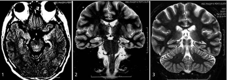
1 2 3
Figs 1, 2 and 3. Axial FLAIR and coronal STIR T2WI show hyper intensity in all left hippocampus and amygdala, with volumetric reduction of them.
patients (25.2%) and 10 (34.5%) of these had ipsila-teral MB alteration. There were no cases of hippo-campal damage with fornix or MB alteration on the normal side.
The duration of the epileptic symptoms were bet-ween 2 days and 44 years (mean age 19.7 years); One patient with MR performed 2 days after the first episode had fornix and MB alteration.
There were 23 (53.4%) epileptic patients with PHGWM abnormalities at the left side, 17 (39.5%) at the right and 3 (7.1%) bilateral; 15 (34.8%) and 5 (11.6%) had fornix and MB reduction, respectively (table). There was no gender preference. The dura-tion of the epileptic symptoms was known in 18 (41.8%) patients and 11 (61.1%) of them with sei-zures for more than 20 years.
PHGWMA, parahippocampal gyrus white matter abnormalities; ES, epilepsy symptoms; F, fornix; MB, mammilary body.
Fig 6 and 7. Right hippocampal sclerosis with mild PHGWM volumetric reduction (arrow).
Fig 8. Coronal STIR T2WI - Bilateral hippocampal atrophy.
Table. Hippocampal sclerosis x PHGWMA
N Right Left Bilateral ES F MB
HS 115 51 (44.3%) 53 (46.0%) 11(9.7%) 19.7 anos 29 (25.2%) 10 (34.5%) 53 (46.1%) 62 (53.9%) PHGWMA 43 (37.3%) 17 (39.5%) 23 (53.4%) 3 (7.1%) 22.2 anos 15 (34.8%) 55 (11.6%) 22 (~50%) 21 (~50%)
DISCUSSION
The importance of a abnormal MR imaging in the diagnosis of HS is its higher rate of surgical cure in temporal lobe epilepsy (70-90%) in contraposition to the 50% in patients with normal MR imaging. The most important take home message to the radiologist is the use of the correct name of this entity, because HS is not mesial temporal sclerosis (MTS) synonymous. We also suggest that the term MTS should be used only in cases that present also PHGWM affection.
There were 43 (37.3%) patients with PHGWM abnormalities in this group of 115 cases. Another important fact was the correlation between parahippocampal gyrus findings and the duration of the epileptic syndrome that was more common in patients that had over 20 years of symptoms.
There was not significant difference in HS side (44.3% at right and 46% at left). PHGWM atrophy was slightly more common on the left side (53.4%). This does not necessarily indicate that side predis-position to PHGWM abnormalities exists. The average duration time of epilepsy in patients with abnormal PHGWM on the left side was longer (26 years) than on those with affection of the right side (16.7 years). Bronen and col.5 studied 9 patients and found
77% of PHGWM abnormalities, what is not in accordance with our findings (37.3%). It has to be considered the small size of their studied population (9 cases) and also the fact that the duration of symptoms is not specified in their paper. Meiners and col.9 studied 80 patients with HS and found 68%
of abnormal PHGWM. In their paper the duration of symptoms is not specified either. The percentages found by Oppenhein and col.8 are in accordance with
our own, despite the smaller number of cases (59)
Fig 10. Right hippocampal sclerosis and parahyppocampal gyrus hyper intensity in T2WI.
Fig 9 A and B. Right hippocampal sclerosis in association with mild parahippocampal gyrus hyper intensity on T2WI (short arrow) and ipsilateral fornix atrophy (thin arrow).
presented by them. Kim and col.7 studied 33 patients
CONCLUSION
It was shown that hippocampal sclerosis was more common in women and the parahippocampal gyrus white matter involvement occurred in 37.3% of the cases. The knowledge about parahippocampal gyrus white matter involvement in temporal lobe epilepsy cases is important and we suggest the use of the term mesial temporal sclerosis only in cases where this gyrus is involved.
J Neuradiol 1995;16:509-515
8. Oppenheim C, Dormont D, Biondi A, et al. Lost of digitations of the hippocampal head on high-resolution fast spin-echo MR: a sign of mesial temporal sclerosis. Am J Neuroradiol 1998;19:457-463. 9. Meiners LC, Witkamp TD, deKort GA, et al. Relevance of temporal
lobe white matter changes in hippocampal sclerosis. Magnetic resonance imaging and histology. Invest Radiol. 1999;34:38-45. 10. Berkovic SF, McIntosh AM, Kalnins RM, et al. Preoperative MRI predicts
outco-me of temporal lobectomy: an actuarial analysis. Neurology 1995;45:1358-1363. 11. Wyler AR, Herman BP, Richey ET, et al. Results of reoperation for failed
epilepsy surgery. J Neurosurg 1989;71:815-819.
12. Oliver A, Tanaka T, Andermann F. Reoperations in temporal lobe epilepsy [abstract]. Epilepsia 1988;29:678.