Coronal and apical leakage analysis
of two different root canal obturation
systems
Abstract: This study compared the coronal and apical leakage of AH Plus with gutta-percha to that of Epiphany with Resilon. Twenty-four single rooted teeth were instrumented and divided into 2 groups accord-ing to the solutions for smear layer removal and the obturation materi-als employed: Group A - 17% EDTA-T and AH Plus with gutta-percha; Group B – primer and Epiphany with Resilon. The Group B specimens were light-cured in the coronal area for 20 s. The external root surfaces were covered with a double layer of ethyl cyanoacrylate, except for the apical foramen and the cavity access. The teeth were immersed in 0.5% methylene blue for 48 h. The specimens were rinsed, dried and axially split for dye penetration measurement with the ImageLab 2.3 software. The t-test showed no signiicant differences for coronal leakage between the groups, but there were signiicant differences for apical leakage be-tween the groups (P < 0.05). AH Plus with gutta-percha and Epiphany with Resilon provided the same coronal seal, whereas Epiphany with Re-silon provided the best apical seal.
Descriptors: Root canal obturation; Dental leakage. Patricia Gimenez Oddoni(a)
Isabel Mello(b) Jeffrey Martin Coil(c)
João Humberto Antoniazzi(d)
(a) DDS, private practice, São Paulo, SP, Brazil.
(b) PhD, private practice, São Paulo, SP, Brazil.
(c) PhD, Assistant Professor, School of Dentistry,
University of British Columbia, Vancouver, BC, Canada.
(d) PhD, Chairman, Department of
Endodontics, School of Dentistry, University of São Paulo, São Paulo, SP, Brazil.
Corresponding author:
Isabel Mello Rua Iliria, 203 São Paulo - SP - Brazil CEP: 04284-060
E-mail: [email protected]
Introduction
The goals of modern endodontic therapy are cleaning, shaping, disinfection and three-dimen-sional obturation of the root canal system that does not allow leakage and promotes periapical heal-ing.1,2 The sealing ability of the sealers used plays an
important role in achieving this goal.
There are different sealers available such as zinc oxide-eugenol, calcium hydroxide, resin, glass iono-mer or silicone.3 Resin-based root canal sealers
as-sociated to gutta-percha have been used for many years with clinical success.4 AH Plus is a well
es-tablished resin-based sealer and has good sealing properties.5-11 Recently, Resilon, a new
thermoplas-tic, illed polymer has been introduced and has the potential to challenge gutta-percha as a root illing material.12,13 This material is used with Epiphany,
a dual-cured resin sealer and a self-etching primer and forms a single monoblock in the root canal sys-tem.14
Several studies have examined whether apical or coronal leakage may adversely affect the success of root canal therapy.4,7,13,15-18 Therefore, leakage
stud-ies on sealers remain important and necessary to de-termine the most suitable obturation materials for achieving therapeutic success. Limited data is avail-able about the sealing properties of Epiphany/Resi-lon. Because of the ability to provide an immediate light-cured seal, Epiphany/Resilon, and purportedly a monoblock obturation, could offer some advan-tages over the traditional obturating materials. The aim of this study was to compare both coronal and apical leakages of a traditional obturating material to those of a new obturation system.
Material and Methods
Sample selection
Twenty-four freshly extracted single-rooted maxillary and mandibular human teeth were radio-graphed to conirm the presence of one canal, ma-ture apex, and absence of resorption or endodontic obturation. They were immersed in saline solution for 72 h to maintain hydration.
Sample preparation
After access preparation, a #10 K-type ile was
used to determine the working length by penetrat-ing the apical foramen and pullpenetrat-ing back 1 mm. The coronal thirds were prepared with #2 and #3 Gates Glidden drills, and the middle and apical thirds were prepared with K-iles (Dentsply-Tulsa Dental, Tulsa, OK, USA) up to ile #45. During instrumentation, 1% sodium hypochlorite and Endo-PTC (urea per-oxide + Tween 80 + carbowax - Farmácia de Ma-nipulação Fórmula e Ação, São Paulo, SP, Brazil) were used as chemical adjuncts. After instrumenta-tion, the canals were rinsed with 5 ml of 1% sodium hypochlorite followed by a inal rinse with 5 ml of distilled water.
Experimental procedures
The teeth were randomly divided into 2 experi-mental groups according to Table 1. For the meth-odology control, two teeth were completely covered with cyanoacrylate and two teeth had their foramen and access cavity sealed.
For smear layer removal in Group A, a inal rinse with 10 ml of 17% EDTA-T (Farmácia de Manipu-lação Fórmula e Ação, São Paulo, SP, Brazil) for 3 min was performed; for Group B, the primer (Pen-tron Clinical Technologies, LLC Wallingford, CT, USA) was applied with a speciic microbrush for 3 min and the canals were dried with paper points. The Epiphany (Pentron Clinical Technologies, LLC Wallingford, CT, USA) and AH Plus (Dentsply-Tul-sa Dental, Tul(Dentsply-Tul-sa, OK, USA) sealers were manipu-lated according to the manufacturers’ instructions. The sealers were placed into the canals using the master point: for group A, gutta-percha (Dentsply-Tulsa Dental, (Dentsply-Tulsa, OK, USA) was used and for group B, Resilon (Pentron Clinical Technologies, LLC Wallingford, CT, USA). The lateral condensa-tion technique was used. The specimens of Group B were light-cured on the coronal area for 20 s. No
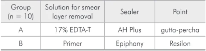
Table 1 - Experimental groups, solutions for smear layer removal and obturation materials used.
Group (n = 10)
Solution for smear
layer removal Sealer Point
A 17% EDTA-T AH Plus gutta-percha
temporary or permanent illing was placed over the root canal obturations.
Microleakage test
The external root surfaces were covered with a double layer of ethyl cyanoacrylate, except for the apical foramen and the access cavity. The teeth were immersed in 0.5% methylene blue for 48 h. After-wards, they were washed in running water for 1 h and dried. The crowns were sectioned from the roots and the roots were split axially into two halves for dye penetration measurement.
Material analysis
The two halves of each specimen were scanned and saved at 300 dpi each. The digital images were analysed by the ImageLab 2.3 software, using a tool that measures the distance between two points. The dye microleakage was evaluated by selecting the deepest longitudinal dye penetration towards both coronal and apical directions. The data was record-ed in a chart and statistically analyzrecord-ed at the 5% level of signiicance.
Results
The means and standard deviations of the groups tested are shown in Table 2. The t-test showed no differences for coronal leakage between the groups, but there were differences for apical leakage be-tween the groups, where Group B exhibited a better sealing (P < 0.05).
Discussion
In the present study, both groups had the same coronal leakage. In spite of the light curing pro-cedure of the coronal area recommended by the
Epiphany manufacturer being right after obtura-tion, Epiphany with Resilon did not provide a better coronal seal than AH Plus with gutta-percha. The manufacturer’s instruction to immediately light-cure the coronal root illing to create a coronal seal may limit the low of the resin sealer for stress re-lief.19 Furthermore, manipulation of the partially
polymerized sealer during condensation may dis-rupt developing bonds between self-etching primer and root dentin.20 Group B exhibited a better apical
sealing than Group A. Other authors, using a silver tracer penetration protocol also found that the qual-ity of the apical seal achieved with the Epiphany with Resilon root illing material was not superior to that of gutta-percha with AH Plus.21 However,
studies employing bacteria showed that gutta-per-cha used with different sealers exhibited a lower sealing ability when compared to that of Epiphany with Resilon.12,13 Such discrepancies are probably
due to differences in methodology and sample size. Analysis of the sealing ability of a new material un-der different conditions is therefore very important. Dye penetration is a methodology widely used for leakage studies and easy to reproduce.5,8,11 The
con-trol groups had expectable results and validated the methodology employed.
To achieve a successful endodontic treatment, the root canal illing material must seal the canal space both apically and coronally to prevent the in-gress of microorganisms or tissue luids into the ca-nal space. Apical and coroca-nal leakages are reported to be important reasons for root canal treatment failure.4,7,13,15-18 The sealing quality of a root canal
illing depends much on the sealing ability of the sealer. Excellent sealing has been achieved by resin-based sealers.5,7-9,11,18 AH Plus, an epoxy-resin based
sealer used with gutta-percha, was selected for this study based on the good sealing results found in the literature.5-11 This sealer also has good adhesion to
dentin which could enhance its sealing ability.3,22
The release of new materials demands research about their different properties. Epiphany is a dual-cured resin sealer which should be used with a self-etching primer. Resilon is a thermoplastic, illed polymer material that could replace gutta-percha. The combination of Resilon, Epiphany and the Table 2 - Means (in pixels) and standard deviations of dye
leakage.
Group Mean and Standard Deviation
Coronal Leakage Apical Leakage
A 27.3000 ± 25.5476a 84.6000 ± 37.7071b
B 46.6000 ± 28.5354a 53.3000 ± 23.1087c
primer has been reported to create a monoblock ob-turation, where the Resilon is chemically bonded to the Epiphany and the Epiphany is bonded to
den-tin.12-14 Neither the gutta-percha or the AH Plus are
capable of chemical bonding.
It is relevant to remember that after root canal preparation, the smear layer should be removed.15,17,23
EDTA-T is a chelator + detergent used for smear lay-er removal and it was used in Group A.24 For Group
B, a speciic self-etching primer was used according to the manufacturer’s instructions. A review of a large number of published leakage studies points to general agreement that leakage occurs between the root illing and the root canal wall.1 Therefore,
any-thing that may inluence the adaptation of the root illing to the canal wall is of great signiicance in de-termining the degree and the extent of leakage, and ultimately the prognosis of the endodontic therapy.
This study reports short-term leakage values, but the results may change with time. Further studies should be done regarding microleakage and other properties of Epiphany/Resilon.
Conclusions
Under the conditions of the present study, both obturation systems did not provide a perfect seal. AH Plus with gutta-percha and Epiphany with Re-silon provided the same coronal sealing, whereas Epiphany with Resilon provided the best apical seal.
Acknowledgements
This research was supported by grants from The State of São Paulo Research Foundation (FAPESP), São Paulo, Brazil, as part of a scientiic initiation program.
References
1. Wu MK, De Gee AJ, Wesselink PR. Leakage of AH26 and Ketac-Endo used with injected warm gutta-percha. J Endod. 1997;23(5):331-6.
2. Yared GM, Bou Dagher F. Sealing ability of the vertical condensation with different root canal sealers. J Endod. 1996;22(1):6-8.
3. Saleh IM, Ruyter IE, Haapasalo M, Orstavik D. The effects of dentine pretreatment on the adhesion of root-canal sealers. Int Endod J. 2002;35(10):859-66.
4. Cohen S, Hargreaves KM. Pathways of the pulp. 9th ed. St.
Louis: Mosby Elsevier; 2006.
5. De Almeida WA, Leonardo MR, Tanomaru Filho M, Silva LAB. Evaluation of apical sealing of three endodontic sealers. Int Endod J. 2000;33(1):25-7.
6. Kayaoglu G, Erten H, Alaçam T, Orstavik D. Short-term an-tibacterial activity of root canal sealers towards Enterococcus faecalis. Int Endod J. 2005;38(7):483-8.
7. Kopper PM, Figueiredo JA, Della Bona A, Vanni JR, Bier CA, Bopp S. Comparative in vivo analysis of the sealing ability of three endodontic sealers in post-prepared root canals. Int Endod J. 2003;36(12):857-63.
8. Mello I, Robazza CR, Antoniazzi JH. Influence of Er:YAG laser irradiation on apical sealing of four different sealers. Braz Dent J. 2004;15(3):190-3.
9. Miletic I, Ribaric SP, Karlovic Z, Silvana J, Bosnjac A, Anic I. Apical leakage of five root canal sealers after one year of storage. J Endod. 2002;28(6):431-2.
10. Saleh IM, Ruyter IE, Haapasalo M, Orstavik D. Survival of Enterococcus faecalis in infected dentinal tubules after root
canal filling with different root canal sealers in vitro. Int En-dod J. 2004;37(3):193-8.
11. Sevimay S, Kalayci A. Evaluation of apical sealing ability and adaptation of two resin-based sealers. J Oral Rehabil. 2005;32(2):105-10.
12. Shipper G, Orstavik D, Teixeira FB, Trope M. An evaluation of microbial leakage in roots filled with a thermoplastic synthetic polymer-based root canal filling material (Resilon). J Endod. 2004;30(5):342-7.
13. Shipper G, Teixeira FB, Arnold RR, Trope M. Periapical inflam-mation after coronal microbial inoculation of dog roots filled with gutta-percha or Resilon. J Endod. 2005;31(2):91-6. 14. Versiani MA, Carvalho-Junior JR, Padilha MI, Lacey S,
Pascon EA, Sousa-Neto MD. A comparative study of physi-cochemical properties of AH Plus and Epiphany root canal sealants. Int Endod J. 2006;39(6):464-71.
15. Cobankara FK, Adanr N, Belli S. Evaluation of the influence of smear layer on the apical and coronal sealing ability of two sealers. J Endod. 2004;30(6):406-9.
16. Cobankara FK, Adanir N, Belli S, Pashley DH. A quantitative evaluation of apical leakage of four root-canal sealers. Int Endod J. 2002;35(12):979-84.
17. Economides N, Kokorikos I, Kolokouris I, Panagiotis B, Go-gos C. Comparative study of apical sealing ability of a new resin-based root canal sealer. J Endod. 2004;30(6):403-5. 18. Leonardo MR, Salgado AA, Da Silva LA, Tanomaru Filho
19. Davidson CL, De Gee AJ. Relaxation of polymerization con-traction stresses by flow in dental composites. J Dent Res. 1984;63(2):146-8.
20. Tay FR, Loushine RJ, Lambrechts P, Weller RN, Pashley DH. Geometric factors affecting dentin bonding in root canals: a theoretical modeling approach. J Endod. 2005;31(8):584-9. 21. Tay FR, Loushine RJ, Weller RN, Kimbrough WF, Pashley
DH, Mak YF et al. Ultrastructural evaluation of the apical seal in roots filled with a polycaprolactone-based root canal filling material. J Endod. 2005;31(7):514-9.
22. Pecora JD, Cussioli AL, Guerisoli DM, Marchesan MA, Sousa-Neto MD, Brugnera Jr A. Evaluation of Er:YAG laser and EDTA-C on dentin adhesion of 6 endodontic sealers. Braz Dent J. 2001;12(1):27-30.
23. Pommel L, About I, Pashley D, Camps J. Apical leakage of four endodontic sealers. J Endod. 2003;29(3):208-10. 24. Scelza MF, Antoniazzi JH, Scelza P. Efficacy of final