http://www.uem.br/acta ISSN printed: 1679-9283 ISSN on-line: 1807-863X
Doi: 10.4025/actascibiolsci.v34i1.7473
Effects of the cafeteria diet on the salivary glands of trained and
sedentary Wistar rats
Aline Rosa Marosti1, Felipe Natali de Almeida2, Solange Marta Franzói de Moraes2, Sonia Lucy Molinari1 and Maria Raquel Marçal Natali1*
1
Departamento de Ciências Morfológicas, Universidade Estadual de Maringá, Av. Colombo, 5790, 87020-900, Maringá, Paraná, Brazil.
2
Departamento de Ciências Fisiológicas, Universidade Estadual de Maringá, Maringá, Paraná, Brazil. *Author for correspondence. E-mail: [email protected]
ABSTRACT. The objective of this work was to study the effect of the aerobic physical training and the cafeteria diet introduced after weaning of Wistar rats and on the morphology of the main salivary glands (parotid, submandibular, sublingual). Male rats after weaning were subjected to the cafeteria diet or the standard rodent chow, and either performed aerobic physical training in a treadmill for 100 days, or did not performed any physical activity. Analyses were done considering the response in body weight, adipose tissues and salivary glands, and the data were submitted to statistical treatment (p < 0.05). The morphological and morphometric analyses of the salivary glands were performed through histological sections stained with hematoxylin and eosin. Despite the normophagic behavior, the rodents fed with the cafeteria diet became obese, with repercussions on parotid gland weight. However, this obesity and/or physical training did not influence the histological organization of the salivary glands. The morphometric analysis of the submandibular glands pointed out a reduction in the levels of serous acinar cells as an effect of the diet and physical training. In conclusion, the parotid and the submandibular glands alter themselves due to the nature and consistency of food present in the cafeteria diet as well as due to the aerobic physical training.
Keywords: salivary gland, cafeteria diet, aerobic physical training, obesity.
Efeitos da dieta de cafeteria sobre as glândulas salivares de ratos Wistar sedentários e
treinados
RESUMO. O objetivo deste trabalho foi avaliar as características morfológicas das glândulas salivares (parótida, submandibular, sublingual) apresentadas por ratos submetidos a treinamento físico aeróbio e dieta de cafeteria após o período de lactação. Ratos machos após a lactação consumiram dieta de cafeteria ou ração-padrão para roedores e realizaram treinamento físico aeróbio em esteira rolante por um período de 100 dias, ou não realizaram nenhuma atividade física. Foram feitas análises sobre a resposta do peso corporal, dos tecidos adiposos e das glândulas salivares e os dados submetidos a tratamento estatístico (p < 0,05). A análise morfológica e morfométrica das glândulas salivares foi realizada a partir de cortes histológicos corados com Hematoxilina e Eosina. Apesar do comportamento normofágico, os roedores alimentados com dieta de cafeteria apresentaram maior quantidade de gordura corporal, com repercussão sobre o peso da glândula parótida. A análise morfométrica das glândulas submandibulares indicou redução na proporção dos ácinos serosos como efeito da dieta de cafeteria e do treinamento físico. No entanto, excesso de gordura corporal, dieta de cafeteria e/ou treinamento físico não influenciou a organização histológica das glândulas salivares. Concluiu-se que as glândulas parótidas e submandibulares são mais influenciáveis em função da natureza e consistência dos alimentos presentes na dieta de cafeteria, assim como do treinamento físico aeróbio.
Palavras-chave: glândulas salivares, dieta de cafeteria, treinamento físico aeróbio, obesidade.
Introduction
Obesity is currently regarded as worldwide public health problem, and is clinically associated with several health problems, such as cardiovascular diseases, orthopedic disorders, cancer, among others, resulting in increased mortality (KRETSCHEMER et al., 2005).
The significant increase in obesity rates indicates that behavioral and environmental aspects play an
associated complications (ESTADELLA et al., 2004).
Adults are able to maintain a constant body weight thanks to a complex system of neural, hormonal and chemical mechanisms that keep the balance between intake and energy loss, within closely regulated limits, with possible contributions to individual differences from genetic factors (ROSADO; MONTEIRO, 2001).
Exposure to fat-rich diets for extended periods results in a positive energy balance and obesity in experimental models with rodents, and is considered a model for human obesity. A diet with high levels of fat and carbohydrates similar to fast food (cafeteria diet) has considerable implications for the onset of obesity, leading to body weight gain, body fat deposition and associated problems, reflecting a non-genetic model of animal obesity (CESARETTI; KOLHMANN JR., 2006).
This diet, in addition to producing obese rodents, imposes on them a distinct eating habit, both in the nature and consistency of the diet - from dry feeds (pellet ration) to a high-fat diet with varied consistency (chocolates, eggs, marshmallow, mocotó, sausages, soft drinks, French bread and bacon).
Saliva production is involved in a wide variety of biological and physiological processes essential to the beginning of digestion in the upper gastrointestinal tract, including lubrication, purification, enzyme digestion, as well as preservation of the teeth and oral mucosa. Any reduction in food particle size and changes in its consistency can significantly alter the salivary secretion process (GAVIÃO; BILT, 2004).
Saliva is derived from secretions of the mucous and serous cells that make up the secretory portion of the salivary glands. The mucous cells produce a viscous liquid rich in mucin, and serous cells produce a secretion that contains enzymes and other proteins. According to Banks (1992), the type of diet influences the amount and nature of secretion.
Dawes (1981) believes there is an influence of physical activity over salivary secretion according to the intensity of the exercise, altering the levels of water, ions and hormones present in saliva. However, reports on morphologic changes in the structures that secrete saliva are scarce.
The objective of this work was to evaluate the morphological characteristics of salivary glands (parotid, submandibular, sublingual) as observed in rats subjected to aerobic physical training and cafeteria diet after weaning.
Material and methods
Twenty-eight male Wistar rats, 21 days old, were adapted to a treadmill and randomly divided into four groups (with no significant difference in initial body weight for the different groups), according to diet and physical training conditions: sedentary control (SC), sedentary cafeteria (SCa), trained control (TC), and trained cafeteria (TCa), following the experimental protocol until 120 days. The animals were housed in the vivarium of the Department of Physiological Science at the State University of Maringá, in polypropylene boxes under a 12/12 photoperiod (light cycle beginning at 6 a.m. and dark cycle beginning at 6 p.m.), controlled using a Brasfort® timer and with
temperature (20 to 24ºC) controlled as well. The entire experimental protocol that involved the use of animals was approved by the Ethics Committee for Animal Research at the State University of Maringá.
Diet
The animals in the control groups (SC and TC) were fed with Nuvilab® chow (composition
according to the recommendations of the National Research Council and National Institute of Health - USA) and water ad libitum. The animals in the cafeteria groups (SCa and TCa) received a high-fat diet containing food items such as: potato chips, chocolates, marshmallow, mocotó, sausages, French bread, bacon, cookies and soft drinks, in addition to regular feed and water. Food intake by both groups was subject to daily control, featuring analyses of intake in grams, calories and macronutrient percentage - the latter two items were calculated according to information provided by the product manufacturers.
Physical training
Animals in groups TC and TCa were subjected to aerobic physical training starting right after weaning (21 days) and lasting for 14 weeks, during which the animals ran on a treadmill (Inbrasport®). The training
protocol, adapted from Iwamoto et al. (1999), consisted of five sessions of physical training a week, starting at an initial speed of 8.6 m min.-1 for 15 minutes, with
increments in running time during each session until reaching one hour, after which only the running speed was changed, reaching approximately 20 m min.-1
Mean intensity was approximately 55 - 75% of maximum VO2 of the animals. We observed the effect
Tissue sampling
At 120 days of age, the animals were weighed and euthanized using a lethal dose of the anesthetic sodium thiopental - Thionembutal® (40 mg kg-1,
intraperitoneal injection), followed by measurement of naso-anal length to determine the Lee index [body weight1/3(g)/naso-anal length (cm) x 1000]
and removal and weighing of the periepididymal and retroperitoneal fat pads, as well as collection and weighing of the parotid, submandibular and sublingual glands.
The salivary glands were fixed in Bouin’s solution, dehydrated in a series of increasing alcohol concentrations, diaphanized in xylol and included in paraffin to be sliced in 6-μm thick semi-serial histological sections stained with hematoxylin and eosin (H.E). For the morphometric analysis of the submandibular and sublingual glands, the areas occupied by mucous and serous were estimated from 25 images/gland/animal, totaling 175 images/gland/group, captured and analyzed in a system of computer image analysis (Image-Pro Plus 4.5 - Media Cibernetics®). The parotid glands were
submitted only to morphological analyses, as their area was occupied predominantly by serous acinar cells. Acini differentiation was based on morphological and staining characteristics of those cells - mucous acinar cells were characterized by the presence of cells featuring a nucleus peripheral and flattened and apical cytoplasm vacuolized, whereas serous acinar cells featured cells with nucleus rounded and central and basolateral cytoplasm basophilic.
The statistical evaluation of the results was performed using Tukey’s test with a significance level of 95% (p < 0.05) using Prism, v.3.0 software (GraphPad, USA).
Results and discussion
Table 1 shows the percentage values of macronutrients (carbohydrates, proteins and lipids) and diet intake of the control groups (sedentary and trained) and cafeteria groups (sedentary and trained), showing that the cafeteria diet was high-fat and high-protein compared to the control diet, which characterizes the eating pattern adopted by a large part of the world’s population and is widely related to excess body fat (MONTEIRO et al., 2000).
Given that the animals ingested the same amount of food, we observed that the fundamental factor for excess weight and adiposity shown by group SCa was the significantly higher caloric intake by the cafeteria groups vs. the control groups (p < 0.05), as shown in Table 1.
Table 1. Food intake and caloric intake values, with values expressed as mean ± SEM and percentage of macronutrients (carbohydrates, proteins and lipids) present in the diets of animals belonging to the control groups (SC and TC) and cafeteria groups (SCa and TCa) (n = 7).
Parameters SC SCa TC TCa
Carbohydrates (%) 61 43.4 61 43.8
Proteins (%) 26.8 12.14 26.8 12.5
Lipids (%) 12.2 44.46 12.2 43.7
Intake (g) 26.44 ± 0.48a 24.29 ± 0.38a 27.10 ± 0.28a 23.65 ± 0.36a
Intake (Kcal) 73.40 ± 1.47a 85.76 ± 1.52b 75.89 ± 0.78a 82.49 ± 1.27b
*different letters in the same row indicate significant differences (p < 0.05).
However, caloric intake is not the only factor to influence body weight and fat. These are dependent on the balance between the amount of energy consumed and spent (KRETSCHEMER et al., 2005), giving significant importance to physical training, as it makes the body spend energy. This would be the fundamental aspect that led group TCa to show control over the increase in body weight, which remained similar to that of the control groups (SC and TC), as well as over adiposity, which was higher than the control groups but lower than that of group SCa (Table 2). Studies show that physical training increases the mobilization capability of the existing fat through the increase in lipolysis, which could explain the reduction in relative weight of the adipose tissue of the animals subjected to the cafeteria diet, without changes in diet intake (EGUCHI et al., 2008).
But, because the control groups (SC and TC) did not show significant changes with regard to body weight and adiposity (Table 2) and showed reduced adiposity compared to group TCa, we can infer that the diet appears as a primary factor in the energy balance. Thus, we can say that the relationship between inadequate diet and excess adiposity is confirmed.
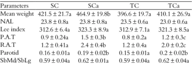
Table 2. Lee index [body weight1/3(g)/naso-anal length (cm) x
1000]; weight (g) of the retroperitoneal adipose tissue (R.A.T.) and periepididymal adipose tissue (P.A.T.) corrected for 100 g of body weight and weight of the parotid, submandibular and sublingual (SbMd/SbLg) glands of rats from the control groups (SC and TC) and cafeteria groups (SCa and TCa) (n = 7).
Parameters SC SCa TC TCa
Mean weight 421.5 ± 21.7a 464.9 ± 19.8b 396.6 ± 19.7a 410.1 ± 26.9a NAL 23.8 ± 0.8a 23.8 ± 0.8a 23.5 ± 0.6a 23.0 ± 0.6a Lee index 312.6 ± 6.4a 323.3 ± 8.9a 312.9 ± 7.1a 321.3 ± 8.5a P.A.T 0.9 ± 0.24a 1.5 ± 0.3b 0.8 ± 0.2a 1.2 ± 0.3c R.A.T 1.2 ± 0.41a 2.4 ± 0.4b 1.2 ± 0.4a 2.0 ± 0.2c Parotid 0.16 ± 0.01a 0.19 ± 0.02b 0.15 ± 0.01a 0.2 ± 0.02b SbMd/SbLg 0.59 ± 0.04a 0.62 ± 0.01a 0.59 ± 0.04a 0.62 ± 0.04a
*different letters in the same row indicate significant differences (p < 0.05).
high-fat diet did not show changes in the weight of any of the glands. The change in weight of the parotids in the cafeteria groups (SCa and TCa) was likely caused by the change in diet, as changes in the consistency and nature of the food items influence salivary secretion (BANKS, 1992). According to Dodds et al. (1991) and Leal et al. (2003), rats subjected to a liquid diet show atrophied parotids, and the parotids hypertrophy in animals on diets that require more chewing, followed by an increase in salivary flow with higher protein concentration. The cafeteria diet imposed on those animals possibly altered the chewing process, leading to higher production of serous secretion and consequently to an increase in gland size, and/or stimulated saliva production because of higher palatability.
Despite the significant increase in the weight of the parotid gland, there were no changes to its morphology, which remained as described in the literature, regardless of diet and/or training (Figure 1). The parotid is a compound acinar gland, with a secretory portion formed only by serous cells in pets, humans and rodents (BANKS, 1992; JUNQUEIRA; CARNEIRO, 2008). Given that the acinar cells were exclusively serous, it was not necessary to evaluate this gland morphometrically.
Figure 1. Parotid gland of Wistar rats from the Sedentary Control (SC) group. H.E.
However, because the gland was larger for the cafeteria group (SCa and TCa), we can infer that the diet caused higher production of serous secretion,
but studies on salivary flow and composition are necessary to confirm this hypothesis.
The submandibular glands of the animals showed a characteristic pattern in all analyzed groups (Figure 2), with the presence of serous and mucous acinar cells, ducts, connective tissue and blood vessels. According to Junqueira and Carneiro (2008), the submandibular gland is classified as compound tubulo-acinar gland, because its secretory portion is made up of mucous and serous cells.
Figure 2. Submandibular gland of Wistar rats from the Sedentary Control (SC) group. H.E.
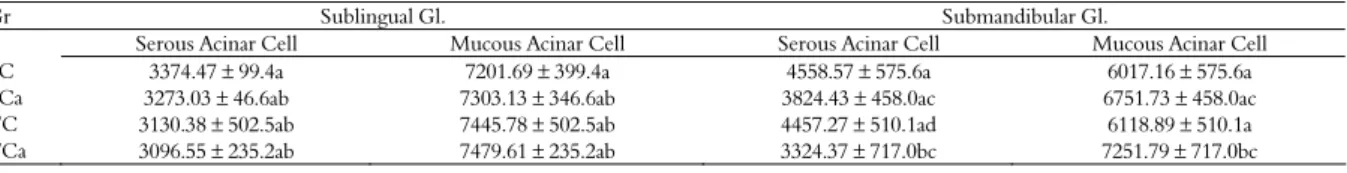
The area occupied by mucous and serous acinar cells in the submandibular and sublingual salivary glands is shown on Table 3.
The morphometric analysis of the submandibular gland revealed that group TCa differed from groups SC and TC in the makeup of serous and mucous acinar cells, as shown on Table 3, which allowed us to infer that the cafeteria diet in conjunction with physical training has a reducing effect in the quantity of serous acinar cells for that gland. Serous acinar cells are responsible for protein synthesis, storage and secretion, especially of amylase. Considering that type of secretion, the reduction in serous acinar cells can be explained by the lower amount of carbohydrates present in the diet of animals from the cafeteria group. The decrease in the intake of these foods combined with physical training led to the production of a more mucous secretion, which can be explained by the increase in mucous acinar cells. These acinar cells have lower enzyme content and high levels of glycoproteins.
Table 3. Morphometrics of the submandibular and sublingual salivary glands of rats from the control groups (SC and TC) and cafeteria groups (SCa and TCa) (n = 7). The area value of the acinar cells was measured from the total area of the image (10576.172 µm2).
Gr Sublingual Gl. Submandibular Gl.
Serous Acinar Cell Mucous Acinar Cell Serous Acinar Cell Mucous Acinar Cell
SC 3374.47 ± 99.4a 7201.69 ± 399.4a 4558.57 ± 575.6a 6017.16 ± 575.6a
SCa 3273.03 ± 46.6ab 7303.13 ± 346.6ab 3824.43 ± 458.0ac 6751.73 ± 458.0ac
TC 3130.38 ± 502.5ab 7445.78 ± 502.5ab 4457.27 ± 510.1ad 6118.89 ± 510.1a
TCa 3096.55 ± 235.2ab 7479.61 ± 235.2ab 3324.37 ± 717.0bc 7251.79 ± 717.0bc
Physical exercise can lead to changes in several saliva components, including immunoglobulins, hormones, lactate, proteins and electrolytes, mediated especially by the influence that exercise has on the autonomous nervous system, while the sympathetic nervous system tends to stimulate mucous secretion, and the parasympathetic nervous system stimulates serous secretion (AIRES, 2008; CHICHARRO et al., 1998).
Saliva tends to present different aspects according to secretory flow, becoming more fluid with a higher secretory flow. Thus, it is expected that during physical exercise, because the sympathetic system is predominant, saliva will tend to be more fluid due to the stimulation of the secretory flow; but in a second moment, due to the resulting vasoconstriction, secretory flow is reduced and saliva tends to become more viscous, especially because it is rich in mucus, leading to the sensation of “dry mouth”. However, another morphological aspect was expected, as one of the effects of continuous physical exercise (chronic effect) would be an increase in parasympathetic activity of the body at rest and because that part of the autonomous nervous system is responsible for the stimulus of fluid salivary secretion (AIRES, 2008).
Thus, apparently the morphological aspect of the submandibular gland underwent a change in its serous levels as a compensatory mechanism to control salivary fluidity, but new studies should be performed with the objective of improving data on the influence of aerobic physical exercise on the salivary glands, both in their morphological and functional aspects, mediated by the autonomous nervous system.
The sublingual gland maintained its characteristic morphology in all groups, with the presence of mucous and serous acinar cells (Figure 3).
Figure 3. Sublingual gland of Wistar rats from the Sedentary Control (SC) group. H.E.
Mucous acinar cells were predominant in all groups, regardless of diet and/or physical training.
This gland is classified as compound tubulo-acinar, but does not feature exclusively serous acinar cells, as its cells are always grouped in curved positions at the edge of the mucous acinar cells (JUNQUEIRA; CARNEIRO, 2008). The sublingual gland also has well-evidenced striated ducts, intercalated and excretory ducts, and a main excretory duct (TAGA; SESSO, 2002).
The morphometric analysis of this gland did not show any significant difference among the groups, which indicates that neither variable (diet or physical training) had any influence on the levels of serous and mucous acinar cells in the gland.
Conclusion
The cafeteria diet is an experimental model that results in a positive energy balance, leading to obesity in rats. The animals subjected to this diet showed an increase in body weight and adipose tissue. Thus, this model is quite useful, as it resembles obesity in humans.
Moreover, we observed that changes in the nature and consistency of foods interfere in the morphological response of the parotid gland, as did physical training, which led to changes in the submandibular glands, which can result in altered patterns of salivary secretion.
References
AIRES, M. M. Fisiologia. 3. ed. Rio de Janeiro: Guanabara Koogan, 2008.
BANKS, W. J. Histologia veterinária aplicada. 2. ed. São Paulo: Manole, 1992.
CESARETTI, M. L. R.; KOLHMANN JR., O. Modelos experimentais de resistência a insulina e obesidade: lições aprendidas. Arquivos Brasileiros de Endocrinologia e Metabologia, v. 50, n. 2, p. 190-197, 2006.
CHICHARRO, J. L.; LUCIA, A.; PEREZ, M.; VAQUERO, A. F.; URENA, R. Saliva composition and exercise. Sports Medicine, v. 26, n. 1, p. 17-27, 1998. DAWES, C. The effects of exercise on protein and electrolyte secretion in parotid saliva. Journal of Physiology, v. 320, p. 139-148, 1981.
DODDS, M. W. J.; HSIEH, S. C.; JOHNSON, D. A. The effect of increased mastication by daily gum-chewing on salivary gland putput and dental plaque acidogenicity.
Journal of Dental Research, v. 70, n. 12, p. 1474-1478, 1991.
ESTADELLA, D.; OYAMA, L. M.; DAMASO, A. R.; RIBEIRO, E. B.; OLLER DO NASCIMENTO, C. M. Effect of patalable hyperlipidic dieta on lipid metabolism of sedentary an exercised rats. Nutrition, v 20, n. 2, p. 218-224, 2004.
FENNING, A.; HARRISON, G.; DWYER, D.; ROSE´MEYER, R.; BROWN, L. Cardiac adaptation to endurance exercise in rats. Molecular Cell Biochemestry, v. 251, n. 1-2, p. 51-59, 2003.
GAVIÃO, M. B. D.; BILT, A. V. Salivary secretion and chewing: stimulatory effects from artificial and natural foods. Journal of Applied Oral Science, v. 12, n. 2, p. 159-163, 2004.
INOUE, S.; CAMPFIELD, L. A.; BRAY, G. A. Comparison of metabolic alterations in hypothalamic and high fat diet-induced obesity. American Journal of Physiology, Regulatory, Integrativy and Comparative Physiology, v. 233, n. 3, p. 162-168, 1977. IWAMOTO, J.; YEH, J. K.; ALOIA, J. F. Differential effect of treadmill exercise on three cancellous bone sites in the young growing rat. Bone, v. 24, n. 3, p. 163-169, 1999. JUNQUEIRA, L. C.; CARNEIRO, J. Histologia básica. 11. ed. Rio de Janeiro: Guanabara Koogan, 2008.
KRETSCHEMER, B. D.; SCHELLING, P.; BEIER, N.; LIEBSCHER, C.; TREUTEL, S.; KRUGER, N. Modulatory role of food, feeding regime and phsycal
exercise on body weight and insulin resistance. Life Sciences, v. 76, n. 14, p. 1553-1571, 2005.
LEAL, S. C.; TOLEDO, O. A.; BEZERRA, A. C. B. Morphological alterations of the parotid gland of rats maintained on a liquid diet. Brazilian Dental Journal, v. 14, n. 3, p. 172-176, 2003.
MONTEIRO, C. A.; MONDINI, L.; COSTA, R. B. L. Mudança na composição e adequação da dieta familiar nas áreas metropolitanas do Brasil (1988-1996). Revista Saúde Pública, v. 34, n. 3, p. 251-258, 2000.
ROSADO, E. L.; MONTEIRO, J. B. R. Obesidade e a substituição de macronutrientes da dieta. Revista de Nutrição, v. 14, n. 2, p. 145-152, 2001.
TAGA, R.; SESSO, A. Ultrastructure of the rat sublingual gland during period of high proliferative activity in postnatal development. Brazilian Journal of Morphological Sciences, v. 19, n. 2, p. 55-62, 2002.
Received on June 27, 2009. Accepted on November 18, 2009.