Increased Brain Lactate During Depressive Episodes
and Reversal Effects by Lithium Monotherapy in
Drug-Naive Bipolar Disorder
A 3-T
1H-MRS Study
Rodrigo Machado-Vieira, MD, PhD,*
†‡
Marcus V. Zanetti, MD, PhD,*
†
Maria C. Otaduy, PhD,§
Rafael T. De Sousa, MD, PhD,*
†
Marcio G. Soeiro-de-Souza, MD, PhD,* Alana C. Costa,*
Andre F. Carvalho, MD, PhD,|| Claudia C. Leite, PhD,§ Geraldo F. Busatto, MD, PhD,¶
Carlos A. Zarate Jr, MD,
‡
and Wagner F. Gattaz, MD, PhD*
†
Abstract:
Objective:Mitochondrial dysfunction and energy metabolism impair-ment are key components in the pathophysiology of bipolar disorder (BD) and may involve a shift from aerobic to anaerobic metabolism. Mea-surement of brain lactate in vivo using proton magnetic resonance spectros-copy (1H-MRS) represents an important tool to evaluate mitochondrial and metabolic dysfunction during mood episodes, as well as to monitor treatment response. To date, very few studies have quantified brain lactate in BD. In ad-dition, no study has longitudinally evaluated lactate using1H-MRS during depressive episodes or its association with mood stabilizer therapy. This study aimed to evaluate cingulate cortex (CC) lactate using 3-T1H-MRS
during acute depressive episodes in BD and the possible effects induced by lithium monotherapy.
Methods:Twenty medication-free outpatients with short length of BD (80% drug-naive) in a current major depressive episode were matched with control subjects. Patients were treated for 6 weeks with lithium monotherapy at therapeutic doses in an open-label trial (blood level, 0.48 ± 0.19 mmol/L). Cingulate cortex lactate was measured before (week 0) and after lithium therapy (week 6) using1H-MRS. Antidepres-sant efficacy was assessed with the 21-item Hamilton Depression Rating Scale as the primary outcome.
Results:Subjects with BD depression showed a significantly higher CC lactate in comparison to control subjects. Furthermore, a significant de-crease in CC lactate was observed after 6 weeks of lithium treatment com-pared with baseline (P= 0.002). CC Lactate levels was associated with family history of mood disorders and plasma lithium levels.
Conclusions:This is the first report of increased CC lactate in patients with bipolar depression and lower levels after lithium monotherapy for 6 weeks. These findings indicate a shift to anaerobic metabolism and a role for lactate as a state marker during mood episodes. Energy and redox dysfunction may represent key targets for lithium’s therapeutic actions. Key Words:bipolar disorder, depression, imaging, lactate, lithium, treatment
(J Clin Psychopharmacol2017;37: 00–00)
E
nergy metabolism dysfunction has been considered a key com-ponent of the pathophysiology of bipolar disorder (BD).1,2 Ev-idence for this metabolic dysfunction includes lower brain intracellular pH and phosphocreatine in BD.3Brain lactate has a key role in neural energy metabolism, and its altered concentra-tions may represent a less severe form of mitochondrial and met-abolic dysfunction.4,5In addition, a different line of evidence showing increased oxidative stress6,7and impaired expression of mitochondrial genes in BD8,9provides further support to the role of mitochondrial and redox dysfunction leading to a shift to-ward anaerobic metabolism and associated elevated risk for cellular injury.10,11Proton magnetic resonance spectroscopy (1H-MRS)
pro-vides a noninvasive technique to measure brain lactate in vivo in psychiatric research. In BD, previous studies during mood episodes (manic and depressed) and euthymia have shown mixed results.5,12,13 In a cross-sectional study, Dager et al5
found an increase in gray matter (GM) lactate concentrations in medication-free BD patients with long-term illness and pre-dominantly in a depressed or mixed state. Brady et al13found similar lactate levels in the anterior cingulate cortex (CC) dur-ing mania (n = 15) compared with control subjects. Seven med-icated patients underwent a subsequent follow-up scan (average follow-up time, 21.1 months) while in euthymia and showed lower anterior CC lactate compared with matched control sub-jects. This is a key area involved in mood regulation and is a tar-get for lithium's biological effects.14A recent study emphasized that lactate is primarily localized in the CC (and caudate) of sub-jects with BD.15
Lithium is the mainstay of pharmacotherapy for acute mood episodes, prophylactic treatment, and suicide prevention in BD.16
Data from the European drug surveillance program describe that lithium is the most frequently prescribed agent for bipolar depres-sion in combined therapy (33%).17Lithium modulates key
mito-chondrial proteins and prevents and/or reverses DNA damage, free radical formation, and lipid peroxidation.16,18,19 Lithium
has also been shown to improve synaptic strength, cellular From the *Laboratory of Neuroscience (LIM-27), Institute and Department of
Psychiatry, University of São Paulo;†Center for Interdisciplinary Research on Applied Neurosciences (NAPNA), University of São Paulo, São Paulo, Brazil; ‡Experimental Therapeutics and Pathophysiology Branch, National Institute of Mental Health, NIH, Bethesda, MD; and §Laboratory of Magnetic Resonance in Neuroradiology (LIM-44), Institute and Department of Radiology, University of São Paulo, São Paulo;‖Department of Clinical Medicine and Translational Psychiatry Research Group Faculty of Medicine, Federal University of Ceara, Fortaleza; and ¶Laboratory of Psychiatric Neuroimaging (LIM-21), Institute and Department of Psychiatry, University of São Paulo, São Paulo, Brazil. Received February 29, 2016; accepted after revision October 11, 2016. Reprints: Rodrigo Machado-Vieira, MD, PhD, National Institute of Mental
Health, National Institute of Health, 10 Center Dr, Clinical Center, Unit 7SE, Rm. 7-5341, Bethesda, MD (e‐mail: [email protected]; [email protected]).
This study was sponsored by São Paulo Research Foundation (FAPESP, Brazil, 2009/14891-9, RM-V). The Laboratory of Neuroscience (LIM-27) is also supported by the Associação Beneficente Alzira Denise Hertzog da Silva (ABADHS) and JNK Empreendimentos e Incorporações.
Copyright © 2016 Wolters Kluwer Health, Inc. All rights reserved. ISSN: 0271-0749
resilience, and glial function.11,20 Interestingly, in a cross-sectional MRS study, Friedman et al21described similar brain lactate concentrations in BD subjects taking lithium compared with healthy controls. In mania, Kim et al12observed a
signifi-cant decrease in midfrontal cortex lactate in subjects with BD treated with quetiapine for 12 weeks; this finding was associated with the antimanic response.
In the present study, CC lactate was evaluated in medication-free BD subjects in a major depressive episode before and after 6 weeks of lithium monotherapy using 3-T 1H-MRS.
Further-more, although a few studies evaluated brain lactate in BD, no study has either longitudinally evaluated lactate using1H-MRS during a specific mood state (mania or depression) or assessed its association with lithium monotherapy treatment response in drug-naive BD subjects.
We hypothesized that BD subjects would have increased lactate in the CC, as measured by multivoxel 3-T1H-MRS dur-ing bipolar depression compared with control subjects and that reductions in lactate with lithium treatment would be associated with treatment response.
METHODS
Participants
Fifty-nine individuals prescreened by phone interviews were evaluated at the outpatient clinic of the Mood Disorders Group (LIM-27), Department and Institute of Psychiatry, University of São Paulo. Twenty-six subjects fulfilled criteria for the present study and were enrolled. Six BD subjects were excluded because of the following reasons: 2 patients dropped out before the com-pletion of a 6-week follow-up, whereas 4 subjects had technical is-sues (data collection) at either baseline or follow-up 1H-MRS scan, which limited a complete postprocessing data analysis. Thus, our final sample consisted of 20 medication-free subjects (80% treatment naive) with BD in a major depressive episode (16 women and 4 men, aged 22-43 years old) who completed the 6-week lithium trial.
Subjects with BD in a depressive episode were included ac-cording to the following criteria: (a) age between 18 and 45 years, (b) diagnosis of BD I or II in a current major depressive episode according to the Diagnostic and Statistical Manual of Mental Disorders, Fourth Edition(DSM-IV), (c) score greater than 17 in the 21-item Hamilton Depression Rating Scale (HDRS),22
(d) no previous use of lithium (lifetime) and absence of the use of any psychiatric medication or drugs with central nervous sys-tem (CNS) effects for at least 6 weeks, and (e) illness duration of no more than 5 years. Exclusion criteria included rapid cycling BD; current substance (including alcohol) abuse or dependence, with the exception of tobacco; previous electroconvulsive ther-apy; presence of neurological disorders or any medical disorder that could affect the CNS; mental retardation; medications with CNS effects; vitamins; and contraindications to magnetic reso-nance imaging scanning.23
Patients were evaluated with a semistructured clinical inter-view and blood tests (including complete blood count, electro-lytes, and renal and thyroid function). All patients were started on lithium carbonate (450 mg/d), and a systematic follow-up was carried out for 6 weeks. Visits for clinical assessment and plasma lithium monitoring in the plasma were performed at weeks 1, 2, 4, and 6. Subsequent dose adjustment was allowed, based on individual clinical efficacy and aiming to reach therapeutic levels of lithium (0.4–1.0 mmol/L). The1H-MRS scanning was
performed at baseline (week 0) and endpoint (week 6) in BD patients and only at baseline in the matched control group.
The healthy volunteers group included 16 subjects (9 women and 7 men, aged 20-43 years old), all free of any psychiatric dis-order (based onDSM-IVcriteria) and with no history of mental disorders among first-degree relatives. Volunteers were recruited through advertisement in the local community. The local institu-tional review board approved this study, and all subjects provided informed written consent prior to participating in the study. This study was part of a multimodal investigation to evaluate central and peripheral therapeutic targets of lithium’s antidepressant actions in BDs.
Clinical Measures
Experienced psychiatrists performed all clinical assessments. Psychiatric evaluation was performed in both patients and control subjects using the Structured Clinical Interview forDSM-IV.24A
medical history was obtained directly from each participant and/ or family member. At each follow-up visit (weeks 1, 2, 4, and 6), the severity of depressive and manic symptoms was assessed using, respectively, the HDRS and the Young Mania Rating Scale.25
Clin-ical response was defined as a decrease of 50% or greater in the HDRS, whereas remission was defined as an HDRS and Young Mania Rating Scale score of 8 or less. The global functioning at baseline and endpoint (improvement) was also assessed using the Clinical Global Improvement (CGI) scale.26Interrater reliability was greater than 0.9 for all rating scales.
1H-MRS Scan and Data Processing
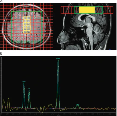
All1H-MRS sequences were performed using a 3-T magnet (Intera Achieva; Philips Medical Systems, Best, the Netherlands) and an 8-channel head coil. Metabolite concentrations of lactate were obtained using a 2-dimensional (2D) MRS imaging (MRSI) Point Resolved Spectroscopy sequence (time to echo [TE]/repetition time = 288/1500 milliseconds) with a slice thickness of 2 cm, field of view = 20 cm, and a matrix of 2020, resulting in indi-vidual voxel sizes of 2 cm3. Twelve individual voxels (2 columns of 6 voxels each) placed on the CC, as shown in Figure 1, were selected for analysis. The MRS grid was placed on an axial slice just above the corpus callosum, following the anterior commissure-posterior commissure angulation. The 12 individual spectra within the selected region of interest (ROI) were averaged and processed as a single spectrum. This method was shown to obtain significantly improved lactate measurement as compared with av-eraging chemical concentrations derived from the fitting of indi-vidual voxels in the ROI.27 Spectral peak quantification was
performed using the Spectroscopy Tool of the Extended MR Workspace R.2.6.3.5 (Philips Medical Systems) after phase ad-justment, exponential line broadening of−1 Hz, and zero filling. The spectrum was fitted to the sum of 4 signals: choline (Cho), creatine (Cr), N-acetyl-aspartate (NAA), and the lactate double peak (Fig. 2). Proton magnetic resonance spectroscopy was ac-quired at a long TE (288 milliseconds), to ensure that lipid and macromolecule signal were fully relaxed and did not overlap with the lactate signal. Lactate was quantified by the integral ratio of the lactate peak with the creatine peak. Quality criteria used for spectra inclusion were based on signal-to-noise ratio (SNR) mea-surements. Because lactate appears as an isolated peak with no overlap with other metabolite signals on a spectrum with a TE of 288 milliseconds, SNR is a convenient rejection criterion.28To
in-vestigate the brain tissue composition of the ROI, 3D volumetric im-ages were obtained using the 3D-T1FFE technique (FA = 8°; TE/ repetition time/TI = 3.2/7/900 milliseconds) with isotropic resolution of 1 mm3. Briefly, the brain tissue was evaluated using the brain
brain segmentation tool FAST, both apart of the FSL suite (http:// www.fmrib.ox.ac.uk/fsl). Finally, the ROI was overlaid on the seg-mented image using a Python-based script developed in-house, and percentages of WM, GM, and CSF were calculated for each ROI. The scanner used for this study is submitted to a weekly MRS quality control performed using a phantom with an aqueous solution of brain metabolites. The focus of this study was lactate. The long-TE MRSI acquisition used was chosen on purpose to measure a significantly high lactate signal, which otherwise is not observable in a single-voxel conventional MRS spectrum. The disadvantage of this long-TE acquisition is that short T2 me-tabolites, such as myo-inositol and glutamate + glutamine, are not measurable. For this purpose, we conducted a different MRS acquisition in the same population, which has already been published29and reporting results on all other metabolites. Statistical Analysis
The Kolmogorov-Smirnov test was used to verify the normality of the data within each study group. Between-group comparisons of continuous variables with a Gaussian distribution were performed with unpaired 2-tailed Student t test, whereas the Mann-WhitneyUtest was used for nonparametric data. Theχ2
test was used for the comparison of categorical variables. Changes in CC lactate and depressive symptoms over time were assessed with the paired-samplesttest (normal distribution) or with the nonparametric Wilcoxon rank sum test (nonnormal distribution). Specifically for the comparison of CC lactate between BD
patients both at baseline and at week 6 as well as control subjects, the analysis of covariance was carried out controlling the CSF fraction of the voxel as a covariate. Mixed-effects models were also used to investigate whether clinical variables and outcomes were associated with changes in brain lactate (dependent variable) after 6 weeks of lithium treatment. Finally, the correlation between brain lactate and clinical variables was evaluated using partial cor-relations controlling for the CSF fraction of the voxel. Pearson correlation coefficients or Spearman tests were used for additional correlations. All statistical analyses were performed using SPSS 21.0 (Chicago, IL) with a significance level set atP< 0.05 (2-tailed).
RESULTS
Demographic and Clinical Data
Demographic and clinical data for subjects with BD and con-trol subjects are summarized in Table 1. No significant difference in age (P= 0.29) or sex (P= 0.12) was observed when comparing BD subjects and control subjects. All patients were free of any psychotropic medication for at least 6 months prior to the enroll-ment in the study, and 16 (80%) had never received treatenroll-ment with any mood stabilizer or antipsychotic agent (treatment naive). Mean lithium levels at week 6 were 0.48 ± 0.19 mmol/L. All pa-tients were using therapeutic doses of lithium (600–900 mg/d; mean dose, 671.1 ± 115 mg). All patients had less than 5 years of illness duration (mean, 36.0 ± 18.7 months).
Furthermore, there were significant changes in HDRS (baseline, 22.40 ± 3.01; endpoint, 7.90 ± 6.23;P< 0.001) and CGI scores (baseline, 4.00 ± 0.32; endpoint, 2.15 ± 0.87; P< 0.001) (Table 1). Twelve BD patients (60%) achieved remis-sion at endpoint. No treatment-emergent affective switches or dropouts were observed.
CC Lactate in BD Versus Control Subjects
Subjects with BD in a depressive episode showed at base-line increased lactate in the CC compared with control subjects (F1= 4.32,P= 0.04; general linear model with the CSF fraction
of the voxel as a covariate) (Table 2). Cerebrospinal fluid fraction of the ROI also did not significantly vary across groups (BD pa-tients = 0.17 ± 0.05 vs control subjects = 0.19 ± 0.06;t34= 1.16,
P= 0.25). No difference between groups was also observed for WM (t23= 0.9,P= 0.37) and GM (t23= 1.32,P= 0.2) fractions
of the ROI. No changes in CC lactate levels were observed when comparing drug-naive versus drug-free BD subjects (data not shown). Cingulate cortex lactate at baseline was not associ-ated with any clinical outcome (total improvement, response, or remission).
CC Lactate in BD at Baseline Compared With Post–Lithium Treatment
A significant reduction in CC lactate was observed in BD patients after 6 weeks of lithium treatment compared with base-line (t19=−3.52,P= 0.002) (Table 2, Fig. 2). After lithium treat-ment, BD patients showed no difference in CC lactate when compared with control subjects (F1 = 2.58,P= 0.11). When
comparing patients pre–versus post–lithium treatment, no changes in CSF (t19=−0.98,P= 0.33), GM (t19= 1.64,P= 0.11), or WM
(t19=−1.145,P= 0.16) were observed.
Predictors of Response
Baseline CC lactate did not correlate with any demographic and clinical variable described in Table 1 (partial correlation con-trolled for the CSF fraction of the voxel) (all not statistically sig-nificant), except for a positive association with family history of mood disorders (P= 0.02,r= 0.46). No significant correlations were observed between changes in CC lactate and HDRS and CGI scores over time or at endpoint (all not statistically signifi-cant). Mixed-effects model analysis revealed no significant asso-ciation between remission status at endpoint and the change in CC lactate over time (F = 1.62,P= 0.211).
Associated Biological Findings
Changes in lactate were not associated with clinical improve-ment (HDRS score change) (P= 0.5). Plasma lithium at endpoint was not associated with lactate at endpoint (week 6) (P= 0.2) but was negatively associated with changes in lactate levels over time (P= 0.04,r=−0.43).
DISCUSSION
To the best of our knowledge, our study is the first longitudi-nal 1H-MRS study to evaluate CC lactate concentrations in
medication-free patients with acute bipolar depression before and after treatment with lithium. As hypothesized, based on evi-dence supporting a key role for brain energy metabolism dysfunc-tion with a shift toward anaerobic metabolism in BD,2,20we found
increased CC lactate levels in subjects with BD during a depres-sive episode compared with matched control subjects. The CC is part of the frontal-subcortical circuit that has a critical role in mood regulation in BD. Similar to our findings, Dager et al5 de-scribed an increase in GM lactate in an axial section focused around the anterior cingulate in BD, which was not associated with improvement in depression.5 Likewise, in a recent study using 2D 1H-MRS, a significant increase in brain lactate in medication-free BD subjects in mania (n = 12) and bipolar
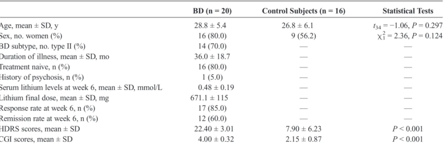
TABLE 1. Demographic and Clinical Information of Patients With BD and Control Subjects
BD (n = 20) Control Subjects (n = 16) Statistical Tests
Age, mean ± SD, y 28.8 ± 5.4 26.8 ± 6.1 t34=−1.06,P= 0.297 Sex, no. women (%) 16 (80.0) 9 (56.2) χ21= 2.36,P= 0.124
BD subtype, no. type II (%) 14 (70.0) — —
Duration of illness, mean ± SD, mo 36.0 ± 18.7 — —
Treatment naive, n (%) 16 (80.0) — —
History of psychosis, n (%) 1 (5.0) — —
Serum lithium levels at week 6, mean ± SD, mmol/L 0.48 ± 0.19 — —
Lithium final dose, mean ± SD, mg 671.1 ± 115 — —
Response rate at week 6, n (%) 17 (85.0) — —
Remission rate at week 6, n (%) 12 (60.0) — —
HDRS scores, mean ± SD 22.40 ± 3.01 7.90 ± 6.23 P< 0.001 CGI scores, mean ± SD 4.00 ± 0.32 2.15 ± 0.87 P< 0.001
TABLE 2. Brain Lactate Before (Week 0) Compared With (A) After (Week 6) Lithium Monotherapy in Patients With Depression in BD
and (B) Matched Control Subjects
A) BD Week 0 BD Week 6 (Li) PairedtTest
Brain lactate, mean ± SD, mmol/L (/Cr) 0.223 ± 0.09 0.143 ± 0.05 t19=−3.52,P= 0.002
B) BD Week 0 Control Subjects UnpairedtTest
depression (n = 12) was found regardless of the brain region when compared with control subjects.30In the same context, decreased
frontal lobe pH was observed in BD patients and has been directly associated with lactic acidosis.1Indeed, cytosolic production of
lactate is regulated by mitochondrial oxidative processes occur-ring duoccur-ring pathological conditions such as acute mood episodes. The elevated conversion of glucose to lactate implies that little glucose is available for biosynthesis or mitochondrial oxidation and that lactate may serve as a metabolic substrate to neurons. In this circumstance, lactate is taken up and used in highly ox-idative neurons because of changes in mitochondrial redox sta-tus. This shift aims to provide supplementary fuel source to neural cells.
In contrast to a previous study showing no changes in brain lactate levels in BD subjects taking lithium over time,21our study found a significant decrease in CC lactate concentrations after 6 weeks of lithium monotherapy. In Friedman et al,21subjects with BD were not evaluated under a specific duration of treatment or mood state, which may account for the difference with the present study in terms of the longitudinal effects of lithium. However, sim-ilar to our findings, they showed no differences in brain lactate levels in BD subjects taking lithium compared with control sub-jects. Despite no statistical significance, lactate posttreatment was lower (0.14) compared with control subjects (0.18), which could be due to lithium-inducing hypernormalization of brain lactate. The observed beneficial actions of lithium on reducing anaerobic metabolism may contribute to the well-known neuro-protective effects of this agent.31In addition, evidence supports the role of lithium as a key modulator of mitochondrial function and cellular energy metabolism,18–20which was supported by the present findings. Lithium also seems to normalize these changes by shifting back to glycolysis, limiting lactate production and im-proving mitochondrial respiration.11It may be hypothesized that the effects of lithium on brain energy production underlie its anti-depressant and mood-stabilizing properties. Our results suggest that lithium acts by reducing the shift from aerobic to anaerobic metabolism observed in BD11,32; however, this shift did not
corre-late with clinical improvement. One explanation is the consider-able number of good responders to lithium in the present sample, based on a potential enriched sample selected, which aimed to allow the use of lithium monotherapy and to avoid pos-sible dropouts. The small number of nonresponders may have pre-cluded the ability to detect differences in mean lactate levels.
The strengths of this study include the longitudinal design with both clinical and imaging measurements, and this is one of
the largest within-subjects study in BD. The study had homoge-neous duration of treatment, specific use of lithium monother-apy, exclusion of comorbid conditions (eg, substance abuse/ dependence), and all BD patients were drug-free (80.0% of whom were treatment naive). In addition, the fact that we recruited a sam-ple with a relatively short duration of illness (mean, 36 months) and no concurrent substance use disorders limits the possibility that our results could have been influenced by confounding factors, such as illness staging and/or co-occurring long-term comorbidi-ties. Differently from the previous studies that assessed brain lac-tate levels in subjects with BD,5,12,13 we used a within-subject
longitudinal design with a systematic clinical follow-up in which the1H-MRS examinations were repeated after a specific period. Fi-nally, the use of a long echo time improves the resolution of the lac-tate resonance, and summing across voxels improves the SNR. It needs to be pointed that we are reporting relative ratios of lactate to Cr signals and not absolute concentrations, so any changes in these ratios could be theoretically also related to changes in Cr or metabolite relaxation times. However, this is very unlikely. An ex-tensive and thorough study5measuring absolute metabolite con-centrations and T2 relaxation times in different brain regions with different tissue compositions concluded that there were no dif-ferences between BD medication-free patients and control subjects in Cr, Cho, NAA, and myo-inositol concentrations or relaxation times. The only differences were found for lactate and glutamate + glutamine concentrations. Therefore, to measure lactate relative to Cr is a common practice and has been used previously to mea-sure longitudinal lactate changes in BD patients.13Some
limita-tions in our study merit discussion. First, the duration of lithium treatment was relatively short. However, most potential good re-sponders to lithium had achieved a response, and most had mild to moderate depression at baseline. In addition, this was an open-label trial, which might limit potential conclusions that can be drawn from clinical outcomes (which could be related to lithium’s effects or natural course of illness). Second, we enrolled a conve-nience sample that could have selected good or excellent lithium responders. Lastly, our study was uncontrolled, and healthy volun-teers had only 1 scan.
Overall, our findings reinforce the presence of altered met-abolic activity in the brain leading to a shift toward lactic acido-sis during mood episodes. They also support a key role for lithium in normalizing this dysfunctional brain energy state through direct effects at mitochondrial function. The present findings reinforce that lactate may be a state biomarker in BD and that mitochondrial modulators might offer promising treat-ment targets in the illness, especially in long-term treattreat-ment. Fur-ther studies with longer duration and larger samples are required to provide further evidence for the key mechanisms of lithium’s modulatory effects on brain mitochondrial and redox activity in BD and its association with clinical outcomes.
AUTHOR DISCLOSURE INFORMATION
A patent for the use of ketamine in depression has been awarded that lists C.A.Z. among the inventors; he has assigned his rights on the patent to the US government, but will share a per-centage of any royalties that may be received by the government. All other authors declare no conflicts of interest.
REFERENCES
1. Quiroz JA, Gray NA, Kato T, et al. Mitochondrially mediated plasticity in the pathophysiology and treatment of bipolar disorder.
Neuropsychopharmacology. 2008;33:2551–2565.
2. de Sousa RT, Zarate CA Jr, Zanetti MV, et al. Oxidative stress in early stage bipolar disorder and the association with response to lithium.J Psychiatr Res. 2014;50:36–41.
3. Hamakawa H, Murashita J, Yamada N, et al. Reduced intracellular pH in the basal ganglia and whole brain measured by31P-MRS in bipolar
disorder.Psychiatry Clin Neurosci. 2004;58:82–88.
4. Lin DD, Crawford TO, Barker PB. Proton MR spectroscopy in the diagnostic evaluation of suspected mitochondrial disease.AJNR Am J Neuroradiol. 2003;24:33–41.
5. Dager SR, Friedman SD, Parow A, et al. Brain metabolic alterations in medication-free patients with bipolar disorder.Arch Gen Psychiatry. 2004;61:450–458.
6. Machado-Vieira R, Andreazza AC, Viale CI, et al. Oxidative stress parameters in unmedicated and treated bipolar subjects during initial manic episode: a possible role for lithium antioxidant effects.Neurosci Lett. 2007;421:33–36.
7. Andreazza AC, Kauer-Sant'anna M, Frey BN, et al. Oxidative stress markers in bipolar disorder: a meta-analysis.J Affect Disord. 2008;111: 135–144.
8. Konradi C, Eaton M, MacDonald ML, et al. Molecular evidence for mitochondrial dysfunction in bipolar disorder.Arch Gen Psychiatry. 2004; 61:300–308.
9. Sun X, Wang JF, Tseng M, et al. Downregulation in components of the mitochondrial electron transport chain in the postmortem frontal cortex of subjects with bipolar disorder.J Psychiatry Neurosci. 2006;31:189–196. 10. de Sousa RT, Machado-Vieira R, Zarate CA Jr, et al. Targeting
mitochondrially mediated plasticity to develop improved therapeutics for bipolar disorder.Expert Opin Ther Targets. 2014;18:1131–1147. 11. de Sousa RT, Streck EL, Zanetti MV, et al. Lithium increases leukocyte
mitochondrial complex I activity in bipolar disorder during depressive episodes.Psychopharmacology (Berl). 2015;232:245–250.
12. Kim DJ, Lyoo IK, Yoon SJ, et al. Clinical response of quetiapine in rapid cycling manic bipolar patients and lactate level changes in proton magnetic resonance spectroscopy.Prog Neuropsychopharmacol Biol Psychiatry. 2007;31:1182–1188.
13. Brady RO Jr, Cooper A, Jensen JE, et al. A longitudinal pilot proton MRS investigation of the manic and euthymic states of bipolar disorder. Transl Psychiatry. 2012;2:e160.
14. Drevets WC, Savitz J, Trimble M. The subgenual anterior cingulate cortex in mood disorders.CNS Spectr. 2008;13:663–681.
15. Chu WJ, Delbello MP, Jarvis KB, et al. Magnetic resonance spectroscopy imaging of lactate in patients with bipolar disorder.Psychiatry Res. 2013; 213:230–234.
16. Licht RW. Lithium: still a major option in the management of bipolar disorder.CNS Neurosci Ther. 2012;18:219–226.
17. Haeberle A, Greil W, Russmann S, et al. Mono- and combination drug therapies in hospitalized patients with bipolar depression. Data from the
European drug surveillance program AMSP.BMC Psychiatry. 2012; 12:153.
18. Quiroz JA, Machado-Vieira R, Zarate CA Jr, et al. Novel insights into lithium's mechanism of action: neurotrophic and neuroprotective effects. Neuropsychobiology. 2010;62:50–60.
19. Khairova R, Pawar R, Salvadore G, et al. Effects of lithium on oxidative stress parameters in healthy subjects.Mol Med Rep. 2012;5: 680–682.
20. Machado-Vieira R, Soeiro-de-Souza MG, Richards EM, et al. Multiple levels of impaired neural plasticity and cellular resilience in bipolar disorder: developing treatments using an integrated translational approach. World J Biol Psychiatry. 2014;15:84–95.
21. Friedman SD, Dager SR, Parow A, et al. Lithium and valproic acid treatment effects on brain chemistry in bipolar disorder.Biol Psychiatry. 2004;56:340–348.
22. HAMILTON M. A rating scale for depression.J Neurol Neurosurg Psychiatry. 1960;23:56–62.
23. Machado-Vieira R, Zanetti MV, DE Sousa RT, et al. Lithium efficacy in bipolar depression with flexible dosing: a six-week, open-label, proof-of-concept study.Exp Ther Med. 2014;8:1205–1208.
24. First MB, Spitzer RL, Gibbon M, et alStructured Clinical Interview for DSM-IV Axis I Disorders, Patient Edition (SCID-I/P). New York, NY: Biometrics Research, New York State Psychiatry Institute; 1995.
25. Young RC, Biggs JT, Ziegler VE, et al. A rating scale for mania: reliability, validity and sensitivity.Br J Psychiatry. 1978;133:429–435.
26. Guy W.Early Clinical Drug Evaluation Unit (ECDEU) Assessment Manual for Psychopharmacology—Revised. National Institute of Mental Health: Rockville, MD; 1976:218–222.
27. Corrigan NM, Richards TL, Friedman SD, et al. Improving1H MRSI measurement of cerebral lactate for clinical applications.Psychiatry Res. 2010;182:40–47.
28. Kreis R. Issues of spectral quality in clinical1H-magnetic resonance spectroscopy and a gallery of artifacts.NMR Biomed. 2004;17:361–381. 29. Machado-Vieira R, Gattaz WF, Zanetti MV, et al. A Longitudinal (6-week)
3 T (1)H-MRS study on the effects of lithium treatment on anterior cingulate cortex metabolites in bipolar depression.
Eur Neuropsychopharmacol. 2015;25:2311–2317.
30. Xu J, Dydak U, Harezlak J, et al. Neurochemical abnormalities in unmedicated bipolar depression and mania: a 2D1H MRS investigation. Psychiatry Res. 2013;213:235–241.
31. Diniz BS, Machado-Vieira R, Forlenza OV. Lithium and neuroprotection: translational evidence and implications for the treatment of
neuropsychiatric disorders.Neuropsychiatr Dis Treat. 2013;9:493–500. 32. Hashimoto T, Brooks GA. Mitochondrial lactate oxidation complex and an
![FIGURE 2. Individual changes in brain lactate from baseline (depressive episode) to endpoint (after 6 weeks of lithium [Li]](https://thumb-eu.123doks.com/thumbv2/123dok_br/15310554.550722/5.863.75.420.122.356/figure-individual-changes-lactate-baseline-depressive-episode-endpoint.webp)
