Effe cts o f le ad and/o r zinc e xpo sure
during the se co nd stage o f rapid
po stnatal brain gro wth o n de
lta-am ino le vulinate de hydratase and
ne gative ge o taxis o f suckling rats
1Curso de Farmácia, Universidade do Sul de Santa Catarina,
Tubarão, SC, Brasil
2Departamento de Bioquímica, Centro de Ciências Biológicas,
Universidade Federal de Santa Catarina, Florianópolis, SC, Brasil E.C. Goulart1,2,
C.A.T. Pereira1, R.C. Garcia1,
M.B.O . Giacomelli1
and A.L.S. Rodrigues2
Abstract
Lead has been shown to produce cognitive and motor deficits in young rats that could be mediated, at least in part, by inhibition of the zinc-containing heme biosynthetic enzyme d-aminolevulinate dehydratase (ALA-D). In the present study we investigated the effects of lead and/ or zinc treatment during the second stage of rapid postnatal brain development on brain, kidney and blood ALA-D specific activity, as well as the negative geotaxis behavior of rats. Eight-day-old Wistar rats were injected intraperitoneally with saline, lead acetate (8 mg/kg) and/or zinc chloride (2 mg/kg) daily for five consecutive days. Twenty-four hours after treatment, ALA-D activity was determined in the absence and presence of DL-dithiothreitol (DTT). The negative geo-taxis behavior was assessed in 9- to 13-day-old rats. Treatment with lead and/or zinc did not affect body, brain or kidney weights or brain-or kidney-to-body weight ratios of the animals. In spite of the absence of effect of any treatment on ALA-D specific activity in brain, kidney and blood, the reactivation index with DTT was higher in the groups treated with lead or lead + zinc than in the control group, in brain, kidney and blood (mean ± SEM; brain: 33.33 ± 4.34, 38.90 ± 8.24, 13.67 ± 3.41; kidney: 33.50 ± 2.97, 37.60 ± 2.67, 15.80 ± 2.66; blood: 63.95 ± 3.73, 56.43 ± 5.93, 31.07 ± 4.61, respectively, N = 9-11). The negative geotaxis response behavior was not affected by lead and/or zinc treatment. The results indicate that lead and/or zinc treatment during the second stage of rapid postnatal brain growth affected ALA-D, but zinc was not sufficient to protect the enzyme from the effects of lead in brain, kidney and blood.
Co rre spo nde nce
A.L.S. Rodrigues
Departamento de Bioquímica CCB, UFSC
88040-900 Florianópolis, SC Brasil
Fax: + 55-48-331-9672 E-mail: analucia@ mbox1.ufsc.br
Received April 12, 2000 Accepted March 5, 2001
Ke y wo rds
·Lead acetate ·Zinc chloride ·Delta-aminolevulinate
dehydratase ·Negative geotaxis ·Postnatal brain growth
Lead is an environmental pollutant that accumulates with toxic effects in blood, liver, kidney and in the central nervous system of exposed mammals. The neurotoxicity of lead is of special interest, since cognitive and
the kidneys, where it may cause proximal tubular dysfunction after acute exposure, or irreversible nephropathy that may lead to renal failure after chronic exposure (1).
Most of the studies concerning the toxic effects of lead are conducted in young ro-dents, which are generally exposed to the metal during the gestational and/or suckling periods (3). During the postnatal period ro-dents present three stages of rapid brain growth (4) in which they are especially prone to neurotoxicants (5,6). However, there are few studies in the literature concerning the effects of lead on specific stages of postnatal brain development (6). Zinc has been shown to prevent hematological alterations induced by lead exposure (7) and has been proposed to prevent the neurotoxicity of lead (8). Ad-ditionally, a great deal of evidence has shown that zinc is a signaling molecule in the brain released by neural activity at many excita-tory synapses (9). However, data on the ef-fects of the administration of zinc, either alone or in combination with lead, on specif-ic stages of rapid postnatal brain develop-ment are lacking.
There are many molecular targets for the action of lead. Lead has great affinity for sulfhydryl groups of endogenous biomol-ecules, which may contribute to its toxicity. It has been proposed that the toxicity of lead may be due, at least in part, to the inhibition of the sulfhydryl-containing enzyme d -aminolevulinate dehydratase (ALA-D) (1). This enzyme catalyzes the condensation of two d-aminolevulinic acid (ALA) molecules with the formation of porphobilinogen, which is a heme precursor. Consequently, ALA-D inhibition may impair heme biosynthesis and can lead to the accumulation of ALA, which may derange aerobic metabolism and may also have some pro-oxidant activity (10). ALA-D activity in erythrocytes is currently the most sensitive indicator of human expo-sure to lead (1). However, divergent results have been reported for ALA-D activity in the blood of lead-exposed animals, with data
showing a reduction (7) or an increase (6,11,12). Mammalian ALA-D has the pecu-liarity of having zinc as its cofactor (13). It has been demonstrated that zinc, as well as DL-dithiothreitol (DTT) in vitro, is able to restore the lead-inhibited ALA-D activity in rat and human blood (1,12-14). In addition, an antagonistic effect of zinc on the inhibi-tion of ALA-D by lead in vivo has been reported (15), as well as the absence of an antagonistic effect (16). However, data are lacking regarding the ability of zinc to re-store the normal activity of ALA-D inhibited by lead in vivo during a specific stage of rapid postnatal brain development.
In the present investigation, we exam-ined the effects of lead and/or zinc exposure during the second stage of rapid postnatal brain development on brain ALA-D specific activity, the reactivation index of cerebral ALA-D with DTT, an indirect indicator of the ALA-D inhibition by lead and/or by the oxidation of critical sulfhydryl groups, and the negative geotaxis behavior of rats. ALA-D activity and the reactivation index with DTT were also assayed in blood and kidney of lead- and/or zinc-exposed rats.
Wistar rats of both sexes from our own breeding colony were used. Rats were main-tained in an air conditioned room (22-25o
C) on a 12-h light/dark cycle with water and food available ad libitum. The breeding regi-men consisted of grouping one virgin female (90-120 days of age) with one male for 20 days. Pregnancy was checked, and the preg-nant rats were separated from the males. On the day of birth, litters were reduced to eight. When pups were 8 days old they started to receive one daily intraperitoneal (ip) injec-tion of 8.0 mg/kg (CH3COO)2Pb or 2.0 mg/
kg ZnCl2 alone or in combination. The
post-natal brain growth (6). The zinc dose was selected since it has been reported that zinc protects rats against the effects of lead on ALA-D at a dose 4-fold lower than the lead dose (7).
Rats were killed by decapitation under light ether anesthesia approximately 24 h after the end of treatment and blood was collected into heparinized tubes. Brain and kidney were quickly removed, placed on ice and then homogenized in 4 and 10 volumes of 150 mM NaCl, respectively. The homog-enate was centrifuged at 4000 g at 4o
C for 10 min to yield a low-speed supernatant frac-tion to be used for enzyme assay.
ALA-D activity was assayed by measur-ing the rate of product (porphobilinogen) formation, according to the method of Sassa (17) except that 40 mM sodium phosphate buffer, pH 6.4, and 4.0 mM ALA were used. The reaction product was determined using modified Ehrlichs reagent at 555 nm, with a molar absorption coefficient of 6.2 x 10⁴
M-1
cm-1
for the Ehrlich-porphobilinogen salt. The incubation was initiated by adding 36 µl of the tissue preparation (0.200-0.350 mg of protein for brain and 0.150-0.350 mg of protein for kidney) or blood (hemolyzed 1:4 in deionized water) to a final volume of 226 µl. The incubation times were 3, 2 and 1 h, respectively. The temperature of incubation was 39o
C for the cerebral and renal enzyme and 37o
C for the blood enzyme. Simulta-neously, a set of tubes was assayed under similar incubation conditions, except that 15 mM DTT was added in order to obtain the reactivation index as previously reported (14). Briefly, this index indicates the extent of the reactivation of ALA-D activity by DTT and is defined as follows:
(ALA-D activity with DTT - ALA-D activity without DTT) x 100
ALA-D activity with DTT
Protein was determined by the method of Lowry et al. (18) using bovine serum
albu-min as standard. Hematocrit was deteralbu-mined using a microcapillary centrifuge. ALA-D activity is reported as nmol porphobilinogen formed h-1
mg protein-1
for cerebral and renal ALA-D activity and as nmol porpho-bilinogen formed h-1
ml erythrocytes-1
for the blood enzyme.
The negative geotaxis response was as-sessed from 24 h after the beginning of treat-ment (9-day-old rats) to 24 after the end of treatment (13-day-old rats), as described by Rocha et al. (5). After body weight measure-ment and just before daily lead acetate and/ or zinc chloride administration, pups were placed head down on an inclined plane (30o
) and the latency to complete the geotaxis response was recorded. This test was re-peated three times for each trial. If the pup did not satisfy this criterion within 60 s, the trial was interrupted.
saline, lead, zinc and lead + zinc were as follows: 31.3 ± 0.89, 28.9 ± 0.61, 30.6 ± 0.78 and 27.2 ± 0.54, respectively (mean ± SEM; N = 10). One-way ANOVA followed by the Newman-Keuls test showed a reduction in hematocrit values in the groups treated with lead and with lead + zinc as compared to the control group (F(3,42) = 6.38, P<0.01). A reduction in hematocrit has also been previ-ously reported in adult rats chronically ex-posed to lead (19), but, to our knowledge, has not been reported in young rats exposed to the metal during a specific stage of brain development which also corresponds to a period of intense tissue development. The reduction in the hematocrit of rats exposed to lead early during development seems to be a sensitive indicator of lead exposure and is not affected by zinc.
One-way ANOVA indicated no signifi-cant effect of lead and/or zinc treatment on specific ALA-D activity in brain (F(3,37) = 0.44, P>0.05), kidney (F(3,38) = 0.055, P>0.05) and blood (F(3,36) = 1.52, P>0.05) (Table 1). The results in brain and kidney are in agreement with the absence of an effect of lead exposure on cerebral and renal ALA-D activity reported by Rocha et al. (6). Our data
also demonstrated that zinc treatment did not change ALA-D activity in any of the tissues examined, in contrast to the reported activa-tion of renal ALA-D in adult rats treated with zinc (20). Blood ALA-D activity was slightly reduced in the lead-exposed group as com-pared to the control group, although this reduction was not statistically significant. This result contrasts with the increase in blood ALA-D activity in rats exposed to lead acetate during the second stage of rapid post-natal growth (6). In fact, both a reduction and an increase in blood ALA-D activity in lead-exposed rodents have been reported (7,11,12). The studies that report an increase in blood ALA-D elicited by lead exposure have attributed it to an increase in ALA-D synthesis (11,12). The group exposed to lead + zinc did not exhibit any decrease in blood ALA-D activity. However, considering that the absence of a significant effect of lead alone on the enzyme activity was observed, we cannot conclude that zinc had an antago-nistic effect. The ability of zinc to prevent ALA-D inhibition by lead has been a matter of extensive study, and contradictory find-ings have been reported. Considering that zinc is able to induce the synthesis of metallothionein, it is possible that pretreat-ment with zinc would be more efficient in preventing the effects of lead administration on ALA-D activity (20). The induction of this zinc-binding protein in target tissues such as brain and kidney, but not blood, during development may play a role in the effects observed in our study. In fact, com-petition between zinc and lead for binding to proteins such as metallothionein and ALA-D has been demonstrated and lead appears to be more potent than zinc in binding to ALA-D (2).
One-way ANOVA revealed a significant effect of lead and/or zinc treatment on the reactivation index with DTT of ALA-D ac-tivity in brain (F(3,37) = 5.15, P<0.01), kid-ney (F(3,38) = 21.34, P<0.01) and blood (F(3,36) = 10.39, P<0.01) (Table 1). Post
Table 1. Brain, kidney and blood ALA-D specific activity and reactivation index w ith DTT in 13-day-old rats exposed to lead acetate and/or zinc chloride.
Saline Pb2+ Zn2+ Pb2+ + Zn2+
Brain N = 10 N = 11 N = 11 N = 9
Specific activity 9.53 ± 0.98 9.77 ± 1.36 9.29 ± 1.30 7.79 ± 1.46
Reactivation index 13.67 ± 3.41 33.33 ± 4.34* 19.29 ± 4.05 38.90 ± 8.24* *
Kidney N = 11 N = 11 N = 10 N = 10
Specific activity 38.79 ± 5.93 36.31 ± 2.93 37.30 ± 4.09 37.01 ± 4.63
Reactivation index 15.80 ± 2.66 33.50 ± 2.97* * 14.80 ± 1.43 37.60 ± 2.67* *
Blood N = 9 N = 11 N = 11 N = 9
Specific activity 171.59 ± 13.83 123.23 ± 17.51 154.55 ± 14.86 173.13 ± 28.48 Reactivation index 31.07 ± 4.61 63.95 ± 3.73* * 32.30 ± 5.49 56.43 ± 5.93* *
hoc Newman-Keuls comparisons showed that the reactivation index with DTT in brain, kidney and blood was significantly higher in the groups exposed either to lead alone or to lead + zinc as compared to the control group. This is an interesting finding that contrasts with the absence of effect of lead and lead + zinc on ALA-D specific activity. The reacti-vation index with DTT has been shown to be a sensitive indicator of ALA-D inhibition by lead and/or by oxidation of sulfhydryl groups of the enzyme which are important in its catalytic activity (13). One possibility which would account for the higher reactivation as for index with DTT in lead-exposed rats, as well as for the absence of lead effect on ALA-D specific activity, is that lead treat-ment may have produced a dual effect on ALA-D, i.e., an inhibition of the enzyme associated with an increase in its synthesis as a compensatory mechanism. The doses of lead acetate that are able to inhibit ALA-D activity in 13-day-old rats have been shown previously, with IC50 for inhibition of
ALA-D by lead being 3.9, 4.4 and 0.8 µM for brain, kidney and blood, respectively (6). We do not have the lead concentrations at-tained in 13-day-old rats. However, our group has data showing that 21-day-old rats ex-posed to lead acetate (8.0 mg/kg, ip) during the second stage of rapid postnatal brain growth have a lead concentration of approxi-mately 0.5, 4.0 and 0.8 µM for brain, kidney and blood, respectively. In view of these data, the lead concentrations attained in our study seem to have reached inhibitory levels. Moreover, even if the lead concentration attained in the tissues was lower than the one required to inhibit ALA-D, we should bear in mind that lead may accumulate in specific cellular microenvironments and that lead has more direct access to proteins located in the cytosol, e.g., ALA-D, compared to other cellular compartments (20). The reduction in heme concentration is believed to have a great impact on young rats in the phase of development in which lead/zinc treatments
were imposed, which corresponds to a stage of rapid postnatal brain development and intense cellular metabolism. Finally, inde-pendently of the mechanism underlying the enhancement in the reactivation index with DTT in lead-exposed rats, zinc was not able to protect against the effects of lead on ALA-D in the regimen employed in this study.
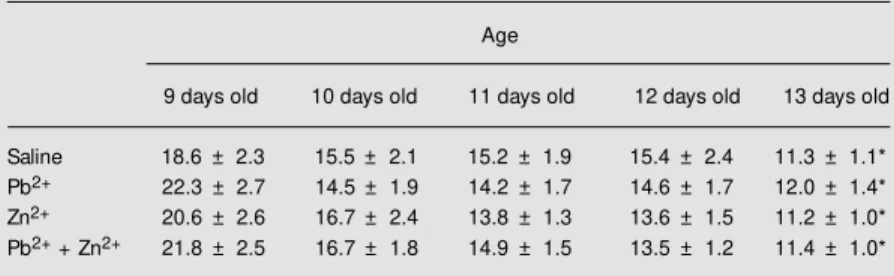
Two-way ANOVA for repeated meas-ures used to analyze the latency to complete the negative geotaxis showed a significant effect of age (F(4,176) = 21.93, P<0.01). No significant effect of treatment (F(3,44) = 0.03, P>0.05) or of treatment x age was observed (F(12,176) = 0.58, P>0.05) (Table 2). Post hoc analysis by the Newman-Keuls test showed a significant reduction in la-tency in 13-day-old rats compared to 9-day-old rats in all groups. The absence of an effect of treatment on negative geotaxis sug-gests that the regimen of lead and/or zinc exposure did not impair the psychomotor function of suckling rats. By contrast, an impairment of negative geotaxis behavior has been reported in young rats exposed to methylmercury (5).
Thus, our lead and/or zinc exposure regi-men was not sufficient to affect physical tissue parameters or motor coordination and also did not affect ALA-D specific activity. However, the reactivation index with DTT was a highly sensitive parameter to indicate
Table 2. Effect of lead acetate and/or zinc chloride administration on the latency to complete a geotaxis response.
Age
9 days old 10 days old 11 days old 12 days old 13 days old
Saline 18.6 ± 2.3 15.5 ± 2.1 15.2 ± 1.9 15.4 ± 2.4 11.3 ± 1.1*
Pb2+ 22.3 ± 2.7 14.5 ± 1.9 14.2 ± 1.7 14.6 ± 1.7 12.0 ± 1.4*
Zn2+ 20.6 ± 2.6 16.7 ± 2.4 13.8 ± 1.3 13.6 ± 1.5 11.2 ± 1.0*
Pb2+ + Zn2+ 21.8 ± 2.5 16.7 ± 1.8 14.9 ± 1.5 13.5 ± 1.2 11.4 ± 1.0*
an effect on the enzyme that we propose might be used to monitor the effects of lead on ALA-D. If we had measured only ALA-D activity, as done in many studies in the
litera-ture, we would have concluded that the treat-ment had no effect on the enzyme, which seems not to be the case.
Re fe re nce s
1. Al-Saleh IAS (1994). The biochemical and clinical consequences of lead poisoning.
M edical Research Review s, 14: 415-486. 2. Bressler J, Kim K, Chakraborti T & Gold-stein G (1999). M olecular mechanisms of lead neurotoxicity. Neurochemical Re-search, 24: 595-600.
3. Davis JM , Otto DA, Weil DE & Grant LD (1990). The comparative developmental neurotoxicity of lead in humans and ani-mals. Neurotoxicology and Teratology, 12: 215-229.
4. Gottlieb A, Keydar I & Epstein HT (1977). Rodent brain grow th stages: An analytical review . Biology of the Neonate, 32: 166-176.
5. Rocha JBT, Freitas AJ, M arques M B, Pereira M E, Emanuelli T & Souza DO (1993). Effects of methylmercury expo-sure during the second stage of rapid postnatal brain grow th on negative geo-taxis and on delta-aminolevulinate dehy-dratase of suckling rats. Brazilian Journal of M edical and Biological Research, 26: 1077-1083.
6. Rocha JBT, Pereira M E, Emanuelli T, Christofari RS & Souza DO (1995). Effect of treatment w ith mercuric chloride and lead acetate during the second stage of rapid postnatal brain grow th on d -amino-levulinic acid dehydratase (ALA-D) activity in brain, liver and blood of suckling rats.
Toxicology, 100: 27-37.
7. Satija NK & Vij AG (1995). Preventive ac-tion of zinc against lead toxicity. Indian Journal of Physiology and Pharmacology, 39: 377-382.
8. Winneke GM D (1996). Zinc to prevent lead poisoning. Canadian M edical Asso-ciation Journal, 154: 1622-1623. 9. Choi DW & Koh JY (1998). Zinc and brain
injury. Annual Review of Neuroscience, 21: 347-375.
10. Bechara EJH (1996). Oxidative stress in acute intermittent porphyria and lead poi-soning may be triggered by 5-aminolevu-linic acid. Brazilian Journal of M edical and Biological Research, 29: 841-851. 11. Fujita H, Orii Y & Sano S (1981). Evidence
of increased synthesis of d-aminolevulinic acid dehydratase in experimental lead-poi-soned rats. Biochimica et Biophysica Acta,
678: 39-50.
12. Kajimoto M M , Kondo M , Niw a M , Suzuki T, Kimura H, Sasaki A & Urata G (1983). Increase of d-aminolevulinic acid dehydra-tase (ALAD) in rat erythrocytes in lead poisoning. Archives of Toxicology, 52: 1-11.
13. Tsukam oto I, Yoshinaga T & Sano S (1979). The role of zinc w ith special refer-ence to the essential thiol groups in delta-aminolevulinic acid dehydratase of bovine liver. Biochimica et Biophysica Acta, 570: 167-178.
14. Rodrigues ALS, Rocha JBT, Pereira M E &
Souza DO (1996). d-Aminolevulinic acid dehydratase activity in w eanling and adult rats exposed to lead acetate. Bulletin of Environmental Contamination and Toxi-cology, 57: 47-53.
15. Haeger-Aronson B & Schutz A (1976). An-tagonistic effect in vivo of zinc on inhibi-tion of delta-aminolevulinic acid dehydra-tase by lead. Archives of Environmental Health, 31: 215-220.
16. Chiba M & Kikuchi M (1984). The in vivo
effects of manganese and zinc on delta-aminolevulinic acid dehydratase activity inhibited by lead. Toxicology Letters, 20: 143-147.
17. Sassa S (1982). Delta-aminolevulinic acid dehydratase assay. Enzyme, 28: 133-145. 18. Low ry OH, Rosebrough NJ, Farr AL & Randall RL (1951). Protein measurement w ith the Folin phenol reagent. Journal of Biological Chemistry, 193: 265-275. 19. Rodrigues ALS, Rocha JBT, M ello CF &
Souza DO (1996). Effect of perinatal lead exposure on rat behaviour in open field and tw o-w ay avoidance tasks. Pharmacol-ogy and ToxicolPharmacol-ogy, 79: 150-156. 20. Goering PL & Fow ler BA (1987). M etal