Letters to the Editor
Radiol Bras. 2015 Mar/Abr;48(2):126–130
127
Dear Editor,
A female, 80-year-old, diabetic, hypertensive and obese pa-tient reported sudden onset of colicky pain in the hypogastrium for two days. Also, there was association with nausea, and the pa-tient was unable to eliminate feces and pass gas, besides present-ing with abdominal distension. Physical examination demon-strated increased abdominal volume, pain to deep palpation of the hypogastrium, abdominal tympanism to percussion and signs of mild dehydration. The clinical hypothesis of obstructive syndrome was raised.
Abdominal radiographic and sonographic images acquired in other service were not conclusive as regards the cause of the condition. Anteroposterior radiography in orthostasis and horizon-tal dorsal decubitus position demonstrated the presence of
obstruc-tion with predominantly gaseous distension of small bowel loops and part of the large bowel, with no signs of pneumoperitoneum. Ultrasonography demonstrated gaseous distension with no signs of free fluid at the moment of the study. With such studies indi-cating low obstruction with no defined cause, one has opted for performing abdominal computed tomography (CT) in order to clarify the diagnosis and define the therapeutic approach.
Due to the clinical suspicion of neoplastic obstruction, the patient was referred to the authors’ institution. With the CT study, the diagnosis could be established, and the several image recon-struction techniques – multiplanar, curve and 3D reconrecon-struction – were utilized so as the findings could be presented to the re-questing physician in a more easily understandable way.
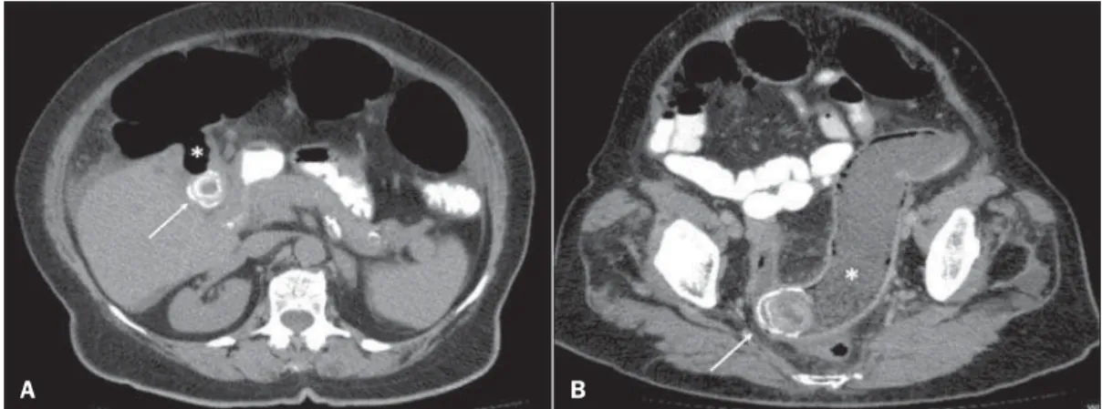
CT demonstrated the fistulous tract communicating the gall-bladder lumen with the lumen of the transverse colon and the presence of a residual calculus in the gallbladder (Figure 1A). Curve reconstruction of the rectal and sigmoid colon region dem-onstrated an impacted calculus obstructing the sigmoid colon and Biliary colon: an unusual case of intestinal obstruction
Cólon biliar: um caso incomum de obstrução intestinal
3. Salvadori PS, Costa DMC, Romano RFT, et al. What is the real role of the equilibrium phase in abdominal computed tomography? Radiol Bras. 2013;46:65–70.
4. Costa DMC, Salvadori PS, Monjardim RF, et al. When the noncontrast-enhanced phase is unnecessary in abdominal computed tomography scans? A retrospective analysis of 244 cases. Radiol Bras. 2013;46:197– 202.
5. Teixeira ACV, Torres US, Westin CEG, et al. Multidetector-row com-puted tomography in the preoperative diagnosis of intestinal complica-tions caused by clinically unsuspected ingested dietary foreign bodies: a case series emphasizing the use of volume rendering techniques. Radiol Bras. 2013;46:346–50.
6. Kierszenbaum ML, von Atzingen AC, Tiferes DA, et al. Colonografia por tomografia computadorizada na visão do médico encaminhador: qual o seu valor segundo a visão de especialistas? Radiol Bras. 2014;47:135– 40.
7. Francisco FAF, Araújo ALE, Oliveira Neto JA, et al. Contraste hepato-biliar: diagnóstico diferencial das lesões hepáticas focais, armadilhas e outras indicações. Radiol Bras. 2014;47:301–9.
8. Terceiro MG, Faria IM, Alfenas R, et al. Hérnia de Amyand com apen-dicite perfurada. Radiol Bras. 2014;47(6):xi–xiii.
9. Cunha EFC, Rocha MS, Pereira FP, et al. Necrose pancreática delimi-tada e outros conceitos atuais na avaliação radiológica da pancreatite aguda. Radiol Bras. 2014;47:165–75.
10. Kadow JS, Fingerhut CJP, Fernandes VB, et al. Peritonite encapsulante: tomografia computadorizada e correlação cirúrgica. Radiol Bras. 2014; 47:262–4.
11. Pedrassa BC, Rocha EL, Kierszenbaum ML, et al. Tumores hepáticos incomuns: ensaio iconográfico – Parte 1. Radiol Bras. 2014;47:310– 6.
12. Pedrassa BC, Rocha EL, Kierszenbaum ML, et al. Tumores hepáticos
incomuns: ensaio iconográfico – Parte 2. Radiol Bras. 2014;47:374– 9.
13. Shinakai-Yasuda MA, Telles Filho FQ, Mendes RP, et al. Consenso em paracoccidioidomicose. Rev Soc Bras Med Trop. 2006;39:297–310. 14. Campos EP, Padovani CR, Cataneo AMJ. Paracoccidioidomicose: estudo
radiológico e pulmonar de 58 casos. Rev Inst Med Trop São Paulo. 1991; 33:267–76.
15. Costa MAB, Carvalho TN, Araújo Júnior CR, et al. Manifestações ex-trapulmonares da paracoccidioidomicose. Radiol Bras. 2005;38:45–52. 16. Tucker ON, Healy V, Jeffers M, et al. Granulomatous appendicitis.
Surgeon. 2003;1:286–9.
17. AbdullGaffar B. Granulomatous diseases and granulomas of the appen-dix. Int J Surg Pathol. 2010;18:14–20.
18. Bronner MP. Granulomatous appendicitis and the appendix in idiopathic inflammatory bowel disease. Semin Diagn Pathol. 2004;21:98–107. 19. Carr NJ. The pathology of acute appendicitis. Ann Diagn Pathol. 2000;
4:46–58.
20. Chojniak R, Vieira RAC, Lopes A, et al. Intestinal paracoccidioidomycosis simulating colon cancer. Rev Soc Bras Med Trop. 2000;33:309–12. 21. Birnbaum BA, Wilson SR. Appendicitis at the millennium. Radiology.
2000;215:337–48.
Priscila Gava1, Alessandro Severo Alves de Melo1, Edson Marchiori1, Márcia Henriques de Magalhães Costa1, Eric Pereira1, Raissa Dantas Batista Rangel1
1. Hospital Universitário Antônio Pedro (HUAP), Rio de Janeiro, RJ, Brazil. Mailing Address: Dra. Priscila Gava. Rua Vítor Meireles, 198, Condomínio Green Country, Mata Paca. Niterói, RJ, Brazil, 24322-110. E-mail: [email protected].
http://dx.doi.org/10.1590/0100-3984.2014.0035
Figure 1. A: Axial CT image demonstrating the presence of a calculus in the gallbladder (ar-row) and fistulous tract commu-nicating the gallbladder with the large bowel (asterisk). B: Curve reconstruction of CT image dem-onstrating impacted biliary calcu-lus in the sigmoid colon (arrow) and bowel distension upstream
Letters to the Editor
Radiol Bras. 2015 Mar/Abr;48(2):126–130
128
Ernesto Lima Araujo Melo1, Francisco Thiago Martins de Paula1, Rainne André Siqueira1, Sariane Coelho Ribeiro2
1. Universidade Estadual do Ceará (UECE), Fortaleza, CE, Brasil. 2. Hospital Israelita Albert Einstein, São Paulo, SP, Brazil. Mailing Address: Dr. Ernesto Lima Araujo Melo. Universidade Estadual do Ceará, Centro de Ciências da Saúde – Curso de Medicina. Avenida Paranjana, 1700, Campus do Itaperi. Fortaleza, CE, Brasil, 60740-000. E-mail: ernesto. [email protected].
http://dx.doi.org/10.1590/0100-3984.2014.0073
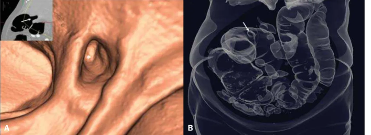
causing abdominal distension upstream due to fecal buildup (Fig-ure 1B). Endoluminal reconstruction of virtual colonoscopy im-ages demonstrated the fistulous orifice in the transverse colon (Figure 2A). Tridimensional reconstruction for gaseous material demonstrated diffuse gaseous distension of the right, transverse and left colons, and also the site of the choledochal-colonic fistula (Figure 2B). Once the diagnosis was established, the patient was successfully submitted to surgery.
Acute obstructive cholecystitis may approach the serosas of the biliary and intestinal tracts due to the gallbladder and/or com-mon biliary duct dilatation. With the repetition of inflammatory episodes and adherence of the serosas, choledochal-colonic fistularization may occur, allowing for the passage of biliary cal-culi into the intestinal lumen(1), besides calculi impaction at some
point in the tract, causing significant pain, severe local irritation, edema or gangrene(2). Amongst cholecystointestinal fistulas,
cholecystoduodenal fistulas represent more than 70%, while cholecystocolonic fistulas represent 8% to 26% of them(3). Rigler
et al. established four criteria (presence of air or contrast medium in the biliary tract; direct or indirect identification of calculus in the bowel; alteration in the position of a previously identified cal-culus; radiological signs of either partial or total occlusion of the intestinal lumen) which corroborate a diagnosis of bowel obstruc-tion caused by a calculus(4). Three findings determined by such
criteria constitute the Rigler’s triad: signs of small bowel dilation, pneumobilia and ectopic calculi.
Despite the rarity of this condition, one should be attentive to the possibility of biliary colon in acute onset of lower intestinal obstruction, in order to allow for a prompt and correct diagnosis and institution of an appropriate treatment.
REFERENCES
1. Wang JK, Foster SM, Wolff BG. Incidental gallstone. Perm J. 2009;13: 50–4.
2. Costi R, Randone B, Violi V, et al. Cholecystocolonic fistula: facts and myths. A review of the 231 published cases. J Hepato Biliary Pancreat Surg. 2009;16:8–18.
3. Del Gaizo A, Raval B. Cholecystocolonic fistula. Applied Radiology. 2006; 35:21–2.
4. Smyth J, Dasari BV, Hannon R. Biliary-colonic fistula. Clin Gastroenterol Hepatol. 2011;9:A26.
Catamenial pneumothorax
Pneumotórax catamenial
Dear Editor,
A previously healthy 29-year-old woman presented at the emergency service complaining of sudden onset dyspnea. At physi-cal examination the vesicular murmur was absent in the entire right hemithorax. Chest radiography demonstrated the presence of pneumothorax at right (Figure 1) and chest computed tomog-raphy (CT) did not demonstrate any other alteration besides the already mentioned pneumothorax. Thoracotomy with underwa-ter seal chest drainage was performed. As the pneumothorax pre-sentation coincided with the patient’s menstrual period, pelvic ultrasonography was performed and identified an image compat-ible with endometrioma in the left ovary. In three months, the patient evolved with a new spontaneous pneumothorax at right, and a pig-tail drainage tube was inserted. Later, thoracocoscopy was performed, and endometriotic foci were identified and resected (Figure 2). The chest wall was repaired with a Marlex mesh. After three months, the patient remains asymptomatic.
Figure 2. A: Endoluminal 3D reconstruction of virtual colono-scopy image demonstrating the fistulous orifice (asterisk) in the large bowel. The reference 2D image of the orifice positioning is highlighted (arrow). B: 3D re-construction demonstrating gaseous bowel distention and the fistulous orifice (arrow).
A B