105
Miranda CMNR et al. MSCT in the staging and follow-up of breast cancer
Radiol Bras. 2012 Mar/Abr;45(2):105–112
Is multislice computed tomography an important tool
for breast cancer staging and follow-up?
*
A tomografia computadorizada multislice é ferramenta importante para o estadiamento e seguimento do câncer de mama
Christiana Maia Nobre Rocha de Miranda1, Carla Jotta Justo dos Santos2, Carol Pontes de Miranda Maranhão2, Lucas de Pádua Gomes de Farias3, Igor Gomes Padilha3, Anna Carolina Mendonça de Andrade4, Mayara Stephanie de Araujo Jatobá4
Breast cancer is the most common cancer in women and the leading cause of cancer death among the female population. Extramammary findings related to breast cancer play an important role in the prognosis and treatment of such entity and the correct diagnosis of such alterations is essential for increasing the chance of cure. Most of the times, such manifestations result from complications arising from treatment, and may be associated with lymph node involvement, locoregional recurrence or distant metastasis.
Keywords: Multislice computed tomography; Staging; Breast cancer; Extramammary changes.
O câncer de mama representa o tipo de câncer mais comum em mulheres e constitui a primeira causa de morte por câncer nesta população. As alterações extramamárias relacionadas ao câncer de mama desempenham papel rele-vante no prognóstico e tratamento desta entidade, sendo fundamental a realização do diagnóstico correto e das diver-sas alterações. A maioria desdiver-sas manifestações é proveniente do tratamento adotado ou de suas complicações e pode estar associada a comprometimento linfonodal, recorrência locorregional ou metástases a distância.
Unitermos: Tomografia computadorizada multislice; Estadiamento; Câncer de mama; Alterações extramamárias.
Abstract
Resumo
* Study developed at Clínica de Medicina Nuclear e Radiolo-gia de Maceió (MedRadiUS), Maceió, AL, Brazil.
1. PhD, Coordinator, Unit of Computed Tomography, Clínica de Medicina Nuclear e Radiologia de Maceió (MedRadiUS), Profes-sor of Radiology and Imaging Diagnosis at Universidade Federal de Alagoas (UFAL), Maceió, AL, Brazil.
2. Titular Members of Colégio Brasileiro de Radiologia e Diag-nóstico por Imagem (CBR), MDs, Clínica de Medicina Nuclear e Radiologia de Maceió (MedRadiUS), Maceió, AL, Brazil.
3. Graduate Students of Medicine, Monitors at Division of Radiology and Imaging Diagnosis, Faculdade de Medicina da Universidade Federal de Alagoas (UFAL), Maceió, AL, Brazil.
4. Graduate Students, Faculdade de Medicina da Universi-dade Federal de Alagoas, Maceió, AL, Brazil.
Mailing Address: Dra. Christiana Maia Nobre Rocha de Mi-randa. Rua Hugo Corrêa Paes, 104, Farol. Maceió, AL, Brazil, 57050-730. E-mail: [email protected]
Received October 23, 2011. Accepted after revision Decem-ber 13, 2011.
Miranda CMNR, Santos CJJ, Maranhão CPM, Farias LPG, Padilha IG, Andrade ACM, Jatobá MSA. Is multislice computed tomography an important tool for breast cancer staging and follow-up? Radiol Bras. 2012 Mar/Abr;45(2):105–112.
REVIEW ARTICLE
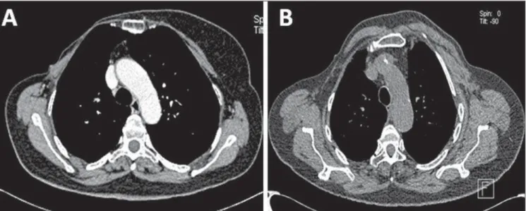
cal technique as follows: 1) radical mas-tectomy (Figure 1A) implies removal of breast, pectoralis major and minor muscles and regional lymph nodes along the axil-lary vein up to the costoclavicular liga-ment; 2) modified radical mastectomy – an alternative method to conservative treat-ment – involves mastectomy and dissec-tion of axillary lymph nodes, sparing the pectoralis major muscle (Patey’s mastec-tomy) (Figure 1B), or sparing both pecto-ralis major and minor muscles (Auchin-closs’ procedure)(6).
Other more conservative reconstruction methods may be adopted. The most com-mon surgical complication is the occur-rence of seroma, besides infections, necro-sis, lymphedema and axillary contrac-ture(6).
POST-RADIOTHERAPY CHEST MANIFESTATIONS
Radiotherapy has been widely utilized following surgery in breast cancer patients to reduce the risk for locoregional recur-phy (MSCT) does not present considerable
evidences of a positive cost-benefit ratio in the evaluation of the breast paren-chyma(3), but its significant role in the stag-ing and follow-up of breast cancer patients should be highlighted considering its use-fulness in the determination of the therapy to be adopted as well as of the patient’s prognosis.
A consensus is still to be achieved on a formal indication for MSCT in the absence os symptoms or clinical indications which justify the request of such study(4), consid-ering the higher sensitivity of positron emission computed tomography(5).
The present study was aimed at demon-strating MSCT findings of extramammary changes detected during breast cancer stag-ing and follow-up of patients submitted to treatment.
POSTOPERATIVE APPEARANCE OF THE CHEST WALL
The postoperative appearance of the chest wall varies with the adopted surgi-INTRODUCTION
Breast cancer is the most frequent type of cancer and is most prevalent in women, accounting for about 22% of new cases of cancer annually (1). Despite the high inci-dence of this disease, the breast cancer mortality rate has been decreasing due to early detection and to the currently avail-able imaging technology(2).
tomogra-Miranda CMNR et al. MSCT in the staging and follow-up of breast cancer
rence and/or increase in tumor volume in cases of advanced disease(7).
Frequently, radiotherapy causes radia-tion pneumonitis, occurring about 4 and 12 weeks after treatment completion, and generally remaining confined to the irra-diation region. Initially, the disease mani-fests with linear opacities or consolidation. Such changes may either gradually disap-pear or result in signs of evolutive fibrotic changes along a period between six months and two years, tending to remain stable after a two-year evolutive period(6,8) (Figure 2).
Three irradiation fields induce radia-tion pneumonitis in breast cancer patients, namely, tangential field, supraclavicular field and internal mammary field. The tan-gential field utilized to irradiate the chest wall results in pneumonitis in the periph-eral lung anterolatperiph-erally, with a typical shape. The supraclavicular field may in-duce changes in the apex of the lung, re-sulting in lesions similar to those observed in pulmonary tuberculosis. The internal mammary field, utilized to irradiate inter-nal mammary lymph nodes, may induce changes in the paramediastinal region. In cases where areas of opacity are seen at post-radiotherapy follow-up radiography, the differential diagnoses include radia-tion pneumonitis, local recurrence, lymphangitic spread and infectious pneu-monitis(6).
POST-CHEMOTHERAPY MANIFESTATIONS
The main chemotherapy agents utilized in the treatment of breast cancer are cyclo-phosphamide, mathotrexate, 5-fluorou-racil and doxorubicin, and the major com-plications to be taken into consideration include pneumonitis, cardiotoxicity and infections.
Chemotherapy-induced cardiotoxicity caused by doxorubicin is generally dose-dependent and is initially asymptomatic, progressing to transient arrhythmia up to fatal cardiomyopathy caused by permanent left ventricular dysfunction. At MSCT, such condition is seen as cardiomegaly and/or pericardial effusion(9) (Figure 3).
Interstitial lung diseases refer to a wide and heterogeneous group of fibrotic pul-monary diseases, including interstitial pneumonitis. In most of cases, the cause of interstitial pneumonitis is unknown. Lung toxicity induced by pharmaceuticals indi-cates a possibly subdiagnosed etiology of interstitial lung diseases. It has been dem-onstrated that currently preconized treat-ments for breast cancer including tamoxifen and taxanes increase the risk for interstitial pneumonitis, particularly in cases of combination with adjuvant radio-therapy. The most frequent pulmonary find-ings resulting from chemotherapy drugs toxicity include interstitial pattern,
ground-glass opacity and consolidation (Figure 4), the latter occasionally simulating a nodule or mass(10).
LOCOREGIONAL RECURRENCE
Local recurrence is defined as reappear-ance of a tumor in the surgical site, occur-ring in 34–84% of cases; and regional re-currence is characterized by the appearance of metastasis in lymph nodes involved in lymphatic drainage, including supraclav-icular, axillary lymph nodes and those of the internal mammary chain(11).
The detection of recurrent breast cancer by mammography represents a challenge due to architectural changes, fibrosis and parenchymal scarring secondary to surgery and radiotherapy which make the interpre-tation of the images more difficult(12).
Generally, patients with involvement of four or more axillary lymph nodes present a significant risk for recurrence which, at MSCT, is seen as multiple lymph nodes with increased diameter. Frequently, me-tastases to lymph nodes of the internal mammary chain (Figure 5) and mediasti-num are clinically occult because of their size, producing equivocal findings at MSCT(13).
Miranda CMNR et al. MSCT in the staging and follow-up of breast cancer
chest wall, and contours irregularity and pectoralis muscles heterogeneity(11).
Normal lymph nodes of the internal mammary chain are less than 5 mm in di-ameter and metastases to this chain cannot be easily detected at clinical examination, mammography or ultrasonography, since they are covered by bony and cartilaginous structures of the chest wall. Normal lymph nodes cannot be routinely identified by computed tomography. Therefore, detect-able lymph nodes of the internal mammary chain with > 6 mm in diameter in breast cancer patients suggest the presence of malignant lymphadenopathy(14).
Disease recurrence in the internal mam-mary chain is rarely observed, occurring with a wide variation in 8–37% of cases, but is concomitantly present in 44% of cases with axillary involvement(11).
LYMPH NODE COMPROMISE
Lymph node metastasis (Figures 6 and 7) is relatively frequent in breast cancer, and an appropriate staging of the tumor is relevant to optimize the diagnostic workup(15).
Despite its proved capacity to reduce morbidity, sentinel lymph node biopsy is an
invasive procedure(16) and, therefore, the acquisition of lymph node images by con-trast-enhanced MSCT seems to be more convenient, despite being an indirect diag-nostic method(17).
Computed tomography is the imaging method of choice for detecting lymph node compromise, many times defining the disease staging according to the TNM classification, in spite of not having value in the evaluation of tumor size. Addition-ally, the performance of this method in cancer detection and quantification is sig-nificantly reduced in cases where the le-sion diameter is < 1 cm(13,18,19).
Figure 2. Post-radiotherapy chest manifestation. Axial MSCT image demonstrating actinic lesion in left lung of a patient submitted to radiotherapy for breast cancer.
Figure 4. Post-chemotherapy manifestation. Axial (A) and coronal (B) MSCT demonstrating pulmonary consolidation resulting from chemotherapy in a breast cancer patient.
Miranda CMNR et al. MSCT in the staging and follow-up of breast cancer
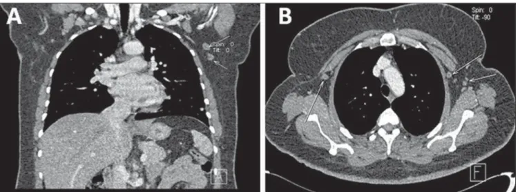
Figure 5. Lymph node enlargement in right internal mammary chain. Regional recurrence six months after conservative surgery. Chest MSCT with volume rendering (A), sagittal (B), axial (C) and coronal (D) images.
Miranda CMNR et al. MSCT in the staging and follow-up of breast cancer
PLEURAL AND PULMONARY METASTASIS
The pleura if a frequent target of me-tastasis in breast cancer patients (Figure 8), and pleural effusion ipsilateral to the pri-mary tumor is the most common sign of such metastasis, probably by lymphatic dis-semination. Breast cancer metastasis is one of the three major causes for malignant ef-fusion(20).
Nodularity, irregular thickening and pleural plaque constitute less common find-ings in pleural metastasis, and rarely occur without association with pleural effusion(6). Multiple nodules occurring by hemato-genic tumor dissemination (Figure 9) are common findings in cases of pulmonary metastasis from breast cancer. Generally, metastatic lesions present a spherical/ovoid shape, with variable sizes, well defined margins, and most of times located periph-erally to the lungs, sometimes found as calcified nodules(6,20,21).
The detection of a solitary lung nodule in patients previously treated for breast can-cer does not necessarily represent meta-static disease. In many cases, a solitary lung nodule is originated from a primary pulmo-nary carcinoma. However, in patients with extrathoracic malignancy, the chance for metastasis corresponds to 25%(6,21).
Other manifestations of pulmonary me-tastasis are carcinomatous lymphangitis and centrolobular nodules, both resulting from endobronchial dissemination(6).
BONE MESTASTASIS
Bone metastasis is the second most common type of breast cancer distant me-tastasis, causing high morbidity because of pain, mobility compromise, hypercalcemia, pathological fracture, compression of the dural sac, spinal cord or nerve roots, and bone marrow infiltration. Breast cancer is the most common cause for medullary compression in women(6).
Several imaging methods are available for evaluating bone metastasis in breast cancer patients. Bone scintigraphy is sug-gested as a first imaging technique for as-ymptomatic patients, considering the high sensitivity of this method in the detection of bone metastasis, allowing excellent skel-etal evaluation(22).
Radiologically, the majority of bone me-tastases are multiple and may be osteolytic, osteoblastic or a combination of both types(6, 23,24) (Figure 10). It is important to highlight
that after radiotherapy, chemotherapy or hormone-based therapy, initially osteolytic metastases may become osteosclerotic.
LIVER METASTASIS
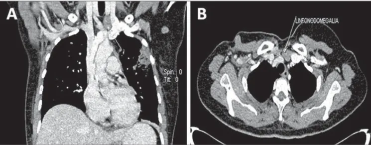
Approximately 50% of women with metastatic breast cancer present liver me-tastasis in the course of the disease. Radio-graphically, liver metastases present several appearances as follows: “target” lesions at ultrasonography, and hypoattenuating at portal phase computed tomography, be-cause of their hypovascularization(25,26) (Figure 11). Generally, they are non-calci-Figure 7. Lymph node compromise. Coronal (A) and axial (B) MSCT images demonstrating supraclavicular and axillary lymph nodes enlargement at left.
Miranda CMNR et al. MSCT in the staging and follow-up of breast cancer
fied nodular lesions, and may present cal-cifications after chemotherapy treatment (Figure 8).
Studies have demonstrated that liver metastases can be identified as hypervas-cularized at contrast-enhanced arterial-phase CT. Hypervascularized lesions are less common and may appear isoattenu-ating during the portal phase(26,27).
The typical enhancement pattern of breast cancer metastases corresponds to a subtle peripheral enhancement during the arterial phase, and a more intense enhance-ment during the portal phase. Other typi-cal pattern of contrast-enhancement of breast cancer metastasis is that of a lesion with minimal or no peripheral
enhance-ment after intravenous contrast injection in the arterial phase and no enhancement in the portal phase, becoming noticeable in this phase for presenting hypoattenuation in relation to the hepatic parenchyma.
BRAIN METASTASIS
Breast cancer is responsible for approxi-mately 10–15% of cases of brain metasta-sis (Figure 12), which in 70–80% of cases present as multiple lesions many times di-agnosed after alterations in other systemic alterations. The most frequent locations of brain metastases are the grey-white matter junction and vascular borders followed by deep parenchymal structures and
posteri-orly in the cerebral trunk, with supratento-rial regions being more frequently affected than infratentorial regions. Brain me-tastases may also occur in the leptom-eninges (2–5%) and dura-mater(28).
In most cases, brain metastasis mani-fests as isoattenuating or subtly hyperat-tenuating lesions, or even as hypoattenu-ating lesions with perilesional edema and intravenous contrast enhancement. Hemor-rhages, cystic changes and necrosis are commonly seen in all types of metastatic tumors. However, there is no pathogno-monic finding differentiating brain me-tastasis from primary malignant tumors from those originating from non-neoplas-tic conditions(29).
Figure 9. Lung metastasis. Axial MSCT images demonstrating metastatic pulmonary nodules in a patient submitted to right total mastectomy for breast can-cer.
Miranda CMNR et al. MSCT in the staging and follow-up of breast cancer
CONCLUSION
Despite the fact that MSCT is not the imaging method of choice to evaluate the breast parenchyma, its utilization allows the identification of relevant data in symp-tomatic patients, in patients with
radio-graphic alterations and in patients who had lesions detected at abdominal ultrasonog-raphy, and also in the follow-up of re-sponse to chemotherapy treatment. The early diagnosis still remains as the best method to enhance the chances of cure for breast cancer.
REFERENCES
1. Brasil. Ministério da Saúde. Instituto Nacional de Câncer. Estimativa 2010: incidência de câncer no Brasil. Rio de Janeiro, RJ: INCA; 2009. 2. Lee CH, Dershaw DD, Kopans D, et al. Breast
cancer screening with imaging: recommendations from the Society of Breast Imaging and the ACR on the use of mammography, breast MRI, breast
Figure 11. Liver metastasis. Axial, arterial phase (A) portal phase (B) and coronal, portal phase (C), images demonstrating hypovascularized liver metastasis in a breast cancer patient.
Miranda CMNR et al. MSCT in the staging and follow-up of breast cancer
ultrasound, and other technologies for the detec-tion of clinically occult breast cancer. J Am Coll Radiol. 2010;7:18–27.
3. Chala LF, Barros N. Avaliação das mamas com métodos de imagem. Radiol Bras. 2007;40(1):iv– vi.
4. Brasil. Instituto Nacional de Câncer. Controle do câncer de mama. Documento de consenso. Rio de Janeiro, RJ: INCA; 2004.
5. Soares Junior J, Fonseca RP, Cerci JJ, et al. Lista de Recomendações do Exame PET/CT com 18
F-FDG em Oncologia. Consenso entre a Sociedade Brasileira de Cancerologia e a Sociedade Brasi-leira de Biologia, Medicina Nuclear e Imagem Molecular. Radiol Bras. 2010;43:255–9. 6. Jung JI, Kim HH, Park SH, et al. Thoracic
mani-festations of breast cancer and its therapy. Radiographics. 2004;24:1269–85.
7. Formenti SC, Demaria S. Local control by radio-therapy: is that all there is? Breast Cancer Res. 2008;10:215.
8. Krengli M, Sacco M, Loi G, et al. Pulmonary changes after radiotherapy for conservative treat-ment of breast cancer: a prospective study. Int J Radiat Oncol Biol Phys. 2008;70:1460–7.
9. Bird BR, Swain SM. Cardiac toxicity in breast cancer survivors: review of potential cardiac prob-lems. Clin Cancer Res. 2008;14:14–24.
10. Christensen S, Pedersen L, Grijota M, et al. Inci-dence of interstitial pneumonitis among breast cancer patients: a 10-year Danish population-based cohort study. Br J Cancer. 2008;98:1870– 5.
11. Chen L, Gu Y, Leaw S, et al. Internal mammary lymph node recurrence: rare but characteristic
metastasis site in breast cancer. BMC Cancer. 2010;10:479.
12. Bénard F, Turcotte E. Imaging in breast cancer: single-photon computed tomography and posi-tron-emission tomography. Breast Cancer Res. 2005;7:153–62.
13. Yang SK, Cho N, Moon WK. The role of PET/CT for evaluating breast cancer. Korean J Radiol. 2007;8:429–37.
14. Cody HS 3rd, Urban JA. Internal mammary node status: a major prognosticator in axillary node-negative breast cancer. Ann Surg Oncol. 1995;2: 32–7.
15. Jager JJ, Keymeulen K, Beets-Tan RG, et al. FDG-PET-CT for staging of high-risk breast cancer pa-tients reduces the number of further examina-tions: a pilot study. Acta Oncol. 2010;49:185–91.
16. Chae BJ, Bae JS, Kang BJ, et al. Positron emis-sion tomography-computed tomography in the detection of axillary lymph node metastasis in pa-tients with early stage breast cancer. Jpn J Clin Oncol. 2009;39:284–9.
17. Takahashi M, Sasa M, Hirose C, et al. Clinical efficacy and problems with CT lymphography in identifying the sentinel node in breast cancer. World J Surg Oncol. 2008;6:57.
18. Yamashita K, Shimizu K. Evaluation of sentinel lymph node metastasis alone guided by three-di-mensional computed tomographic lymphography in video-assisted breast surgery. Surg Endosc. 2009;23:633–40.
19. Bowen SL, Wu Y, Chaudhari AJ, et al. Initial char-acterization of a dedicated breast PET/CT scan-ner during human imaging. J Nucl Med. 2009;50: 1401–8.
20. Avdalovic M, Chan A. Thoracic manifestations of common nonpulmonary malignancies of women. Clin Chest Med. 2004;25:379–90.
21. Seo JB, Im J, Goo JM, et al. Atypical pulmonary metastases: spectrum of radiologic findings. Radiographics. 2001;21:403–17.
22. Costelloe CM, Rohren EM, Madewell JE, et al. Imaging bone metastases in breast cancer: tech-niques and recommendations for diagnosis. Lan-cet Oncol. 2009;10:606–14.
23. Koizumi M, Yoshimoto M, Kasumi F, et al. Com-parison between solitary and multiple skeletal metastatic lesions of breast cancer patients. Ann Oncol. 2003;14:1234–40.
24. Mundy GR. Metastasis to bone: causes, conse-quences and therapeutic opportunities. Nat Rev Cancer. 2002;2:584–93.
25. Diamond JR, Finlayson CA, Borges VF. Hepatic complications of breast cancer. Lancet Oncol.
2009;10:615–21.
26. Roach H, Whipp E, Virjee J, et al. A pictorial re-view of the varied appearance of atypical liver me-tastasis from carcinoma of the breast. Br J Radiol. 2005;78:1098–103.
27. Blake MA, McDermott S, Rosen MP, et al. Ex-pert panel on gastrointestinal imaging. ACR Ap-propriateness Criteria® suspected liver me-tastases. [online publication]. Reston, VA: Ameri-can College of Radiology; 2011.
28. Wadasadawala T, Gupta S, Bagul V, et al. Brain metastases from breast cancer: management ap-proach. J Cancer Res Ther. 2007;3:157–65.
29. Soffietti R, Rud~ R, Mutani R. Management of