TIPS FOR CONTROLLING PORTAL HYPERTENSION
COMPLICATIONS: EFFICACY, PREDICTORS OF OUTCOME
AND TECHNICAL VARIATIONS*
Néstor Hugo Kisilevzky1
OBJECTIVE: To evaluate the efficacy of TIPS (transjugular intrahepatic portosystemic shunt) for resolving clinical complications in patients with portal hypertension. MATERIALS AND METHODS: Forty-four caucasian patients, 30 men and 14 women, with a mean age of 52 years have been evaluated. Indication for TIPS has been gastrointestinal hemorrhage in 28 patients, and refractory ascites in 16. There has been 7 Child-Pugh A patients, 24 Child-Pugh B, and 11 Child-Pugh C. RESULTS: TIPS was successfully performed in all the pa-tients (100%), with a decrease in the mean portosystemic pressure gradient of about 49.69% (from 18.98 mmHg to 9.55 mmHg). A clinical improvement was observed in 35 patients (79.55%). The general postop-erative mortality rate was 13.64%, with higher incidence in Child-Pugh C patients (45.45%). The most relevant factors associated with a poor prognosis were increase in bilirubin and creatinine seric levels. The mean survival time was 11.5 months for Child-Pugh A patients, 10.97 months for Child-Pugh B patients, and just 5.9 months for Child-Pugh C patients. Complications directly related to the procedure have been observed in nine cases (20.44%). CONCLUSION: TIPS is efficient to reduce portosystemic pressure. TIPS-related complications and morbidity-mortality may be considered as acceptable. In the present study, mortality has been directly influenced by some clinical factors such as Child-Pugh class C, and increased bilirubin and creatinin seric levels.
Keywords: Portal hypertension; Cirrhosis; TIPS; Esophageal varices bleeding; Ascites.
TIPS para o controle das complicações da hipertensão portal: eficácia, fatores prognósticos associados e variações técnicas.
OBJETIVO: Avaliar a eficácia do TIPS (transjugular intrahepatic portosystemic shunt) para tratar as compli-cações clínicas em pacientes com hipertensão portal. MATERIAIS E MÉTODOS: Quarenta e quatro pacien-tes, sendo 30 do sexo masculino e 14 do feminino e com idade média de 52 anos foram analisados. A indi-cação para realização de TIPS foi hemorragia gastrintestinal em 28 e ascite refratária em 16. Houve 7 pacientes Child-Pugh A, 24 Child-Pugh B e 11 Child-Pugh C. RESULTADOS: O TIPS foi realizado com sucesso em todos os pacientes (100%), verificando-se queda do gradiente pressórico porto-sistêmico médio de 49,69% (de 18,98 mmHg para 9,55 mmHg). Comprovou-se melhora clínica em 35 pacientes (79,55%). A mortali-dade pós-operatóriaia foi de 13,64%, sendo mais incidente nos pacientes Child-Pugh C (45,45%). Os fato-res mais relevantes de mau prognóstico foram o aumento da bilirrubina e do nível de creatinina. A sobrevida média de pacientes Child-Pugh A foi de 11,5 meses, nos Child-Pugh B foi de 10,97 meses e nos Child-Pugh C foi de apenas 5,9 meses. Foram observadas complicações em nove casos (20,44%). CONCLUSÃO: O TIPS é eficiente para reduzir a pressão portal. As complicações e a morbi-mortalidade relacionadas com o procedimento podem ser consideradas aceitáveis. A mortalidade foi influenciada por alguns fatores clínicos, tais como classe Child-Pugh C e elevação dos níveis séricos de bilirrubina e creatinina.
Unitermos: Hipertensão portal; Cirrose; TIPS; Sangramento por varizes esofagianas; Ascite. Abstract
Resumo
* Study developed at Hospital Santa Catarina – Service of In-terventional Radiology, São Paulo, SP, Brazil.
1. MD, Interventional Radiologist at Hospital Santa Catarina. Mailing address: Dr. Néstor Hugo Kisilevzky. Rua Guararapes, 682, Lapa. São Paulo, SP, Brazil 05077-051. E-mail: kisilevn@ uol.com.br
Received December 7, 2005. Accepted after revision March 24, 2006.
INTRODUCTION
TIPS (transjugular intrahepatic porto-systemic shunt) is a radiological percutane-ous procedure consisting of establishing an intrahepatic communication between a
branch of the portal vein and the inferior vena cava, aiming at decompressing the portal vein, and, therefore, controlling clinical complications resulting from the portal hypertension in patients with chronic hepatopathy. Under the technical point of view, TIPS is considered as one of the most challenging procedures in interventional radiology, for combining several tech-niques such as angiography, parenchymal viscus puncture, angioplasty, handling of stents, embolization, etc. The TIPS idea was initially conceived as an extension of the transjugular cholangiography, late in
the sixties by radiologists at Oregon Uni-versity, in Portland, USA(1,2). However, its
clinical application started to be universally developed only with the publication of the first case in 1989(3). Since then, the
tech-nique has been refined, indications have been defined, and its effectiveness has been well documented in large series(4–8).
Pres-ently, TIPS represents a great contribution developed by Radiologists as a minimally invasive alternative for treating patients with portal hypertension syndrome.
single institution, and discusses, under a technical and clinical viewpoint, indica-tions and factors which might influence the results of the procedure.
MATERIALS AND METHODS
This was a retrospective, descriptive study consisting in a review of all the cases of patients submitted to TIPS performed by a single operator, in a single institution (Hospital Santa Catarina, São Paulo, SP, Brazil), during an eight-year period (1996– 2004). In total, 44 patients (30 men and 14 women), with ages ranging between 34 and 67 years (mean age 52 years), were submit-ted to TIPS.
All of the patients presented with chronic hepatopathy and portal hyperten-sion, the base disease being alcoholic cir-rhosis in 22, chronic hepatitis B in 7, chronic hepatitis C in 4, autoimmune cir-rhosis in 1, biliary circir-rhosis in 2, and cryp-togenic cirrhosis in 8. The main symptom originating the treatment for portal hyper-tension was: gastroesophageal variceal rebleeding in 28 patients, and refractory ascites in 16 — 5 of them with hepatorenal syndrome. Serum bilirubin, albumin, crea-tinine levels and prothrombin time were evaluated. Sixteen (36.36%) patients pre-sented mild ascites, and other 16, tense ascites. Additionally, 11 (25%) patients had a previous history of mild encephalopathy. The hepatic function status of patients evaluated by the Child-Pugh classifica-tion(8) showed that 7 were Child-Pugh A, 24, Child-Pugh B and 11, Child-Pugh C.
All of the TIPS procedures were per-formed in the Sector of Angiography – Center of Diagnostic Imaging at Hospital Santa Catarina, which is equipped with a GE Advantx® digital subtraction angiogra-phy device. Amongst the patients submit-ted to TIPS, 34 were inpatients in the in-stitution and nine came by ambulance from other places, returning to their hospital of origin after being submitted to the proce-dure. In 30 cases (68.18%), the procedure had been previously scheduled and was performed in an elective fashion, but, in 14 (31.82%) cases, the procedure was per-formed with no previous scheduling, given the urgency of the situation. Thirty-seven (84.09%) patients were submitted to the
procedure under local anesthesia and con-scious intravenous sedation with a hypnotic drug (Midazolan) and analgesia (Fentanyl), but 7 (15.91%) patients underwent general anesthesia in assisted respiration because they already were under this condition.
The TIPS technique comprised the fol-lowing steps (Figure 1):
• Puncture and catheterization of the right femoral artery with the purpose of angiographically studying the liver and the portal system;
• puncture and catheterization of the right internal jugular vein with a 10 F in-troducer sheath anti-reflux valve;
• insertion of a multipurpose catheter to perform the central venous pressure mea-surement and venographic study of hepatic veins;
• insertion of a Rosch-Uchida® needle
to perforate the hepatic parenchyma;
• portal catheterization with a multipur-pose catheter to perform the central venous pressure measurement and venographic study of the portal vein (portography);
• intrahepatic route dilatation with an angioplasty balloon (10 mm in diameter); • placement of a self-expandable stent in the intrahepatic route;
• measurement of central and portal venous pressures;
• portal angiography;
• supplementary procedure, if necessary (dilatation, collaterals embolization, etc.). All the patients were clinically fol-lowed-up during their stay in the hospital. Contacts were made with both the patients and their assisting physicians, during the first year following the procedure.
All the information necessary for the research and obtained by means of the medical dossiers review was recorded on
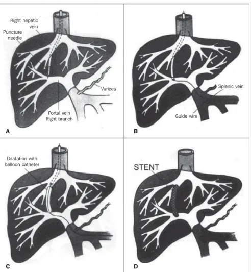
Figure 1. Schemes for TIPS technique comprehension. A: Insertion of an appropriate needle for punc-turing the portal vein from the right hepatic vein. B: Insertion of guide-wire into the portal system. C:
Dilatation of hepatic parenchyma between the portal vein and the hepatic vein. D: Stent placement in the newly formed course.
A B
C D
Right hepatic vein Puncture
needle
Portal vein Right branch
Varices Splenic vein
Guide wire
a precodified form especially designed for the present study. The information register was entirely made by the responsible re-searcher.
For digital data recording of the vari-ables involved in the present study, the Excel worksheet application database man-aging system was employed. The follow-ing softwares were utilized: SAS (Statisti-cal Analysis System) for Windows, version 8.02 (SAS Institute Inc., 1999-2001, Cary, NC, USA); SPSS for Windows, version 10.0.5 (SPSS Inc., 1989-1999, Chicago, IL, USA).
For data analysis, the following statis-tical methodology was employed:
• Data descriptive analysis: statistical, descriptive analysis of continuous variables and frequency of categorical variables;
• the comparison between pre- and post-treatment pressures was performed with the Wilcoxon non-parametric test for paired values;
• in the comparison of survival time to death, the Kaplan-Meyer life table was employed for calculating the survival curves for each class, the death being con-sidered as an event. The Breslow test was employed for comparing survival groups; • factors influencing the post-surgical mortality were investigated by the Cox re-gression model;
• the Fisher’s exact test was employed to analyze the mortality ratio/class;
• the level of statistical significance was 5% (p-value < 0.05).
For the results analysis, the following concepts were employed:
Technical success: Is associated with the TIPS feasibility, allowing the passage of the portal blood to the systemic circula-tion(8).
Clinical success: Is associated with the post-surgical clinical management of the primary symptom which has motivated the procedure.
Hemodynamic success: Is associated with procedure capacity of reducing the portosystemic pressure gradient.
Post-surgical mortality: Is associated with the amount of deaths in the 30-day period following the procedure.
Survival: Is associated with the amount of patients who remain alive during the whole study period.
Censorship time: Represents the 12-month observation period following the procedure.
Complications: Events directly related to the procedure, resulting in hospital stay prolongation or additional therapies.
TIPS occlusion: Is associated with the moment where the absence of portal flow in the TIPS is proved, regardless the pres-ence of symptoms.
RESULTS
Technical success: The procedure has technically succeeded in all of the cases (100%).
In 11 (25%) cases, collaterals emboliza-tion was performed as a supplement to the primary procedure (TIPS) (Figure 2).
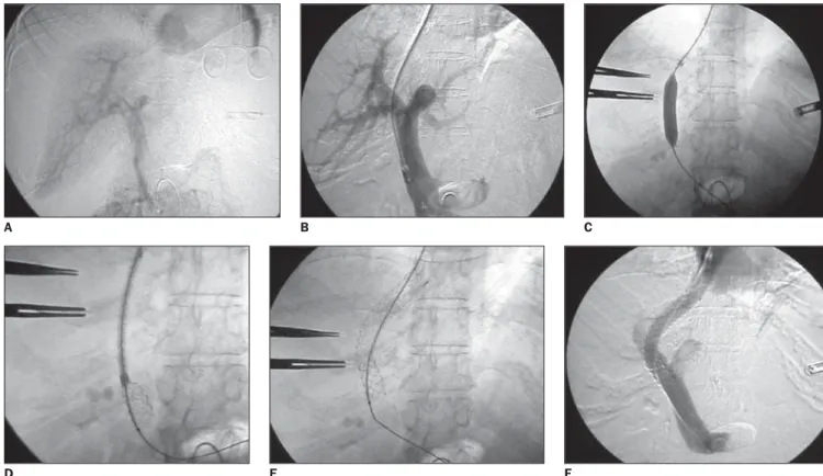
In 31 (70.45%) cases, naked metal stents of several brands were employed, and in 13 (29.55%), a new polytetrafluoroethylene-covered stent-graft (Viatorr®) was utilized
(Figure 3).
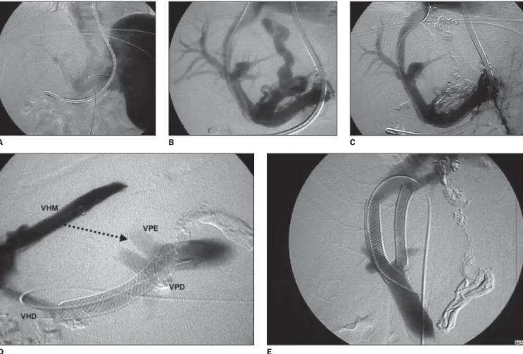
One case (2.27%) required the place-ment of two stents in a parallel TIPS (Fig-ure 4).
Hemodynamic success: The TIPS has caused a change in the pressure values in
all of the patients. Previously to the TIPS placement, the mean portosystemic pres-sure gradient — established by the differ-ence between the portal pressure and the central venous pressure — was 18.98 mmHg, and has fallen, after the TIPS place-ment, to a mean 9.55 mmHg, correspond-ing to a statistically significant 49.69% decrease (Table 1).
Clinical success: Regarding the clinical condition which has motivated the proce-dure, a clinical improvement has been proved in 35 patients (79.55%), and, of the 28 patients (63.64%) with indication of TIPS for treating bleeding, 24 (85.71%) have stopped bleeding; additionally, amongst 11 patients with refractory ascites, 8 (72.72%) had their condition controlled, and three (60%) of five patients with hepatorenal syndrome have achieved rever-sion after TIPS placement (Table 2).
Post-surgical mortality: The 30-day post-surgical follow-up demonstrated the death of six (13.64%) patients, five of them Child-Pugh C, and one Child-Pugh B. Half of these procedures were performed with no previous scheduling, given the urgency of the situation, and the other half were elective (Table 3).
Figure 2. Portography by direct transhepatic puncture. A: Before TIPS performance, an intense portal reflux through collaterals towards the gastroesophageal transition zone. B: As a supplement to TIPS, the collaterals were occluded with metal coils (arrow).
A B
Table 1 Comparison of pre- and post-TIPS P/S gradient (values in mmHg).
Portosystemic gradient
Time
Pre
Post
Post–Pre
Wilcoxon test: p-value < 0.0001. N
44
44
44
Mean
18.98
9.55
–9.43
Standard deviation
3.87
2.14
4.49
Minimum
12.00
5.00
–21.00
Median
19.00
10.00
–9.00
Maximum
29.00
14.00
Figure 3. Angiography images documenting the utilization of a covered stent. A: Portal angiographic study. B: Transhepatic portography. C: Dilatation of the intrahepatic portosystemic shunt. D: Covered stent insertion with the stent naked portion inside the portal vein. E: Stent positioning in the place appropriate for release. F: Portography following complete stent release.
F E
D
A B C
The logistic regression analysis of vari-ables conditioning the post-surgical mortal-ity demonstrated a significant influence of serum bilirubin/creatinine levels and pro-thrombin time (Table 4).
On the other hand, the multivariate analysis showed bilirubin and creatinine levels as the factors most significantly in-fluencing the post-surgical mortality, and patients with hyperbilirubinemia with a 6.7 times higher chance of dying immediately after the procedure, while patients with high serum creatinine levels have a 22.9 times higher chance (Table 5).
Survival: Thirty-three patients (75%) survived during the censorship period — 8 of 9 Pugh A Patients, 20 of 24 Child-Pugh B, and only 5 of 11 Child-Child-Pugh C patients. So, a statistically significant dif-ference in the patients survival associated with the Child-Pugh classification was evidenced. Mean survival time for Child-Pugh A patients was 11.50 months, for Child-Pugh B, 10.97 months, and 5.90 months for Child-Pugh C patients (Figure 5; Table 6).
Complications: Immediate, unexpected events associated with the procedure oc-curred in nine (20.44%) patients (Table 7). One patient has developed a hematoma on the neck, and another, a hematoma on the inguinal region. In both patients, the hematomas were resolved with clinical treatment. One patient presented with hemobilia on the 14th post-surgical day,
which has caused a new hospitalization. An angiography demonstrated an accidental lesion in the right branch of the hepatic artery, requiring metal coil embolization (Figure 6).
Six patients presented with encephal-opathy, five of them with moderate degree which has been resolved with clinical treat-ment. Another patient, besides severe en-Table 2 Evolution of symptoms in patients submitted to TIPS.
Clinical improvement
Improvement
No Yes
High digestive hemorrhage improvement
No Yes
Ascites improvement
No Yes
Hepatorenal syndrome improvement
No Yes
9 35
4 24
3 8
2 3
20.45 79.55
14.29 85.71
27.27 72.72
40.00 60.00
9 44
4 28
3 11
2 5
20.45 100.00
14.29 100.00
27.27 100.00
40.00 100.00 Frequency Percentage
Accumulated frequency
Figure 4. Documentation of some TIPS technical variables according to the strict portosystemic gradient control. A: Indirect portography performed by selective splenic artery catheterization, observing that, in the venous return phase, a great part of the portal flow is deviated towards the gastroesophageal transition zone. B: After TIPS placement, a 14 mmHg portosystemic gradient was observed, yet maintaining a significant reflux through the left gastric vein C:
Then, the left gastric vein embolization was performed with an adhesive substance. Now, the portography shows an absent reflux, but the portosystemic gradient rises to 18 mmHg. D: We have decided to place a second stent to achieve the objective of reducing the portosystemic gradient; on the lateral angiographic image, one may observe the anatomical relation between hepatic veins and the right and left portal vein branch and the direction of the puncture for placement of a second stent (arrow) (VPD, right portal vein branch; VPE, left portal vein branch; VHD, right hepatic vein; VHM, middle hepatic vein). E: After placing a second stent aimed at reducing the pressure gradient to 9 mmHg. On the image, a parallel TIPS is observed, besides a first stent between the right hepatic vein and the right portal vein branch, and a second stent between the middle hepatic vein and the left portal vein branch.
C B
A
D E
Figure 5. Survival curve estimated by the Kaplan-Meyer method, considering the death as an event (for nine Child-Pugh A patients, 24 Child-Pugh B, and 11 Child-Pugh C). Mean survival of patients was of 11.50 months for Child-Pugh A patients, 10.97 months for Child-Pugh B patients, and 5.90 months for the Child-Pugh C patients (p-value = 0.0015).
cephalopathy, presented a significant wors-ening of the liver function which com-pelled, initially, a reduction in the gauge of the TIPS, and later, its occlusion.
Amongst the six patients with encepha-lopathy, four (66.66%) had alcoholic cir-rhosis as the base disease and a history of previous encephalopathy pre-operatively. TIPS occlusion: TIPS occlusion oc-curred in five patients (11.36%), present-ing acutely in two of them (eight and twenty days following the surgery), and in the other three, in a chronic fashion (seven, nine and eleven months post-operatively) (Figure 7).
Four patients have died because of symptoms worsening or recurrence. In one
patient a TIPS revision was performed eight months post-operatively. At that oc-casion, a new stent was placed, allowing a good portal decompression, and extending the communication lifetime and the patient survival.
In all of the cases with TIPS occlusion, naked (uncovered) metal stents had been utilized. However, the relation between TIPS occlusion and stent type has not been statistically significant (Table 8).
DISCUSSION
Interventional radiology has undergone a deep transformation. The last decades technological development, both in
imag-A
c
c
u
m
u
la
te
d
p
ro
b
a
b
ili
ty
ing equipment and interventional instru-ments and materials, in conjunction with the imagination of researchers involved with the progress of medicine, have al-lowed the current utilization of minimally invasive procedures to manage cases of severe conditions. Undoubtedly, TIPS is an example of this progress. Before the advent of TIPS, thousands of patients with portal hypertension have died in the absence of an effective therapy, since, in many situations, neither the endoscopic nor the surgical al-ternative might offer a significant benefit(7).
Most of times, TIPS is indicated for controlling variceal bleeding and/or refrac-tory ascites(6). Less frequent indications
include management of Budd-Chiari syn-drome(9), hepatic hydrothorax(10), hepato-pulmonary syndrome(11) and ectopic
va-rices(12), but the TIPS role in these
situa-Table 3 Post-TIPS mortality rate. General frequency related to Child-Pugh classification or urgency.
Post-surgical mortality rate (up to 30 days)
General
No Yes
Child A
No Yes
Child B
No Yes
Child C
No Yes
Urgency
No Yes
Elective
No Yes
38 6
9 0
23 1
6 5
11 3
27 3
86.36 13.64
100.00 0.00
95.83 4.17
54.54 45.45
78.57 21.42
90.00 10.00
38 44
9 9
23 24
6 11
11 14
27 30
86.36 100.00
100.00 100.00
95.83 100.00
54.54 100.00
78.57 100.00
90.00 100.00 Frequency Percentage
Accumulated frquency
Accumulated percentage
Figure 6. TIPS complication. Unintentional injury of the hepatic artery during the TIPS performance caused post-procedural hemobilia, requiring a new intervention for right hepatic artery branch embolization. A: arteriography evidencing lesion on the right hepatic artery branch (arrow). B: Arteriography after the embolization of the injured branch.
A B
Table 4 Analysis of variables influencing the post-TIPS mortality rate.
Parameters
Age
Sex (F vs. M) Symptom (H vs. A) Condition (U vs. E) Bilirubin
Albumin Prothrombin time
Creatinine
Encefalopathy (Y vs. N) Portosystemic gradient
df
1
1 1
1 1
1 1
1 1
1
Estimate
0.0320
0.8979 0.1541
0.8979 1.6263
–1.7621 –0.0632
2.4055 0.4769
–0.0115
SE
0.0545
0.8914 0.9290
0.8914 0.5812
0.9299 0.0322
0.7810 0.9464
0.1160
χ²
0.3454
1.0147 0.0275
1.0147 7.8292
3.5906 3.8441
9.4856 0.2540
0.0098
p-value
0.5567
0.3138 0.8682
0.3138
0.0051
0.0581
0.0499 0.0021
0.6143
0.9210
OR
1.033
2.455 1.167
2.455 5.085
0.172 0.939
11.084 1.611
0.989
CI 95%
0.928
0.428 0.189
0.428 1.628
0.028 0.881
2.398 0.252
0.788
F, female; M, male; H, hemorrhage; A, ascites; U, urgency; E, elective; Y, yes; N, no; df, degrees of freedom; SE, standard error; OR, odds ratio; CI, confidence interval.
1.149
4.085 7.207
14.085 15.886
1.062 1.000
51.227 10.296
tions has not been accurately defined yet(13).
In the experiment reported by the present study, there were two predominant indications for TIPS: 1) high digestive hemorrhage caused by rupture of gastroe-sophageal varices or congestive gastro-pathy; 2) refractory ascites, patients with hepatorenal syndrome being included in the latest. The clinical improvement ob-served in almost 80% of patients allows us to affirm that TIPS is an efficient method for controlling both situations, mainly when compared with other therapeutical alternatives.
Results from previous experiments with surgical portal decompression were dis-couraging, because of the high mortality rates (31% to 77%), principally when the surgery is performed in compelling urgency circumstances(14).
Also, TIPS has shown to be more effi-cient than the endoscopic treatment for controlling digestive hemorrhage in pa-tients with portal hypertension. In 1999, a meta-analysis study reviewed 11 studies comparing TIPS versus endoscopic treat-ment, and demonstrated a lower incidence of hemorrhage recurrence in patients sub-mitted to TIPS(15). These studies reported
hemorrhage recurrence in 40% to 50% of patients submitted to endoscopic treatment, and 15% to 25% of percutaneously treated patients, during a variable, 15 to 33-month follow-up period.
In relation to refractory ascites, The TIPS has been compared with the treatment by means of repetitive paracentesis. Re-Figure 7. TIPS occlusion. A: Angiography performed seven months following TIPS in a patient with
recurrent bleeding, where the impossibility of TIPS retrograde catheterization from the hepatic vein is demonstrated. B: A new puncture is performed proving the TIPS occlusion and reflux into collaterals, despite the previous embolization.
B A
Table 5 Multivariate analysis for evaluating preponderant factors influencing patients’ death in up to 30 days – utilization of the stepwise selection method.
Parameters
Interceptum
Bilirubin
Creatinine gl
1
1
1
Estimate
–12.7187
1.8973
3.1331 SE
5.2866
0.9539
1.4462
χ²
5.7879
3.9561
4.6936
p-value
0.0161
0.0467
0.0303
OR
—
6.668
22.945
CI 95%
—
1.028 43.242
1.348 390.560
df, degrees of freedom; SE, standard error; OR, odds ratio; CI, confidence interval.
Table 7 Complications resulting from TIPS.
Complication
Cervical hematoma
Inguinal hematoma
Hemobilia
Severe encephalopathy
Moderate encephalopathy
No
Frequency
1
1
1
1
5
35
Percentage
2.27
2.27
2.27
2.27
11.36
79.54
Accumulated frequency
1
2
3
4
9
44
Accumulated percentage
2.27
4.54
6.81
9.08
20.44
100.00
Table 8 Comparison between covered stent ver-sus uncovered stent as cause for TIPS occlusion.
Tipe of stent
Covered
Uncovered
Total
Permeable
13 29.55 100.00 33.33
26 59.09 83.87 66.67
39 88.64
Occlusion
0 0.00 0.00 0.00
5 11.36 16.13 100.00
5 11.36
Total
13 29.55
31 70.45
44 100.00
Fisher’s exact test: p-value = 0.3005. No statistically significant difference.
Table 6 Survival analysis according Child-Pugh classification.
Child A
Child B
Child C
Total
Total
9
24
11
44
Nbr. of events
1
4
6
11
Censored Nbr.
8
20
5
33
Censored %
88.89
83.33
45.45
75.00
Survival equality statistical test – Child distribution
Statistics
13.03
df
2
p-value
0.0015
Breslow
cently, a meta-analysis study gathering five studies and including more than 300 cases, has been published(16). Amongst patients submitted to TIPS the mean symptomatic improvement was of 66%, while in those treated with parencentesis the improvement was of only 24% in a four-month follow-up period. In the one-year follow-follow-up pe-riod, improvement with TIPS compared favorably (55%) with paracentesis (19%). Several factors which could affect nega-tively the post-surgical survival should be taken into consideration for TIPS indica-tion. Child-Pugh classification, serum bi-lirubin level, serum creatinine level, coagu-lation status, and indication in a compelling urgent situation are some of these factors(17).
A multicentric study developed in the USA, has reported bleeding management in 93.6% of patients submitted to TIPS in urgent circumstances, with early recurrent hemorrhage in 12.4% of these patients. However, in-hospital mortality in up to six weeks was considered as high (35,8%)(18). Another study developed in France, has reported that, in 58 patients with uncontrol-lable bleeding and submitted to urgent TIPS, the post-surgical mortality rate up to 30 days was 29%, and 35% up to 60 days(19).
Encarnacion et al., in the USA, have reported 65 consecutively performed TIPS for variceal bleeding management. In this study, 60% of patients were hemodinami-cally unstable, and were submitted to TIPS in compelling urgent circumstances(20). The
authors have clearly documented that pa-tients submitted to TIPS in such circum-stances have a significantly poor progno-sis than those submitted to elective TIPS. Thirty-day mortality rate for the first ones was 28%, while for the latest was only 4% (p = 0.013).
In the experiment reported in the present study, no statistically significant difference was found in mortality of patients submit-ted to TIPS, in the comparison between elective and urgent procedures. This may be due to different interpretations of the words “urgent” or “emergency” by differ-ent services or communities. In fact, there was an evident difference in the patients´ evolution according their clinical charac-terization by the Child-Pugh classification. Additionally, a potentially higher risk of death has been observed in patients with
increased serum bilirubin and creatinine levels, and these factors are the most sig-nificant for determination of a poorer prog-nosis following TIPS. Generally, there is a consensus among the majority of authors on the concept that Child-Pugh C patients scoring 12 or more points, present a high risk of early death when submitted to TIPS(21).
A study developed in the USA with 231 patients submitted to elective TIPS, has identified four factors influencing patient’s survival: serum creatinine level, serum bi-lirubin level, International Normalized Ratio (INR) and cause of cirrhosis(22). The authors have developed a formula for cal-culating the risk applying these four vari-ables, and have observed that patients scor-ing more than 1.8 presented a mean sur-vival of 2.8 months, and patients scoring less than 1.8 presented a mean survival of 1.3 year. Then, it has been established that a score higher than 1.8 implies a poor prog-nosis. The authors have considered this model quite effective, with 77% sensitiv-ity, and 79% specificsensitiv-ity, 63% positive prog-nostic value, and 88% negative progprog-nostic value(22). The original model developed by Malinchoc et al. has been slightly modi-fied: small changes in the formula includ-ing the elimination of the cause of cirrho-sis and multiplication of the score by 10 aiming at facilitating its application(23). The
new model has been called model for end-stage liver disease (MELD) and currently is universally employed for TIPS candi-dates selection.
Invariably, TIPS is performed by an interventional radiologist, i.e., a specialist in minimally invasive imaging-guided per-cutaneous procedures. In experienced hands, this procedure can be successfully completed in more than 95% of cases(7,8,24,25). Notwithstanding the 100% technical success achieved by TIPS in the present study, it is important to note that just a par-tial experience is reported by the author, since it was developed in a single institu-tion, under ideal conditions, and after com-pleting a learning curve with this proce-dure. The author has already performed more than 150 TIPS during the last ten years, in several institutions in Brazil and abroad, also experimenting some technical failures(7).
The main step for a successful TIPS completion is the portal puncture. Some authors have suggested certain anatomical parameters or technical variants aiming at facilitating the portal puncture(26–29).
Nev-ertheless, results are unpredictable when one does not know the degree of hepatic atrophy and displacement of vascular struc-tures in a cirrhotic liver.
The primary goal of TIPS is the portal system decompression to avoid variceal bleeding and/or reduce ascites formation. As regards varices, it is well established that the reduction of the portosystemic gra-dient to a level < 12 mmHg causes a sig-nificant decrease in the bleeding risk. Other concept utilized is the proportional reduc-tion of the portosystemic gradient. Rossle et al. have shown that, after TIPS, the re-bleeding risk was 18%, 7% and 1%, respec-tively, in patients whose the portosystemic gradient had been reduced in 0%, between 25% and 50%, and more than 50%(30).
Another study has reported that a 50% reduction of the initial portosystemic gra-dient is associated with a rebleeding rate of 11%/year, and patients with lower gradient reduction presented a rebleeding rate of 31%(31). In this latest study, the only
abso-lute value preventing rebleeding was a portosystemic gradient lower than 12 mmHg, which in some way equates both concepts. It is important to note that an excessive reduction of the portosystemic gradient may be associated with a higher incidence of post-TIPS encephalopathy.
In 2001, the Society of Interventional Radiology (SIR) developed and published standards for TIPS creation, and a consen-sus has been reached, establishing that the technical success (creation of the commu-nication and reduction of the portosystemic gradient to 12 mmHg) must be achieved in 95% of patients, and the clinical success (resolution of the portal hypertension com-plication) must be achieved in 90% of pa-tients(8).
to-wards the gastroesophageal transition zone (left gastric and/or tributary vein) and natu-ral splenorenal anastomosis. This decision is based on the fact that we have already observed cases where collaterals with con-siderable caliber persist after TIPS with high incidence of persistent bleeding or early rebleeding, even though the post-TIPS portosystemic gradient is below 12 mmHg. On the other hand, natural porto-systemic anastomosis follow the intent to prevent the competence between two portosystemic communications, which could lead to thrombosis and early TIPS dysfunction.
It is important to mention that, usually, when a pathway is occluded, there is a change in the local hemodynamics and, therefore, the portosystemic gradient must be constantly measured following collat-erals embolization to avoid portal hyperten-sion.
TIPS has shown to be a reasonably safe procedure with an acceptable complica-tions level(32).
Most of times, death following TIPS occurs because of the liver disease progres-sion, a situation probably influenced by the portal flow shunt, but not as a result of a procedural complication itself, like a por-tal or hepatic perforation with intraperito-neal hemorrhage. It is estimated that the occurrence of this type of major complica-tion is not superior to 3%(8,32).
The two mostly-feared post-TIPS nega-tive effects are encephalopathy and shunt dysfunction caused by stenosis or occlu-sion.
The central factor in the onset of en-cephalopathy is the presence of a portosys-temic communication, and, by definition, TIPS may cause this complication in up to 30% of patients(33,34).
This complication, most of times, can be clinically controlled with no difficulty, but, in about 5% of cases, encephalopathy may be an extremely limiting condition, compelling a new intervention which may consist in the shunt caliber reduction or occlusion(35–37).
The encephalopathy incidence amongst the patients of the present study casuistic was 25%. However, we have observed that the majority of patients who presented en-cephalopathy had alcoholic hepatopathy or
presented a history of previous encephalo-pathy.
Some risk factors for the onset of en-cephalopathy have already been men-tioned: age higher than 60 years, female patient, alcoholic disease, hypoalbumin-emia, previous history of encephalopathy, the caliber of the created communication, the final pressure gradient, and the base disease severity(33,34).
In patients at high encephalopathy risk, it might be interesting to create a lower caliber 8 mm) communication, or even two parallel communications, and complete the procedure with collateral embolization through the TIPS(38,39).
The major concern of TIPS is its short durability. In 25% to 50% of cases, a > 50% communication stenosis is observed and may lead to portal hypertension recurrence within a period of time between 6 and 12 months following the TIPS creation(40–44).
In 1993, LaBerge et al. reported in de-tail their findings in patients with stenotic and occluded TIPS(45). The authors
pro-posed that small bile pools resulting from rupture of biliary ducts during the portal puncture procedure, caused an inflamma-tory reaction contributing to coagula for-mation and TIPS occlusion(45).
An important aspect that should be ob-served by the time of the TIPS creation is a good coverage of the hepatic vein with stent prolongation to the confluent between the hepatic vein and the inferior vena cava. We have observed that the hepatic vein diameter is reduced in up to 50% as a re-sponse to TIPS, and so the communication outflow is limited, making its occlusion frequent and likely to occur(46). This was
observed in a case where TIPS occlusion was demonstrated seven months after the procedure. By the time of the angiographic follow-up, we found a retraction and defor-mity of the stent placed proximal to the hepatic parenchyma and with poor cover-age of the hepatic vein (Figure 7).
Many researches have been developed indicating several alternatives to extend the TIPS permeability. The majority of inves-tigators have focused their attention on the idea of utilizing new stents covered with biocompatible, impermeable prosthetic material. This has been the origin of the stent-graft concept.
An array of prosthetic materials was evaluated for covering metal stents, includ-ing silicone, polytetrafluoroethylene (PTFE), polyethylene terephthalate (PET), dacron. In 2001, The American company W.L. Gore launched in the market a covered stent specifically developed for TIPS creation and denominated Viatorr®. It is a nitinol (a
nickel and titanium alloy) internally and externally covered with a special type of expanded PTFE that minimizes transmural permeation of bile and mucin (ePTFE).
In 2004, Charon et al. retrospectively analyzed the Viatorr® stent utilization in
Europe(47). The stent produced by Gore was utilized in 100 patients submitted to TIPS for portal hypertension. The primary per-meability in the first year follow-up was of 84%, which represented an evident im-provement in relation to the historical TIPS permeability(47).
In Italy, Rossi et al., utilizing the same material observed a primary permeability of 84% and a secondary permeability of 98% in the first year follow-up (48).
Hausegger et al., in Austria, have cre-ated TIPS with Viatorr® stent in a 71-pa-tient population(49). Four occlusions, and
three stenosis have been found during the first year follow-up, generating 11.3% new interventions. The primary permeability six months and one year after was, respec-tively, 87.4% and 80.8%.
Bureau et al. have compared, in a ran-domized study, the TIPS durability in two groups of patients submitted to the proce-dure with covered stents, or with classical uncovered stents(50). After a mean 300-day
follow-up period, they have observed 13% of TIPS dysfunction in the group with cov-ered stents and 44% in those who received the classical uncovered stents.
CONCLUSION
In the present study, we have observed that TIPS is an excellent method for reduc-ing portal hypertension and controllreduc-ing symptoms in patients with chronic hepat-opathy and portal hypertension.
Additionally, we have observed that the survival of patients submitted to TIPS is acceptable, and can constitute an invalu-able alternative for patients who are wait-ing for a liver transplant as a definite therapy. The complications resulting from the procedure are note frequent and the mortality rate is acceptable, considering the clinical complexity of the patients’ condi-tion. The mortality is directly influenced by some clinical factors, with higher incidence in patients clinically classified as Child-Pugh C, with increased serum bilirubin or creatinine levels. The latest constituted fac-tors implying the worst prognosis for pa-tients submitted to TIPS.
The eventual variation of the technique, with utilization of covered stents, has not altered the result with statistical signifi-cance, although we have observed that in none of these patients there was a commu-nication occlusion, differently from those patients who had received classical metal uncovered stents.
REFERENCES
1. Rösch J, Hanafee W, Snow H, Barenfus M, Gray R. Transjugular intrahepatic portocaval shunt. Am J Surg 1971;121:588–592.
2. Hanafee W, Weiner M. Transjugular percutane-ous cholangiography. Radiology 1967;88:35–39. 3. Richter GM, Palmaz JC, Noeldge G. Intrahepatic stent-assisted portosystemic shunt: a new non-operative transjugular and percutaneous method. Radiologe 1989;29:406–411.
4. Laberge JM, Ring EJ, Gordon RL, et al. Creation of transjugular intrahepatic portosystemic shunt with the wallstent endoprosthesis: results in 100 patients. Radiology 1993;187:413–420. 5. Rossle M, Haag K, Ochs A, Sellinger M, Noldge
G, Perarnau JM. The transjugular intrahepatic portosystemic stent-shunt procedure for variceal bleeding. N Engl J Med 1994;330:165-171. 6. Shiffman ML, Jeffers L, Hoofnalge JH, Tralka TS.
The role of transjugular intrahepatica portosys-temic shunt for treatment of portal hypertension and its complications: a conference sponsored by the National Digestive Diseases Advisory Board. Hepatology 1995;5:1591–1597.
7. Kisilevzky N. TIPS (transjugular intrahepatic portosystemic shunt) no controle das complica-ções da hipertensão portal. Experiência prelimi-nar e revisão da literatura. GED 1999;18:7–18. 8. Haskal ZJ, Martin L, Cardella JF, et al. Quality
improvement guidelines for transjugular
intrahe-patic portosystemic shunts. J Vasc Interv Radiol 2001;12:131–136.
9. Bilbao JI, Pueyo JC, Longo JM, et al. Interven-tional therapeutic techniques in Budd-Chiari syn-drome. Cardiovasc Intervent Radiol 1997;20: 112–119.
10. Spencer EB, Cohen DT, Darcy MD. Safety and efficacy of transjugular intrahepatic portosyste-mic shunt creation for the treatment of hepatic hy-drothorax. J Vasc Interv Radiol 2002;13:385– 390.
11. Selim KM, Akriviadis EA, Zuckerman E, Chen D, Reynolds TB. Transjugular intrahepatic porto-systemic shunt: a successful treatment for hepato-pulmonary syndrome. Am J Gastroenterol 1998; 93:455–458.
12. Haskal ZJ, Scott M, Rubin RA, Cope C. Intesti-nal varices: treatment with the transjugular intra-hepatic portosystemic shunt. Radiology 1994; 191:183–187.
13. Boyer TD. Transjugular intrahepatic portosyste-mic shunt: current status. Gastroenterology 2003; 124:1700–1710.
14. D’Amico G, Pagliaro L, Bosch J. The treatment of portal hypertension: a meta-analytic review. Hepatology 1995;22:332–353.
15. Papatheodoridis GV, Goulis J, Leandro G, Patch D, Burroughs AK. Transjugular intrahepatic portosystemic shunt compared with endoscopic treatment for prevention of variceal rebleeding: a meta-analysis. Hepatology 1999;30:612–622. 16. Deltenre P, Mathurin P, Dharancy S, et al.
Trans-jugular intrahepatic portosystemic shunt in refrac-tory ascites: a meta-analysis. Liver Int 2005;25: 349–356.
17. Ferral H, Patel NH. Selection criteria for patients undergoing transjugular intrahepatic portosyste-mic shunt procedures: current status. J Vasc Interv Radiol 2005;16:449–455.
18. Chalasani N, Kahi C, Francois F, et al. Improved patient survival after acute variceal bleeding: a multicenter, cohort study. Am J Gastroenterol 2003;98:653–659.
19. Azoulay D, Castaing D, Majno P, et al. Salvage transjugular intrahepatic portosystemic shunt for uncontrolled variceal bleeding in patients with decompensated cirrhosis. J Hepatol 2001;35: 590–597.
20. Encarnacion CE, Palmaz JC, Rivera FJ, et al. Transjugular intrahepatic portosystemic shunt placement for variceal bleeding: predictors of mortality. J Vasc Interv Radiol 1995;6:687–694. 21. Coldwell DM, Ring EJ, Rees CR, et al. Multi-center investigation of the role of transjugular intrahepatic portosystemic shunt in management of portal hypertension. Radiology 1995;196:335– 340.
22. Malinchoc M, Kamath PS, Gordon FD, Peine CJ, Rank J, ter Borg PC. A model to predict poor survival in patients undergoing transjugular intra-hepatic portosystemic shunts. Hepatology 2000; 31:864–871.
23. Kamath PS, Wiesner RH, Malinchoc M, et al. A model to predict survival in patients with end-stage liver disease. Hepatology 2001;33:464– 470.
24. Bilbao JI, Quiroga J, Herrero JI, Benito A. Trans-jugular intrahepatic portosystemic shunt (TIPS): current status and future possibilities. Cardiovasc Intervent Radiol 2002;25:251–269.
25. Tripathi D, Helmy A, Macbeth K, et al. Ten years’
follow-up of 472 patients following transjugular intrahepatic portosystemic stent-shunt insertion at a single centre. Eur J Gastroenterol Hepatol 2004;16:9–18.
26. Darcy MD, Sterling KM. Comparison of portal vein anatomy and bony anatomic landmarks. Ra-diology 1996;200:3, 707–710.
27. Shultz SR, Laberge JM, Gordon RL, Warren RS. Anatomy of the portal vein bifurcation: intra-versus extrahepatic location – implications for transjugular intrahepatic portosystemic shunt. J Vasc Interv Radiol 1994;5:457–459.
28. Uflacker R, Rechiert P, D’Albuquerque LC, Silva A. Liver anatomy applied to the placement of transjugular intrahepatic portosystemic shunt. Radiology 1994;191:705–712.
29. Krajina A, Lojik M, Chovanec V, Raupach J, Hulek P. Wedged hepatic venography for target-ing the portal vein durtarget-ing TIPS: comparison of carbon dioxide and iodinated contrast agents. Cardiovasc Intervent Radiol 2002;25:171–175. 30. Rossle M, Siegersterrer V, Olschewski M, Ochs A, Berger E, Haag K. How much reduction in portal pressure is necessary to prevent variceal rebleeding? A longitudinal study in 225 patients with transjugular intrahepatic portosystemic shunts. Am J Gastroenterol 2001;96:3379–3383. 31. Casado M, Bosch J, Garcia-Pagan JC, et al. Clini-cal events after transjugular intrahepatic portosys-temic shunt: correlation with hemodynamic find-ings. Gastroenterology 1998;114:1296–1303. 32. Barton RE, Rosch J, Saxon RR, Lakin PC,
Peter-sen BD, Keller FS. TIPS: short- and long-term results: a survey of 1750 patients. Semin Interv Radiol 1995;12:364–367.
33. Sanyal AJ, Freedman AM, Shiffman ML, Purdum PP, Liketic VA, Cheathman AK. Portosystemic encephalophathy after transjugular intrahepatic portosystemic shunt: results of a prospective con-trolled study. Hepatology 1994;20:46–55. 34. Somberg KA, Riegler JL, Laberge JM, et al.
He-patic encephalophaty after transjugular intrahe-patic portosystemic shunt: incidence and risk fac-tors. Am J Gastroenterol 1995;90:549–555. 35. Haskal ZJ, Midlebrook MR. Creation of a stenotic
stent to reduce transjugular intrahepatic portosys-temic shunt flow. J Vasc Interv Radiol 1994;5: 827–830.
36. Hauenstein KH, Haag K, Ochs A, Langer M, Rossle M. Reducing stent for TIPS-induced re-fractory hepatic encephalophaty and livr failure. Radiology 1994;194:175–179.
37. Kerlan R, LaBerge JM, Baker E, Ring EJ. Suc-cessful reversal of hepatic encephalopathy by in-tentional occlusion of TIPS. SCVIR Proceedings 1995;242.
38. Somberg KA. Transjugular intrahepatic portosys-temic shunt for refractory ascites: shunt diameter-optimizing risks and benefits. Hepatology 1997; 25:1, 254–255.
39. Haskal ZJ, Ring EJ, LaBerge JM, et al. Role of parallel transjugular intrahepatic portosystemic shunt in patients with persistent portal hyperten-sion. Radiology 1992;185:813–817.
40. Haskal Z, Pentecost MJ, Soulen MC, et al. Trans-jugular intrahepatic portosystemic shunt stenosis and revision: early and midterm results. AJR Am J Roentgenol 1994;163:439–444.
in 90 patients. Gastroenterology 1995;108:143– 151.
42. Nazarian GK, Ferral H, Castaneda-Zuniga WR, et al. Development of stenoses in transjugular in-trahepatic portosystemic shunt. Radiology 1994; 192:231–234.
43. Saxon RS, Mendel-Hartvig J, Corless CL. Bile duct injury as a major cause of stenosis and oc-clusion in transjugular intrahepatic portosystemic shunts: comparative histopathologic analysis in humans and swine. J Vasc Interv Radiol 1996;7: 487–497.
44. Sterling KM, Darcy MD. Stenosis of transjugular intrahepatic portosystemic shunt: presentation and management. AJR Am J Roentgenol 1997; 168:239–244.
45. LaBerge JM, Ferrell LD, Ring EJ, Gordon RL. Histopathologic study of stenotic and occluded transjugular intrahepatic portosystemic shunts. J Vasc Interv Radiol 1993;4:779–786.
46. Saxon RR, Ross PL, Mendel-Hartvig J. Trans-jugular intrahepatic portosystemic shunt parency and the importance of stenosis location in the de-velopment of recurrent symptoms. Radiology 1998;207:683–693.
47. Charon JP, Alaeddin FH, Pimpalwar SA, et al. Results of a retrospective multicenter trial of the Viatorr expanded polytetrafluoroethylene-cov-ered stent-graft for transjugular intrahepatic portosystemic shunt creation. J Vasc Interv Radiol 2004;15:1219–1230.
48. Rossi P, Salvatori FM, Fanelli F, et al. Polytetra-fluoroethylene-covered nitinol stent-graft for transjugular intrahepatic portosystemic shunt cre-ation: 3-year experience. Radiology 2004;231: 820–830.
49. Hausegger KA, Karnel F, Georgieva B, et al. Transjugular intrahepatic portosystemic shunt creation with the Viatorr expanded polytetra-fluoroethylene-covered stent-graft. J Vasc Interv Radiol 2004;15:239–248.