Revista Brasileira de Hematologia e Hemoterapia
Brazilian Journal of Hematology and Hemotherapy
w w w . r b h h . o r g
Original article
Clinical complications in pregnant women with
sickle cell disease: prospective study of factors
predicting maternal death or near miss
Patrícia Santos Resende Cardoso
a,b, Regina Amélia Lopes Pessoa de Aguiar
a,
Marcos Borato Viana
a,∗aUniversidade Federal de Minas Gerais (UFMG), Belo Horizonte, MG, Brazil bFundac¸ão Hemominas, Belo Horizonte, MG, Brazil
a r t i c l e
i n f o
Article history:
Received 28 October 2013 Accepted 6 February 2014 Available online 29 May 2014
Keywords:
Anemia, Sickle cell Hemoglobin SC disease Pregnancy
Maternal death
Pregnancy complications
a b s t r a c t
Objective:To evaluate complications in pregnant women with sickle cell disease, especially those leading to maternal death or near miss (severe obstetric complications).
Methods:A prospective cohort of 104 pregnant women registered in the Blood Center of Belo Horizonte (Hemominas Foundation) was followed up at high-risk prenatal units. They belonged to Group I (51 hemoglobin SS and three hemoglobin S/0-thalassemia) or Group II (49 hemoglobin SC and one hemoglobin S/+-thalassemia). Both groups had similar median ages. Predictive factors for ‘near miss’ or maternal death withp-value≤0.25 in the univariate analysis were included in a multivariate logistic model (significance set forp-value≤0.05). Results:Group I had more frequent episodes of vaso-occlusive crises, more transfusions in the antepartum and postpartum, and higher percentage of preterm deliveries than Group II. Infections and painful crises during the postpartum period were similar in both the groups. The mortality rate was 4.8%: three deaths in Group I and two in Group II. One-third of the women in both the groups experienced near miss. The most frequent event was pneumo-nia/acute chest syndrome. Alpha-thalassemia co-inheritance and-gene haplotypes were not associated with near miss or maternal death. In multivariate analysis predictors of near miss or death were parity above one and baseline red blood cell macrocytosis. In Group I, baseline hypoxemia (saturation < 94%) was also predictive of near miss or death.
Conclusion:One-third of pregnant women had near miss and 4.8% died. Both hemoglobin SS and SC pregnant women shared the same risk of death or of severe complications, especially pulmonary events.
© 2014 Associac¸ão Brasileira de Hematologia, Hemoterapia e Terapia Celular. Published by Elsevier Editora Ltda. All rights reserved.
∗ Corresponding author at: Departamento de Pediatria da Faculdade de Medicina, Universidade Federal de Minas Gerais – UFMG, Av. Alfredo
Balena, 190, sala 267, 30130-100 Belo Horizonte, MG, Brazil. E-mail address: [email protected] (M.B. Viana). http://dx.doi.org/10.1016/j.bjhh.2014.05.007
Introduction
The first case description of sickle cell disease (SCD) dates back to over a century ago. Since then, medicine and related areas have built a large body of knowledge on the disease. SCD prob-ably arose on the African continent and in the Middle East with the slave trade being responsible for its spread to the New World. It has become one of the most common genetic diseases in the world.1
Sickle cell anemia, or SS hemoglobinopathy, is the result of sickle cell gene homozygosis. SCD also includes hemoglobin (Hb) S interactions with Hb variants other than normal Hb A. The most common Hb variants are C (SC hemoglobinopathy), D-Punjab, and the co-inheritance of the beta thalassemia trait (Hb S/-thalassemia).1–3
Early diagnosis through newborn screening, prevention and treatment of complications have increased the life expectancy of individuals with SCD. This also accounts for SCD women reaching the fertile age and becoming pregnant.3,4
The extreme variability of SCD clinical phenotypes makes it very difficult to predict the course of most pregnancies.5The
incidence of severe complications is high not only because of biological aspects inherent to SCD, but also because the disease is poorly understood by healthcare staff (‘lack of visi-bility’), it is rarely discussed beyond the academic arena, and there are few well-designed studies that would lead to devel-oping appropriate protocols.
In non-pregnant women, less severe clinical forms such as SC hemoglobinopathy and S/+-thalassemia co-inheritance may pass unnoticed for these individuals are usually asymptomatic or oligosymptomatic and have hemoglobin concentrations near to or within the normal range. During pregnancy, however, these women may undergo compli-cations as severe as those associated with the Hb SS genotype.5,6
Several studies have reported complications in pregnant women with SCD, but focused only upon perinatal outcomes.7–13 This study aims at the following: (i)
gain-ing an in-depth understandgain-ing of the profile of pregnant women assisted by hematologists at the Blood Center of Belo Horizonte (Hemominas Foundation) and by obstetricians at high-risk maternities in Belo Horizonte, Minas Gerais State, Brazil; (ii) describing the morbi-mortality of pregnant women with SCD; and (iii) investigating the causes of the most severe complications that led do death or near miss.
Methods
The study is built on a prospective cohort of pregnant women with SCD followed up at the Blood Center of Belo Horizonte (Hemominas Foundation) from December 2007 to November 2011. The population comprised pregnant women with SCD registered in the ‘Project Aninha’, who were treated at the Hemominas Foundation and high-risk mater-nities in Belo Horizonte. Project Aninha was created in 2007 to provide a multidisciplinary team to support pregnant women with SCD at the Educational and Support Center for Hemoglobinopathies (Cehmob), a center coordinated by the
Unit for Newborn Screening and Genetic Diagnosis (Nupad) of the Medicine School of the Universidade Federal de Minas Gerais (UFMG), and the Hemominas Foundation.
The sample included a total of 104 pregnant women, 51 with the Hb SS genotype, 49 with SC hemoglobinopathy, three with Hb S/0-thalassemia, and one with Hb S/+-thalassemia. All cases were confirmed by hemoglobin electrophoresis. The Hb SS genotype was distinguished from Hb S/0-thalassemia by polymerase chain reaction restriction fragment length polymorphism (PCR-RFLP) using the Ddel restriction enzyme.
Both hematological and obstetrical complications were investigated, but this report focuses on hematological com-plications. The analysis included investigating both maternal death and the most severe complications that could lead to maternal death (near miss). The inclusion criteria required that the patients were pregnant at the medical appointment, had been followed up by one of the Project Aninha partici-pants, and had signed an informed consent to participate in this study. The investigation was approved by Ethics Com-mittees recognized by the Brazilian Committee for Research Ethics (CONEP) and was carried out in accordance with the Declaration of Helsinki (revised as of 2008).
The initial statistical analysis involved data such as age, hemoglobinopathy type (genotype), age at menarche, number of pregnancies, number of abortions, clinical comorbidities, and baseline phenotypic characteristics, including hypoxemia (baseline finger oxygen saturation < 94%) identified during a medical appointment despite a lack of symptoms, painful crises or infections. Asymptomatic hypoxemia was confirmed by more than one evaluation at distinct clinical appointments. Baseline hematologic values were calculated as the average of three measurements in patients without blood transfusions three months prior to the tests. Alloimmunization was inves-tigated in the patients’ transfusion records before pregnancy. The-globin gene haplotypes were determined by PCR-RFLP; alpha-thalassemia genotypes were determined by multiple gap PCR to detect the seven most common alpha-globin gene deletions.
The following clinical events were recorded during preg-nancy and during the first 42 days postpartum: infections, vaso-occlusive crises (exclusively those episodes which required emergency care for intravenous hydration and the administration of analgesia), pulmonary complications/acute chest syndrome (pulmonary symptoms and signs associated with new pulmonary infiltration as observed through chest imaging), number of blood transfusions, and number of hospi-talizations (days of stay). Symptomatic urinary infection was defined as urinary symptoms associated with pyuria and a positive urine culture (>100,000 colony forming units). Posi-tive urine culture without urinary symptoms was interpreted as asymptomatic bacteriuria. Both symptomatic urinary infec-tion and asymptomatic bacteriuria were grouped together for statistical analysis.
Variables that might have contributed to maternal death or severe clinical complications (near miss) were included in the prognostic factors analysis. These variables were classified according to the adapted criteria for near miss.14,15
consequences and need of transfusions, as well as clinical criteria such as critical sickness, acute renal failure, acute chest syndrome/pulmonary complications, sepsis, severe hemolytic crisis, severe painful crisis, and preeclampsia. Maternal death was that which happened during pregnancy or within 42 days after delivery due to any direct or indirect obstetric cause, according to the criteria established in the 10th review of the International Classification of Diseases and Related Health Problems (ICD-10).
Statistical methods
Univariate analysis included testing genotype associations with total hemoglobin level, fetal hemoglobin percentage, baseline white blood cell count, platelet count,-globin hap-lotypes, thalassemia genotype, alloimmunization, pre- and post-partum painful crises, pre- and post-partum blood trans-fusions, infections, and other complications during pregnancy and up to 42 days postpartum. The Pearson chi-squared test or Fisher’s exact test was used to analyze qualitative variables and Student’st-test or the Mann–Whitney test was used for continuous variables with or without normal distributions, respectively.
The analysis included possible factors associated with very severe complications (near miss) or maternal death. A univariate analysis was carried out before proceeding to a multivariate logistic regression analysis. Given the distinct inherent hematological data of the two most common geno-types, Hb SS and SC, hematological variables were analyzed separately from other variables. A genotype-conditioned chi-square test (Cochran statistics) was performed to check the association of maternal death or near miss with values above or below the medians of baseline hemoglobin, mean corpus-cular volume (MCV), and total white blood cell, platelet, and reticulocyte counts. The variables initially included in the multivariate logistic regression were those which were statis-tically significant atp-value≤0.25 in the univariate analysis.
The ‘least significant’ variables (with higher p-values) were removed stepwise until only variables withp≤0.05 remained
in the model.
The Statistical Package for Social Sciences (SPSS version 17.0) was used for the statistical analysis. In this study, ap -value≤0.05 was considered statistically significant for
alpha-type errors.
Results
The SS/S0-thalassemia (Group I) and SC/S+-thalassemia (Group II) groups were similar regarding the age at the begin-ning of pregnancy and gestational age at the first hematology appointment. Menarche was at a significantly older age in patients in Group I (Table 1). The number of pregnancies was not significantly different between the groups, nor were the numbers of full-term pregnancies, stillborn babies, and miscarriages. The prevalence of alloimmunization prior to pregnancy was higher in Group I, albeit not significantly (29.6% vs. 14.0%;p-value = 0.06).
A total of 48 Hb SS women had BEN/CAR (45.8%) as the most common haplotype, the homozygous CAR/CAR women repre-sented 20.8%, and the BEN/BEN reprerepre-sented 18.8%. One patient had the BEN/CAM haplotype, and six others (12.5%) had CAR or BEN heterozygous with an atypical haplotype (three CAR/Atp and three Ben/Atp).
Out of 95 pregnant women, type ␣3.7 alpha-thalassemia was found in 29 (30.5%) patients – 28 in heterozygosis and one in homozygosis (−␣3.7/−␣3.7). Considering the Hb SS and Hb SC genotype distribution, alpha-thalassemia was found in 20 patients with sickle cell anemia, and in only nine patients with SC hemoglobinopathy (p-value = 0.048).
Clinical complications
Most patients needed hospitalization (60.6%) at some time during their pregnancy (Table 2). The between-group differ-ence was statistically significant: 74.1% for Group I and 46% for Group II. Vaso-occlusive crises were the most common cause of hospitalization in the antepartum period; they were significantly more frequent in Group I and occurred mostly in the third trimester and in the puerperium. The median hospital stay was 13 days, ranging from two to 130 days. Most pregnant women with SCD received blood transfusions due to symptomatic anemia (<7.0 g/dL) – increased hemol-ysis or sudden decrease in Hb of over 2 g/dL compared to the baseline value, or a 20% decrease in hematocrit com-pared to the baseline level. A total of 54 (51.9%) pregnant women received blood transfusions at some time during their gestation period: 83.3% in Group I and 18% in Group II (p-value = 0.0001).
Table 1 – Characteristics of the 104 pregnant women with sickle cell disease.
Characteristic Mean Standard deviation Median Minimum Maximum p-valuea
Age at pregnancy
Hb SS/S0-thalassemia 25.4 4.8 25 16 35 0.94
Hb SC/S+-thalassemia 25.4 5.8 26 14 37
Age at menarche
Hb SS/S0-thalassemia 14.8 2.0 15 9 18 <0.001
Hb SC/S+-thalassemia 13.1 1.4 13 11 18
Age at the first hematology appointment
Hb SS/S0-thalassemia 14.6 6.5 14 4 34 0.37
Hb SC/S+-thalassemia 16.1 7.2 14 5 37
Table 2 – Clinical complications of 104 pregnant women with sickle cell disease.
Complications Hb SS/S0-thalassemia Hb SC/S+-thalassemia Total p-value RR 95% CI
n % n % n %
Hospital admission
Yes 40 74.1 23 46.0 63 60.6 0.003 3.4 1.5–7.6
No 14 25.9 27 54.0 41 39.4
Transfusion during pregnancy
Yes 45 83.3 9 18.0 54 51.9 <0.0001 22.7 8.4–62.5
No 9 16.7 41 82.0 50 48.1
Painful crises during pregnancy
Yes 42 77.8 25 50.0 67 64.4 0.003 3.5 1.5–8.2
No 12 22.2 25 50.0 37 35.6
Pulmonary complications/ACS
Yes 16 29.6 16 32.0 32 30.8 0.79 1.1 0.5–2.6
No 38 70.4 34 68.0 72 69.2
Infections
Yes 38 70.4 33 66.0 71 68.3 0.63 1.3 0.5–2.5
No 16 29.6 17 34.0 33 31.7
Urinary infection
Yes 23 42.6 23 46.0 46 44.2 0.73 1.2 0.5–2.5
No 31 57.4 27 54.0 58 55.8
Sepsis
Yes 11 20.4 6 12.0 17 16.3 0.25 2.0 0.6–5.5
No 43 79.6 44 88.0 87 83.7
RR: relative risk; 95% CI: 95% confidence interval; ACS: acute chest syndrome.
The prevalence of pulmonary complications, including acute chest syndrome, was 29.6% in Group I and 32% in Group II (p-value = 0.79). These complications occurred mainly in the third trimester and in the puerperium (Figure 1). Sev-eral patients experienced acute chest syndrome preceded by symptoms of vaso-occlusive crises (back pain, limb pain) or, sometimes, preceded by upper airway symptoms leading to chest pain, hypoxemia, and a new pulmonary infiltration, especially of the right lung. Other clinical complications were rare but included acute renal failure, symptomatic gallstones, and acute splenic sequestration. Because of the small num-bers, these complications were not statistically analyzed.
Hb SS/Sβ0 thalassemia 12
10
8
6
4
2
0
1st tr imester
2nd tr imester
3rd tr imester
Post-pa rtum
Hb SC/Sβ+ thalassemia
Figure 1 – Period of pulmonary complications/acute chest syndrome.
Infections occurred in 68.3% of the cases. Urinary tract infection and/or asymptomatic bacteriuria accounted for 44.2% of the cases. No statistically significant difference was found between Groups I and II. One patient with SC hemoglobinopathy developed pyelonephritis that progressed to severe sepsis, multiple organ failure, and death in the 32nd week of pregnancy, notwithstanding the treatment provided. Infections in other organs or systems occurred less often; they were more common, albeit with no statistically significant difference, in Group I. These infections included endometri-tis, tonsilliendometri-tis, septic arthriendometri-tis, cholecystiendometri-tis, skin infections, leg ulcers, and mastitis. One patient with sickle cell anemia had acute aplastic crises due to parvovirus infection. The diagnosis was confirmed through the detection of viral genome during the acute viral infection period.
Five deaths were registered out of 104 pregnancies (4.8%). Four were due to acute chest syndrome (two in Hb SS, one in Hb S/0-thalassemia and one in Hb SC patients); the fifth death was of a pregnant woman with Hb SC and sepsis origi-nating from the urinary tract. One patient died in the second trimester, and the remaining four in the puerperium, three of them after severe symptoms of pulmonary complications that started in the third trimester and forced delivery.
14 12 10 8 6 4 2 0
ACS/PNM
a
Sepsis ARDS
b
Multiorgan f ailure
Severe pre-eclampsiaSe vere post-par
tum
Severe hemolytic cr isis
Splenic sequestration Strok
e
Severe painful cr isis
Aplastic cr isis
Hb SS/Sβ0 thalassemia
aAcute chest syndrome / pneumonia bAcute respirator y distress syndrome
Hb SC/Sβ+ thalassemia
Figure 2 – Near-miss and maternal death causes according to genotype.
Pulmonary complications were the main cause for ‘near/death’ in 26 of 39 pregnant women (66.7%; Figure 2). Severe acute chest syndrome leading to adult respiratory dis-tress syndrome (ARDS), acute respiratory failure and the need for mechanical ventilation occurred in six patients – four cases in Group I and two in Group II.
Sepsis occurred in 17 cases, including those that suffered septic shock due to pulmonary complications. Two cases evolved to death: one in Group I (septic shock due to pul-monary complications on the 42nd day of puerperium) and one in Group II, secondary to pyelonephritis.
Multiple organ failure including liver impairment, dissem-inated intravascular coagulation, renal failure and pulmonary failure occurred in six cases – three in Group II (one end-ing up in death) and three in Group I (also leadend-ing to death in one case). The latter occurred in a patient with Hb S/0 -thalassemia on the 10th day of puerperium.
In general, patients experienced more than one severe complication, for instance, an association of acute chest syn-drome with sepsis or multiple organ failure. Other causes of near miss were severe preeclampsia in four cases (two in each group) and hemorrhage after birth in four cases (three in Group I and one in Group II). Severe hemolytic crises occurred in two cases in Group II, an acute splenic sequestration in Group II, and a severe painful crisis, also in Group II. Stroke occurred in a patient with Hb SS after fetal death in the 27th week of gestation. An acute aplastic crisis due to parvovirus occurred in one Hb SS patient as mentioned previously.
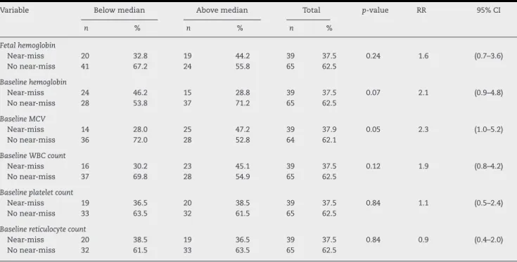
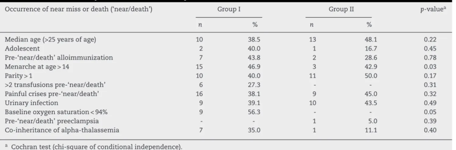
Table 3 shows the univariate analysis for hematologic variables predictive of “near/death”. Following the statistical methodology adopted in this study, only the mean baseline Hb (p-value = 0.07), the mean baseline MCV (p-value = 0.05) and the mean baseline white blood cell count (p-value = 0.12) were included in the multivariate logistic model. No statistically significant association was found between “near/death” and Shaplotypes or co-inheritance of alpha-thalassemia. Table 4 shows the results of other factors predicting “near/death”. Fol-lowing the same statistical criterion, patient’s age greater than the median at the start of pregnancy (p-value = 0.22), menarche at age≥14 years (p-value = 0.03), parity > 1 (p-value = 0.17) and
baseline hypoxemia (p-value = 0.05) were initially included in the multivariate logistic model.
Table 3 – Univariate analysis of hematological variables predictive of maternal death or near miss (near-death).a
Variable Below median Above median Total p-value RR 95% CI
n % n % n %
Fetal hemoglobin
Near-miss 20 32.8 19 44.2 39 37.5 0.24 1.6 (0.7–3.6)
No near-miss 41 67.2 24 55.8 65 62.5
Baseline hemoglobin
Near-miss 24 46.2 15 28.8 39 37.5 0.07 2.1 (0.9–4.8)
No near-miss 28 53.8 37 71.2 65 62.5
Baseline MCV
Near-miss 14 28.0 25 47.2 39 37.9 0.05 2.3 (1.0–5.2)
No near-miss 36 72.0 28 52.8 64 62.1
Baseline WBC count
Near-miss 16 30.2 23 45.1 39 37.5 0.12 1.9 (0.8–4.2)
No near-miss 37 69.8 28 54.9 65 62.5
Baseline platelet count
Near-miss 19 36.5 20 38.5 39 37.5 0.84 1.1 (0.5–2.4)
No near-miss 33 63.5 32 61.5 65 62.5
Baseline reticulocyte count
Near-miss 20 38.5 19 36.5 39 37.5 0.84 0.9 (0.4–2.0)
No near-miss 32 61.5 33 63.5 65 62.5
RR: Relative risk; 95% CI: 95% confidence interval: MCV: mean corpuscular volume; WBC: white blood count.
Table 4 – Univariate analysis of clinical variables predictive of maternal death or near miss.
Occurrence of near miss or death (‘near/death’) Group I Group II p-valuea
n % n %
Median age (>25 years of age) 10 38.5 13 48.1 0.22
Adolescent 2 40.0 1 16.7 0.45
Pre-‘near/death’ alloimmunization 7 43.8 2 28.6 0.78
Menarche at age > 14 15 46.9 3 42.9 0.03
Parity > 1 10 40.0 11 50.0 0.17
>2 transfusions pre-‘near/death’ 6 27.3 - - 0.31
Painful crises pre-‘near/death’ 16 38.1 9 45.0 0.32
Urinary infection 9 39.1 10 43.5 0.49
Baseline oxygen saturation < 94% 9 56.3 - - 0.05
Pre-‘near/death’ preeclampsia - - 1 5.0 0.39
Co-inheritance of alpha-thalassemia 7 35.0 1 11.1 0.40
a Cochran test (chi-square of conditional independence).
Table 5 – Multivariate analysis of prognostic factors for maternal death or near miss in 94 pregnant women with SCD.
Factors Relative risk 95% confidence interval p-value
Genotype 1.8 0.64–5.13 0.26
Baseline mean corpuscular volume 3.1 1.21–7.73 0.02
Parity > 1 2.6 1.03–6.52 0.04
Baseline oxygen saturation < 94% 4.3 1.09–16.97 0.04
This analysis included a total of 94 pregnant women because data were not completely available for ten participants.
Table 5 shows the final multivariate logistic model of the association between “near/death” and the predictors selected through the univariate model. Upon adjusting for genotype, only parity > 1, baseline hypoxemia, and baseline MCV higher than the median remained statistically significant in the model.
Discussion
The high rate of complications in pregnant patients with SCD has already been reported in previous studies. Complications include an increased number of cesarean deliveries, preterm births, restricted intrauterine growth, and low weight babies, especially in pregnant women with the homozygous form of the disease (Hb SS). Increased numbers of vaso-occlusive crises, more frequently in the third trimester and in the puer-perium, as well as increased numbers of cases with acute chest syndrome have also been associated with substantial mater-nal morbidity and mortality.7–13In this study, the number of complications was high among pregnant women with SCD: 60.6% were admitted to hospital at least once for reasons other than assisting the birth, miscarriages or puerperium, more fre-quently in Group I. A higher rate of painful crises was found in this study when compared to others in the literature.7–13One main reason for this might be the concept of painful crises adopted in the present study, which set as criteria the need of administering venous analgesia and hydration and is thus not limited to episodes of drug administration. The prevalence of infections was similar to those reported in previous stud-ies, with no significant differences between the Hb SS and Hb SC genotypes. Urinary tract infections were also more preva-lent in this study as it included patients with asymptomatic bacteriuria.
Blood transfusions during pregnancy were more common in Group I. Symptomatic anemia secondary to various compli-cations was the main reason for blood transfusions. Based on a prospective and randomized study, Koshy et al.16suggested that resorting to prophylactic transfusion does not signifi-cantly decrease the rate of maternal and fetal mortality, but increases the risk of transfusion reactions and alloimmuniza-tion, and leads to hemosiderosis. Even with more stringent indications for blood transfusions, over half of the pregnant women in this study needed at least one transfusion. Among Hb SS patients, the frequency of transfusions (83.3%) was similar to that in other studies.17Exchange transfusion was needed in 20% of the pregnant women, mainly for acute chest syndrome and stroke.
Pulmonary complications included acute chest syndrome and classic pneumonia. The main cause of maternal death or near miss in this study was acute chest syndrome, with similar risks for pregnant women with the Hb SC and Hb SS sub-types. Previous studies have already identified an increased mortality rate in pregnant women with SC hemoglobinopa-thy, especially in the third trimester and in the puerperium.6,18 The literature shows conflicting results regarding the relative severity of complications in pregnant women with Hb SS and SC hemoglobinopathy.7–10
maternal death is higher during the third trimester and the puerperium,22probably due to variations in hormones,
hyper-coagulability and increased susceptibility to infections during these periods.4In the current study, one third of the pregnant
women had pulmonary complications, which was the reason for four deaths due to acute respiratory failure, regardless of the treatment provided.
The haplotypes of theS gene [Benin and Bantu (CAR)] were not significantly associated with near miss death in this study. Similar to the study by Belisário et al.,23 this study
did not find statistically significant differences between hap-lotypes regarding the clinical manifestations in sickle cell anemia. Furthermore, co-inheritance of alpha-thalassemia was not associated with near miss/death. Similarly, other studies24,25 did not find any significant association between
co-inheritance of alpha-thalassemia and protection against acute chest syndrome.
The mortality rate was 4.8%, which is much higher than the figures reported in recent studies, but it should be stressed that the pregnant women in the present study were all followed up at high-risk prenatal units. In addition, three of the deceased women already had severe medical conditions when they were admitted to hospital.
In the univariate analysis, late menarche was associated with near miss/death, probably meaning major organ injury, that is, a delayed sexual maturation could be indicative of the clinical severity of the underlying disease and, therefore, may suggest a higher susceptibility to complications and a higher risk of near miss and maternal death.
Several hematologic variables were associated with near miss/death in the univariate analysis. However, only macro-cytosis (increased MCV) remained as a significant variable in the multivariate analysis. Pregnant women with MCV higher than the median had a three times higher risk of develop-ing near miss or maternal death than those with lower MCV. Macrocytosis may be a surrogate for increased hemolysis, and thus a more severe underlying disease.
Hb SS patients with baseline hypoxemia were found to have a four-time higher risk of facing near miss or maternal death. Patients who have baseline hypoxemia are usually those with a more severe form of the disease.26It is, therefore, important
to determine the baseline oxygen saturation in these women at the beginning of the prenatal period, as it could be a marker of disease severity and complications during pregnancy.
Parity > 1 was also identified as a higher risk (two times) for near miss/death in this study. The reason for this remains unclear. A hypothesis is that pregnancy-driven physiologic changes could pose more risk to the pregnant women with SCD, a risk that is proportionally higher as the number of gestations and accumulated complications deriving from the disease increases.
The major follow-up recommendations for SCD pregnant women consist of carefully monitoring hematologic, obstetrical and fetal complications, and recognizing SCD complications as early as possible.27 The type of delivery
should be indicated by obstetric considerations, but it is crit-ical to take precautions during labor and childbirth in order to prevent vaso-occlusion and pain, such as maintaining the temperature to avoid hypothermia, acidosis and hypoxemia. Blood transfusions are indicated for acute and severe episodes
during pregnancy complications that may be enhanced by SCD.27,28 Partial exchange transfusions should be indicated
as early as possible after the beginning of symptoms of acute chest syndrome, especially when the hematocrit is equal to or higher than 25%.29 Critical care should be delivered by a
concerted team composed by hematologists, obstetricians, general practitioners and intensivists.30
Providing genetic counseling and educating SCD women about their disease and complications some time before a potential pregnancy are crucial items of care for these patients. They should be warned about the need of spe-cialized gynecologic control and follow up with adequate contraception techniques, if they wish. It is critical to provide educational training at government health clinics so that the care staff can follow up patients as early as adolescence and help them plan their pregnancy when they are ready. Decid-ing which contraceptive method is the most adequate is a joint decision between the doctor and the patient.31
Once pregnant, women with SCD should be followed up by a multidisciplinary team at a specialized center for prenatal care. This team should include a hematologist, who will be responsible for detecting signs and symptoms that might flag potentially severe complications that require the patient to be immediately referred to a hospital specialized in emergency care for pregnant women with SCD.
Conflicts of interest
The authors declare no conflicts of interest.
Acknowledgments
To all members of Project Aninha, in particular to the coor-dinator Milza Cintra Januario, M.D, the general director of Nupad, Prof. José Nelio Januario, and Vanessa Fenelon da Costa, M.D., responsible for the obstetrics data collection, and to the Hemominas Foundation Research and Molecular Biol-ogy Section, for carrying out the genetic tests.
r e f e r e n c e s
1. Serjeant GR. The emerging understanding of sickle cell disease. Br J Haematol. 2001;112(1):3–18.
2. Steinberg MH. Pathopysiologically based drug treatment of sickle cell disease. Trends Pharmacol Sci. 2006;27:204–10. 3. Rogers DT, Molokie R. Sickle cell disease in pregnancy. Obstet
Gynecol Clin North Am. 2010;37:223–37.
4. Hassel K. Pregnancy and sickle cell disease. Hematol Oncol Clin North Am. 2005;19:903–16.
5. Charache S, Scott J, Niebyl J, Bonds D. Management of sickle cell disease in pregnant patients. Obstet Gynecol.
1980;55:407–10.
6. Dickinson FT. Sickle-cell hemoglobin C disease in pregnancy: report of a case with review of the literature. J Am Osteopath Assoc. 1980;79:591–4.
8. Sun PM, Wilburn W, Raynor BD, Jamieson D. Sickle cell disease in pregnancy: twenty years of experience at Grady Memorial Hospital, Atlanta, Georgia. Am J Obstet Gynecol. 2001;184:1127–30.
9. Serjeant GR, Loy LL, Crowther M, Hambleton IR, Thame M. Outcome of pregnancy in homozygous sickle cell disease. Obstet Gynecol. 2004;103:1278–85.
10. Yu CK, Stasiowska E, Stephens A, Awogbade M, Davies A. Outcome of pregnancy in sickle cell disease patients attending a combined obstetric and haematology clinic. J Obstet Gynaecol. 2009;29:512–6.
11. Afolabi BB, Iwuala NC, Iwuala IC, Ogedengbe OK. Morbidity and mortality in sickle cell pregnancies in Lagos, Nigeria: a case control study. J Obstet Gynaecol. 2009;29:104–6. 12. Al Jama FE, Gasem T, Burshaid S, Rahman J, Al Suleiman SA,
Rahman MS. Pregnancy outcome in patients with homozygous sickle cell disease in a university hospital. Eastern Saudi Arabia. Arch Gynecol Obstet. 2009;280: 793–7.
13. Nomura RM, Igai AM, Tosta K, Fonseca GH, Gualandro SF, Zugaib M. Resultados maternos e perinatais em gestac¸ões complicadas por doenc¸as falciformes. Rev Bras Ginecol Obstet. 2010;32:405–11.
14. Reichenheim ME, Zylbersztajn F, Moraes CL, Lobato G. Severe acute obstetric morbidity (near miss):a review of the relative use of its diagnostic indicators. Arch Gynecol Obstet. 2009;280:337–43.
15. Souza JP, Cecatti JG, Fagundes A, Morais SS, Villar J, Carroli GA, et al. Maternalnear missand maternal death in the World Health Organization’s 2005 global survey on maternal and perinatal health. Bull World Health Org. 2010;88:
113–9.
16. Koshy M, Burd L, Wallace D, Moawad A, Baron J. Prophylactic red-cell transfusions in pregnant patients with sickle cell disease: a randomized cooperative study. N Engl J Med. 1988;319:1447–52.
17. Al-Farsi SH, Al-Riyami NM, Al-Khabori MK, Al-Hunaini MN. Maternal complications and the association with baseline variables in pregnant women with sickle cell disease. Hemoglobin. 2013;37:219–26.
18. Pritchard JA, Scott DE, Whalley PH, Cunningham FG, Mason RA. The effects of maternal sickle cell hemoglobinopathies
and sickle cell trait on reproductive performance. Am J Obstet Gynecol. 1973;117:662–70.
19. Villers MS, Jamison MG, De Castro LM, James AH. Morbidity associated with sickle cell disease in pregnancy. Am J Obstet Gynecol. 2008;199:125e1–5e.
20. Nomura RM, Igai AM, Tosta K, Fonseca GH, Gualandro SF, Zugaib M. Acute chest syndrome in pregnant women with hemoglobin SC disease. Clinics. 2009;64:927–8.
21. Elsayegh D, Shapiro JM. Sickle cell vasooclusive crisis and acute chest syndrome at term pregnancy. South Med J. 2007;100:77–9.
22. Martin Jr JN, Martin RW, Morrison JC. Acute management of sickle cell crisis in pregnancy. Clin Perinatol. 1986;13: 853–68.
23. Belisário AR, Martins ML, Brito AM, Rodrigues CV, Silva CM, Viana MB.-globin gene cluster haplotypes in a cohort of 221 children with sickle cell anemia or S0-thalassemia and their association with clinical and hematological features. Acta Haematol. 2010;124:162–70.
24. Castro O, Brambilla DJ, Thorington B, Reindorf CA, Scott RB, Gillette P, et al. The acute chest syndrome in sickle cell disease: incidence and risk factors. The Cooperative Study of Sickle Cell Disease. Blood. 1994;84:643–9.
25. Smith JA, Espeland M, Bellevue R, Bonds D, Brown AK, Koshy M. Pregnancy in Sickle Cell Disease: experience of the Cooperative Study of Sickle Cell Disease. Obstet Gynecol. 1996;87:199–204.
26. Quinn CT, Ahmad N. Clinical correlates of steady-state oxyhaemoglobin desaturation in children who have sickle cell disease. Br J Haematol. 2005;131:129–34.
27. Parrish MR, Morrison JC. Sickle cell crisis and pregnancy. Semin Perinatol. 2013;37:274–9.
28. Royal College of Obstetricians and Gynaecologists Green-top, Guideline. Management of sickle cell disease in pregnancy. RCOG. 2011;61:1–20.
29. Bellina JH, Bickers JN. Modern management of sickle cell disease in pregnancy. South Med J. 1974;67:426–30.
30. Danielson CF. The role of red blood cell exchange transfusion in the treatment and prevention of complications of sickle cell disease. Ther Apher. 2002;6:24–31.