rev bras hematol hemoter.2 0 1 4;3 6(4):290–292
Revista Brasileira de Hematologia e Hemoterapia
Brazilian Journal of Hematology and Hemotherapy
w w w . r b h h . o r g
Case report
Pathologic rupture of the spleen in a patient with
acute myelogenous leukemia and leukostasis
Gil Cunha De Santis
∗, Luciana Correa Oliveira, Aline Fernanda Ramos,
Nataly Dantas Fortes da Silva, Roberto Passetto Falcão
Universidade de São Paulo (USP), Ribeirão Preto, SP, Brazil
a r t i c l e
i n f o
Article history:
Received 14 October 2013 Accepted 14 January 2014 Available online 28 May 2014
Keywords: Splenomegaly Leukemia Myeloid Acute Leukostasis
a b s t r a c t
Rupture of the spleen can be classified as spontaneous, traumatic, or pathologic. Patho-logic rupture has been reported in infectious diseases such as infectious mononucleosis, and hematologic malignancies such as acute and chronic leukemias. Splenomegaly is con-sidered the most relevant factor that predisposes to splenic rupture. A 66-year-old man with acute myeloid leukemia evolved from an unclassified myeloproliferative neoplasm, complaining of fatigue and mild upper left abdominal pain. He was pale and presented fever and tachypnea. Laboratory analyses showed hemoglobin 8.3 g/dL, white blood cell count 278×109/L, platelet count 367×109/L, activated partial thromboplastin time (aPTT) ratio 2.10, and international normalized ratio (INR) 1.60. A blood smear showed 62% of myeloblasts. The immunophenotype of the blasts was positive for CD117, HLA-DR, CD13, CD56, CD64, CD11c and CD14. Lactate dehydrogenase was 2384 U/L and creatinine 2.4 mg/dL (normal range: 0.7–1.6 mg/dL). Two sessions of leukapheresis were performed. At the end of the second session, the patient presented hemodynamic instability that culminated in cir-culatory shock and death. The post-mortem examination revealed infiltration of the vessels of the lungs, heart, and liver, and massive infiltration of the spleen by leukemic blasts. Blood volume in the peritoneal cavity was 500 mL. Acute leukemia is a rare cause of splenic rup-ture. Male gender, old age and splenomegaly are factors associated with this condition. As the patient had leukostasis, we hypothesize that this, associated with other factors such as lung and heart leukemic infiltration, had a role in inducing splenic rupture. Finally, we do not believe that leukapheresis in itself contributed to splenic rupture, as it is essentially atraumatic.
© 2014 Associac¸ão Brasileira de Hematologia, Hemoterapia e Terapia Celular. Published by Elsevier Editora Ltda. All rights reserved.
∗ Corresponding author at: Hemocentro de Ribeirão Preto, Universidade de São Paulo – USP, Rua Tenente Catão Roxo 2501, Campus Universitário, Monte Alegre, 14051-140 Ribeirão Preto, SP, Brazil.
E-mail address: gil@hemocentro.fmrp.usp.br (G.C. De Santis). http://dx.doi.org/10.1016/j.bjhh.2014.05.006
rev bras hematol hemoter.2 0 1 4;3 6(4):290–292
291
Introduction
Rupture of the spleen can be classified as spontaneous, trau-matic, or pathologic. Spontaneous rupture is defined when there is no identifiable cause for it. Traumatic rupture occurs more often than not after blunt abdominal injury.1Pathologic rupture has been reported in a large number of infectious diseases (such as malaria,2 dengue fever,3 and infectious mononucleosis4), hematologic malignancies (such as acute and chronic leukemias, and lymphomas5), and in normal indi-viduals who received filgrastim.6Splenomegaly is considered the most relevant factor that predisposes to splenic rupture.7
Case report
The case of a 66-year-old Caucasian man with acute myeloid leukemia (AML) evolved from an unclassified myeloprolifera-tive neoplasm (negamyeloprolifera-tive for BCR/ABL translocation and the JAK2-V617F mutation) is reported. At admission, the patient complained of fatigue and mild upper left abdominal pain. He also presented fever and tachypnea. On examination the patient was pale, his blood pressure was 130×80 mmHg, and
his pulse rate was 125 beats/min.
Laboratory analyses showed hemoglobin 8.3 g/dL, white blood cell count (WBC) 278×109/L, platelet count 367×109/L,
partial thromboplastin time 50.9 seconds (ratio 2.10), pro-thrombin time 18.5 seconds (international normalized ratio – INR 1.60), and fibrinogen 328.9 mg/dL. A blood smear showed 62% of myeloblasts, and a bone marrow examination showed massive infiltration by blasts. The blasts were positive for CD117, HLA-DR, CD13, CD56, CD64, CD11c and CD14 and negative for CD15, CD2, CD19, and CD42a. Lactate dehydroge-nase was 2384 U/L, serum creatinine 2.4 mg/dL (normal range: 0.7–1.6 mg/dL), and blood urea 48 mg/dL. He was positive for thefms-like tyrosine kinase 3(FLT3) mutation when performed at the diagnosis of AML (the AML1-ETO fusion was negative).
Leukapheresis was initiated on the day of diagnosis. Two sessions were performed at an interval of 12 hours; two blood volumes were processed per session. At the end of the sec-ond session, the patient presented worsening of tachypnea and hemodynamic instability that culminated in circulatory shock, refractory to fluid infusion and dopamine. At this time an ST depression was observed on the electrocardiogram. The patient did not undergo cardiopulmonary resuscitation and died two hours later.
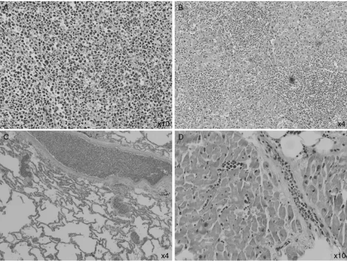
The post-mortem examination revealed infiltration of the vessels of the lungs, heart, and liver, and massive infiltration of the spleen by leukemic blasts (Figure 1) which presented an
292
rev bras hematol hemoter.2 0 1 4;3 6(4):290–292area with hematoma and laceration. Blood volume in the per-itoneal cavity was 500 mL. The adrenal glands were infiltrated by blasts and their cortices had areas of necrosis.
Discussion
The diagnosis of splenic rupture must be considered in patients with splenomegaly due to hematologic malignancies who complain of abdominal pain and present hypoten-sion. Renzulli et al., in a systematic review in which the authors evaluated 632 publications reporting 845 patients with this complication, showed that the major causes of splenic rupture were neoplastic (30.3%), infectious (27.3%), inflammatory (20.0%), drug- and treatment-related (9.2%), and mechanical (pregnancy-related and congestive splenomegaly; 6.8%). In 7.0% of the cases the spleen was normal. More-over, the authors identified that splenomegaly (spleen > 200 g), advanced age and neoplastic disorders were associated with increased mortality.7Acute leukemia is a rare cause of splenic rupture.8 Male patients with AML present splenic rupture more frequently, with a male:female ratio of 3:1.5These fac-tors are in accordance with the case reported here. Curiously, a slightly higher frequency (p-value = 0.08) of leukostasis has been reported in male patients with AML.9This patient was old, male and had splenomegaly; it was later confirmed that he had signs of leukostasis. So, it seems reasonable to hypoth-esize that leukostasis could have played a role in inducing splenic rupture as this complication is related to vessel clog-ging and tissue infiltration by blasts. Only one report of a splenic rupture in a patient with AML and hyperleukocyto-sis (99.2×10⁹/L), and possibly leukostasis, was found in the
literature.8 Recently, it was demonstrated that the spleen is a reservoir of mature and undifferentiated monocytes that assemble in the cords of subcapsular red pulp, which sug-gests that these cells have a tropism for the spleen, and thus contribute to its rupture.10
The patient’s evolution to circulatory shock could be attributed to the splenic rupture, despite the fact that only 500 mL of blood were found in his peritoneal cavity; to leukostasis, as blast cell infiltration was found in the lungs and other organs; to heart failure, as a heart examination revealed areas of myocardial fibrosis and coronary blast cell infiltration;
and perhaps to adrenal insufficiency due to blast infiltration and cortex necrosis. Finally, we do not believe that leukaphere-sis in itself contributed to splenic rupture, as it is essentially atraumatic.
Conflicts of interest
The authors declare no conflicts of interest.
r e f e r e n c e s
1. Kluger Y, Paul DB, Raves JJ, Fonda M, Young JC, Townsend RN, et al. Delayed rupture of the spleen-myths, facts, and their importance: case reports and literature review. J Trauma. 1994;36:568–71.
2. Rabie ME, Hashemey AA, El Hakeem I, Al Hakamy MA, Obaid M, Al Skaini M, et al. Spontaneous rupture of malarial spleen: report of two cases. Mediterr J Hematol Infect Dis.
2010;2:e2010036.
3. Bhaskar E, Moorthy S. Spontaneous splenic rupture in dengue fever with non-fatal outcome in an adult. J Infect Dev Ctries. 2012;6:369–72.
4. Rinderknecht AS, Pomerantz WJ. Spontaneous splenic rupture in infectious mononucleosis: case report and review of the literature. Pediatr Emerg Care. 2012;28:1377–9. 5. Giagounidis AA, Burk M, Meckenstock G, Koch AJ, Schneider
W. Pathologic rupture of the spleen in hematologic malignancies: two additional cases. Ann Hematol. 1996;73:297–302.
6. Brown SL, Dale DC. Spontaneous splenic rupture following administration of granulocyte colony-stimulating factor (G-CSF): occurrence in an allogeneic donor of peripheral blood stem cells. Biol Blood Marrow Transplant. 1997;3:341–3. 7. Renzulli P, Hostettler A, Schoepfer AM, Gloor B, Candinas D.
Systematic review of atraumatic splenic rupture. Br J Surg. 2009;96:1114–21.
8. Tan A, Ziari M, Salman H, Ortega W, Cortese C. Spontaneous rupture of the spleen in the presentation of acute myeloid leukemia. J Clin Oncol. 2007;25:5519–20.
9. De Santis GC, Benicio MT, Oliveira LC, Falcao RP, Rego EM. Genetic mutations in patients with acute myeloid leukemia and leukostasis. Acta Haematol. 2013;130:95–7.
