Rearing five species of Diptera (Calliphoridae) of forensic
importance in Colombia in semicontrolled field conditions
María C. Vélez
1Marta Wolff
1,2AbstRACt
The family Calliphoridae is widely known to lead the colonization of corpses and their development rates are frequently used to estimate the postmortem interval. This study presents the larval growth of five forensically important species of Calliphoridae in Colombia. Rearing took place in semicontrolled field conditions where the egg masses were collected. We show curves of larval growth, larval length and time intervals to reach all immature stages for
Lucilia eximia and Cochliomyia macellaria at two sites with different climatic conditions and for Chrysomya albiceps, Chrysomya megacephala and Calliphora nigribasis at one site. Overall, high temperatures speeded up the development of the species reared at two different sites, whereas low temperatures for C. nigribasis, lengthened the total development time. Differences between this study and others can be explained by the experimental conditions in the field without the possibility of strict laboratory rearing controls.
Keywords: Lucilia eximia, Cochliomyia macellaria, Chrysomya spp., Calliphora nigribasis,
larval growth.
mologist to make more positive species identifica‑ tions, particularly with fly larvae as definitive species determination cannot be made given the lack of dis‑ tinct morphological differences. It also allows a clear‑ er determination of the postmortem interval (PMI) since rearing of subsequent life stages provides a better approximation of the development stage of the insects at the time of collection (Byrd, 2001).
Larval lengths or weights can be used to estimate PMI under two approaches. The first is to measure the accumulated amount of days or hours needed to reach a particular stage of development (Ames & Turner, 2003) and the second is the use of an iso‑
IntRoDuCtIon
The estimation of postmortem interval is the main application of forensic entomology. Therefore, major knowledge requirements are the species associ‑ ated with decomposing corpses in different biogeocli‑ matic zones, their development rates with emphasis on development times of each immature stage (Tur‑ chetto et al., 2001) as well as lengths and weights of the different larval stages (Wells & Lamotte, 2001).
The laboratory rearing of insects collected from a death scene is an integral component of the analysis of entomological evidence. Rearing allows the ento‑
Grupo de Entomología, Universidad de Antioquia. A.A. 1226, Medellín, Colombia.
Corresponding author: Marta Wolff, Dr.Sc., Instituto de Biología, Universidad de Antioquia, A.A. 1226, Medellín, Colombia, Tel. (576)2105662. E‑mail: [email protected]
megalen diagram, i.e. matching the size of the larvae found on the corpse with the development rates for the same blowfly larvae experimentally reared at the seasonal average temperature in which the body was
found (Introna et al., 1989). However, field tempera‑
tures are rarely constant and it has been shown that fluctuating temperature has a great effect on larval development (Introna et al., 1989).
The family Calliphoridae is known as the initial colonizer in the faunal succession on human cadavers (Smith, 1986). Therefore, they are the primary and most accurate forensic indicators of time of death and their development rates are needed to allow more precise PMI estimates (Grassberger & Reiter, 2001). Thus, it is necessary that the development rates for the species involved in decomposition be known for the same conditions of temperature at the possible crime scene (Introna et al., 1989).
The main purpose of this study was the rearing
in situ of the first species to colonize pig corpses in decomposition in four different bioclimatic zones in Colombia. This is the first approach to the applica‑ tion of larval growth as a tool in PMI estimates.
MAteRIAl AnD MethoDs
All of the eggs were collected on pigs (by per‑ mission of the Ethical Committee of Universidad de Antioquia, Medellín) in the early stages of decompo‑ sition and larvae were reared at the same site as the corpses.
The study was carried out at four sites: Tintipán Island, municipality of Gomez Plata, city of Medellín and Chingaza National Park.
Tintipán Island (beach environment) is located off the Caribbean coast within the San Bernardo ar‑ chipelago. The area is designated as tropical very dry forest (bms‑T) (Holdridge, 1987), with mangroves as the predominant flora, it has an altitude of 2 m above sea level, an average rainfall of 1000 mm and an aver‑ age temperature of 27°C.
Gomez Plata (rural environment) is located at 75 km from Medellín. It is designated as tropical pre‑ montane wet forest (bmh‑P) (Espinal, 1985). The site is open pasture used by cattle, at 1080 m above sea level with an average temperature of 25°C, relative hu‑ midity of 80% and an average rainfall of 1800 mm.
The city of Medellín (urban environment) is designated tropical premontane moist forest (bh‑P) (Holdridge, 1987). It is located at 1450 m above sea level with an average temperature of 24°C and an av‑ erage rainfall of 1409 mm.
Chingaza National Park (paramo zone) is at 3035 m above sea level, it has an average temperature of 10°C and relative humidity superior to 80% (IN‑ DERENA, 1986).
The egg masses of approximately 100 to 130 in‑ dividuals were placed in plastic jars with 150 g of beef liver. The jars were placed in containers of polystyrene to avoid contamination with other insects, to mini‑ mize drastic changes in temperature and to eliminate the effects of sun exposure. The larvae did not have a light source within the containers.
Each plastic jar was sampled as follows: the time was noted at the moment of egg collection (we as‑ sumed this moment to be oviposition). During the first 36 hours, we observed the moment of eclosion and removed samples every three hours between 7 am and 7 pm in order to get sufficient larvae at the first stage of development. These larvae growth faster and observations need to be made at shorter intervals (Krüger et al., 2003). After the first 36 hours, sam‑ pling continued every six hours. The samples consist‑ ed of ten larvae per plastic jar at all of the evaluated sites, except Medellín, where each sample consisted of four larvae. When the larvae reached the postfeed‑ ing stage, we placed sawdust in the jars (Dale & Pru‑ dot, 1986) and made observations every 12 hours to verify the presence of pupae and the emergence of adults. During the postfeeding stage we stopped the sampling to prevent handling just before pupation at which time development can be delayed or blocked (Anderson, 2000). The container and environmental temperatures were measured at each sampling time and the average temperature of rearing was calculated from these data.
All of the samples were fixed in 80% ethanol and examined under a binocular stereoscope to iden‑ tify the species with the use of different keys (Shewell,
1981; Dear, 1985; Smith, 1986; Wells et al., 1999).
All larvae were measured (total body lengths) and the accumulated time of development was calculated for all the instars for all the sample jars. Line graphs of larval growth, with intervals for the ages (in hours) and the lengths of each immature stage were plotted using JMP 3.2.2 (SAS Institute Inc., 1997). All insect material was deposited in the Entomological Collec‑ tion of the Universidad de Antioquia in Medellín.
Results
During this study, five species belonging to Cal‑ liphoridae were reared: Lucilia eximia (Wiedemann,
Chrysomya albiceps (Wiedemann, 1819), Chrysomya megacephala (Fabricius, 1794) and Calliphora nigriba-sis Macquart, 1851. L. eximia and C. macellaria were reared at two different sites with different temperature regimes (L. eximia at 23.13 ± 2.45°C, in Medellín
and 25.30 ± 3.26°C, in Gómez Plata; C. macellaria at
25.30 ± 3.26°C, in Gómez Plata and 30.74 ± 0.71°C, in Tintipán). C. albiceps, C. megacephala and C. nigri-basis, were reared at 25.30 ± 3.26°C, in Gomez Plata, 23.13 ± 2.45°C, in Medellín and 10.62 ± 2.51°C, in Chingaza, respectively.
In all cases, development time for the eggs and the moment of eclosion was obtained (Table 1). For
L. eximia, C. macellaria, C. albiceps and C. megaceph-ala, the eclosion was near to 15 hours subsequent to the oviposition, whereas C. nigribasis needed 64 hours.
The larval growth represented by the progressive increase in length is showed in larval growth curves (Figs. 1‑7). The average and standard deviations of the lengths and the development times guarantee the fact of find a larva in a particular stage (Table 2 and 3).
L. eximia reached higher lengths when it was reared at 23.13°C in urban environment (Fig. 1) in compari‑ son with its rearing at 25.30°C in rural environment
(Fig. 2), whereas the lengths of third instar larvae of
C. macellaria showed overlapped intervals between beach environment at 30.74°C and rural environ‑ ment at 25.30°C (Fig. 3, 4 and Table 2). Instead, the postfeeding larvae characterized by a decrease in size (Greenberg & Kunich, 2002) (Figs. 3 and 4), showed narrower and not overlapped intervals for the length (Table 2).
With regard to the accumulated time of devel‑ opment for immature individuals to reach all instars
(Table 3), we observed that L. eximia exhibited suc‑
cessful growth for all larval instars in both rearings, according to the larval growth curves (Figs. 1 and 2). However, we did not obtain pupae or adults. At
tAble 1: Time of eclosion (in hours) of the species at all development temperatures.
Species Temperature (°C) Egg duration (h)
Lucilia eximia 23.13±2.45 15.16±0.77
Lucilia eximia 25.30±3.26 16.42±2.31
Cochliomyia macellaria 30.74±0.71 15.80±2.91
Cochliomyia macellaria 25.30±3.26 15.82±1.50
Chrysomya albiceps 25.30±3.26 15.00±1.73
Chrysomya megacephala 23.13±2.45 15.00±0.71
Calliphora nigribasis 10.62±2.51 64.02±1.79
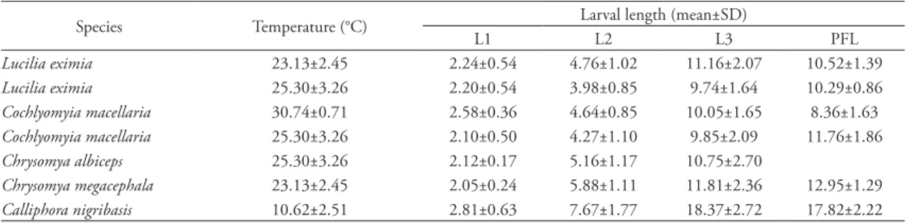
tAble 2: Length (in mm) for the immature stages of the species at all development temperatures.
Species Temperature (°C) Larval length (mean±SD)
L1 L2 L3 PFL
Lucilia eximia 23.13±2.45 2.24±0.54 4.76±1.02 11.16±2.07 10.52±1.39
Lucilia eximia 25.30±3.26 2.20±0.54 3.98±0.85 9.74±1.64 10.29±0.86
Cochlyomyia macellaria 30.74±0.71 2.58±0.36 4.64±0.85 10.05±1.65 8.36±1.63
Cochlyomyia macellaria 25.30±3.26 2.10±0.50 4.27±1.10 9.85±2.09 11.76±1.86
Chrysomya albiceps 25.30±3.26 2.12±0.17 5.16±1.17 10.75±2.70
Chrysomya megacephala 23.13±2.45 2.05±0.24 5.88±1.11 11.81±2.36 12.95±1.29
Calliphora nigribasis 10.62±2.51 2.81±0.63 7.67±1.77 18.37±2.72 17.82±2.22 L1, first larval instar; L2, second larval instar; L3, third larval instar; PFL, postfeeding larvae.
tAble 3: Time to reach the immature stages (in h) for the species at all temperatures.
Species Temp. (°C) Time to reach the immature stage (mean ± DS)
L1 L2 L3 PP P A
Lucilia eximia 23.13±2.45 20.69±4.93 41.82±8.84 57.88±10.09 101.45±19.23 †a †a
Lucilia eximia 25.30±3.26 20.31±4.14 30.93±5.23 55.77±9.77 90.09±9.83 †a †a
Cochliomyia macellaria 30.74±0.71 17.19±3.16 24.69±3.35 54.20±11.08 95.37±11.94 86–120 103±24.04
158–167 162.5±6.36
Cochliomyia macellaria 25.30±3.26 22.24±5.86 44.48±8.87 78.03±11.61 111.25±12.31 116–206 161±63.64
257‑332 294.5±53.03
Chrysomya albiceps 25.30±3.26 19.38±4.26 48.88±8.34 89.75±19.42 192–239 215.5±33.23
332–352 342±14.14
Chrysomya megacephala 23.13±2.45 18.60±3.44 45.04±9.23 65.58±12.22 107.26±13.68 164–309 236.5±102.53
281–357 319±53.74
Calliphora nigribasis 10.62±2.51 111.54±31.41 205.47±24.32 318.11±42.12 551.79±95.00 1324b 2664b a Pupae and adults did not appear for this species.
25.30°C, growth took less time (90.09 ± 9.83 hours) than at 23.13°C (101.45 ± 19.23 hours) (Table 3).
C. macellaria showed successful development to the emergence of the adults at the two different tempera‑ tures (Figs. 3 and 4). At 30.74°C, development was faster, concluding after 162.5 hours. Also, greater synchrony was shown for the accumulated time inter‑ vals of each stage (Table 3). At 25.30°C the time for total development was 294.5 hours. For this species, a difference of 5°C can alter development velocity by
132 hours, according to the present study. C. albiceps
completed its development to the adult stage in 332 hours in rural environment (Table 3). The postfeed‑ ing larvae are not appreciable in the curve, as there was no decrease in its length at the end of the larval
growth (Fig. 5 and Table 2). C. megacephala reached
the adult stage at 23.13°C in urban environment. Appearance of pupae took place in a wide interval (between 164 and 309 hours); therefore we assumed that the pupation was not very synchronic (Table 3). The opposite occurred with the adults, appear‑ ing over a narrower interval (between 281 and 357
FIguRe 1: Larval growth of Lucilia eximia at 23.13 ± 2.45°C. FIguRe 2: Larval growth of Lucilia eximia at 25.30 ± 3.26°C.
FIguRe 3: Larval growth of Cochliomyia macellaria at 30.74 ± 0.71°C.
FIguRe 4: Larval growth of Cochliomyia macellaria at 25.30 ± 3.26°C.
FIguRe 5: Larval growth of Chrysomya albiceps at 25.30 ± 3.26°C.
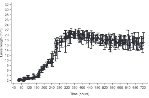
hours). All larval instars were identified, including the postfeeding larvae that are visible in the larval growth curve (Fig. 6). Finally, C. nigribasis showed a different behavior, given that it is a typical species from the Andean zone, in high mountains and low temperatures; hence the development was successful at 10.62°C (Fig. 7). Nevertheless, the colony provid‑ ing the larval growth curve died after several days in a postfeeding larval stage, possibly because they en‑ tered diapause after missing the main conditions for pupation (Krüger et al., 2003). The data presented here (Table 3) was taken from another group of lar‑ vae that were not sampled but were reared under the same conditions.
DIsCussIon
This study is the first report for Colombia on the larval growth of the principal species to arrive at a corpse (Wolff et al., 2001; Perez et al., 2005; Martinez
et al., 2007) in four different bioclimatic zones: L. ex-imia at 23.13°C and 25.30°C in Medellín and Go‑ mez Plata, respectively; C. macellaria at 25.30°C and 30.74°C in Gomez Plata and Tintipán, respectively;
C. albiceps at 25.30°C in Gomez Plata; C. megacepha-la at 23.13°C in Medellín and C. nigribasis at 10.62°C in Chingaza. L. eximia seems to be very sensitive to stress and in the present study this could have been caused by the lack of a successful migration event and posterior burial. This species, as others of the subfam‑ ily Calliphorinae (Greenberg, 1990), has been charac‑ terized by displaying strong intentions to escape from sample jars, showing their need for migration away from the food source and thus explaining the mass movement of larvae during the postfeeding stage in
the jars (Anderson, 2000; Gomes et al, 2003; Gomes
& Zuben, 2004). The absence of this migration re‑ quirement given by the small space, and the absence
of burial given by the kind of substrate to pupate (sawdust instead sand), could explain the failure to reach pupation and the adult stage. For this species, the time taken for maximum larval growth at 25.30°C was similar to the results obtained for Lucilia sericata
(a closely related species), reported by Grassberger & Reiter (2001). The times obtained for its rearing at 23.13°C agree with Anderson (2000) for the first and
second instar of development. However, L. eximia is
different from L. sericata, because the first, prefers tropical and warm environments, becoming an in‑ dicator species of urban and rural premontane biotic zones.
The development of C. macellaria demonstrat‑
ed how the total duration of development decreases with increasing temperature within the optimal range (Chapman, 1982). These results are also in agreement with other studies where an increase in temperature within the optimal range can produce accelerated development (Byrd & Allen, 2001; Grassberger & Reiter, 2002a; Grassberger et al., 2003; Krüger et al.,
2003). Considering that Cochliomyia is a neotropical
genus with a wide distribution (Dear, 1985), that ex‑ hibits preferences for warm and moist environments and it is dominant in rural areas (Prudot & Dale, 1987) it is expected to adapt to the different tempera‑ ture regimes produced by the fluctuations in rural and beach environments like Gomez Plata and Tintipan, respectively.
The times for pupation and emergence of the adults of C. albiceps (8‑10 days and 13‑14 days, re‑ spectively) are similar to that obtained by Grassberger
et al. (2003) at 25°C. They show times from oviposi‑ tion to pupation and emergence of the adults of 8 days and 13 days, respectively. Baumgartner & Greenberg (1985) showed the presence of this species since pre‑ montane to montane biotic zones. This information encloses the area where we found it (Gomez Plata at 1080 m) and reveals an altitudinal adaptation where the temperature is the main factor that influences the rate of development.
The rearing of C. megacephala showed similari‑
ties with Wells & Kurahashi (1994), with regard to the moment of appearance of the pupae. They report‑ ed a maximum time of 144 to 198 hours, which cor‑ responds to the lower value of the interval obtained in the present study. However, this study reported a lon‑ ger time for the emergence of the adults when com‑ pared to the same authors. This discrepancy could be sustained by the difference between the temperatures (27°C in Wells and Kurahashi and 23.13°C in this study). In Medellín, this species is commonly encoun‑ tered, showing a preference for urban areas.
C. nigribasis was reared at 10.62°C, with a de‑ velopment time of 111 days. Studies carried out with other species with the purpose of evaluating the ef‑ fects of temperature on development, clearly show how a decrease in temperature can decrease metabo‑ lism, increasing the time intervals for the immature stages (Anderson, 2000; Byrd & Allen, 2001; Grass‑ berger & Reiter, 2001; Grassberger & Reiter, 2002a; Grassberger & Reiter, 2002b; Ames & Turner, 2003; Grassberger et al., 2003). Hence, it is essential to con‑ sider that this species belongs to the Andean region (including paramo region) (Baumgartner & Green‑ berg, 1985) and it is part of the cadaverous entomo‑ fauna of these regions, becoming an important foren‑ sic indicator.
There are many differences between this study and others treating the same issue. This disparity can be given by the experimental conditions, such as the food source and the photoperiod. Not only do we have to consider differences in the methods (extrinsic factors), but we also have to take into account intrinsic factors such as geographic adap‑ tation, temperature regimes, feeding and density. Variations in these parameters exist between dif‑ ferent geographic populations of the same species (Grassberger & Reiter, 2002a). The extrapolation of results obtained in the laboratory for field condi‑ tions should be carried out with caution given that lab conditions can eliminate competitors and have optimal abiotic conditions, such as controlled tem‑ perature, (Anderson, 2000) which produces higher
viability (Krüger et al., 2003). Because the present
study worked with species commonly found in the studied areas and the semicontrolled conditions allow the influence of the regional temperatures, the development data from this work becomes an important database for the application of forensic entomology, considering that it is important that results used in the solution of forensic cases, be ob‑ tained from field conditions, where temperatures have cyclic fluctuations modifying development times in different ways when compared with con‑ stant temperatures (Introna et al., 1989). Further‑ more, they better resemble the conditions of a body found in decomposition.
It is also important to consider the effects of the preservative solutions. It is known that the use of 70% alcohol produces shrinkage of the larvae, modifying their size and the estimation of age using length (Tan‑ tawi & Greenberg, 1993). Even so, it is important to consider that samples obtained in Colombia from the crime scenes are preserved in alcohol and our results could be applied.
Additionally, this study shows that rearing these species in field conditions can be successful, and that the results obtained could easily be used in forensic entomology, providing valuable information in the determination of the postmortem interval.
ConClusIon
In general, developmental times from oviposi‑ tion to emergence might differ depending on many factors. Geographic variations and climate conditions can influence adaptations explaining the differences in the development at different temperatures. High temperatures accelerate the growth and development, whereas low temperatures slow it down. Alternatively, the rearing in field conditions allows a more precise approximation to the conditions of growth in a real forensic case. However, more studies are necessary to understand the behavior during the development of the immature stages, like the degree‑days estimates and the knowledge of the intrinsic and extrinsic fac‑ tors that could affect the development of the forensi‑ cally important species.
ResuMen
La familia Calliphoridae es ampliamente conocida por liderar la colonización de los cadáveres y sus tasas de desarrollo son frecuentemente utilizadas para estimar el intervalo postmortem. Este estudio presenta el crecimiento larval de cinco especies de Calliphoridae de importancia forense en Colombia, considerando que la cría se dio en condiciones de campo semicontroladas en los lugares donde las masas de huevos fueron colectadas. Mostramos también, los intervalos de longitud y el tiempo empleado en alcanzar todos los estadios inmaduros para Lucilia eximia y Cochliomyia macellaria en dos lugares con diferentes condiciones climáticas y Chrysomya albiceps, Chrysomya megacephala y Calliphora nigribasis, en un solo lugar. En general, las altas temperaturas producen una aceleración en el desarrollo de las especies criadas en dos sitios diferentes, mientras que bajas temperaturas para C. nigribasis, alargaron el tiempo utilizado para completar el desarrollo. Las diferencias entre este estudio y otros, pueden estar dadas por las condiciones experimentales presentadas en campo sin los controles estrictos que son posibles durante la cría en laboratorio.
Palabras‑claves: Lucilia eximia, Cochliomyia
macellaria, Chrysomya spp., Calliphora nigribasis,
ACknoWleDgeMents
This study received financial support from Col‑ ciencias (Instituto Colombiano para el Desarrollo de la Ciencia y la Tecnología), project 1115‑05‑11503 and from the Universidad de Antioquia, Colombia.
ReFeRenCes
Ames, C. & Turner, D. 2003. Low temperature episodes in development of blowflies: implications for postmortem interval estimation. Medical and Veterinary, 17:178‑186. Anderson, G. 2000. Minimum and maximum development of
some forensically important Calliphoridae (Diptera). Journal of Forensic Science, 45(4):824‑832.
Baumgartner, D.L. & Greenberg, B. 1985. Distribution and medical ecology of the blow flies (Diptera: Calliphoridae) of Perú. Annals of the Entomological Society of America,
78:565‑587.
Byrd, J. 2001. Laboratory rearing of forensic insects. In: Byrd, J. & Castner, J. (Editors), Forensic Entomology: The Utility Of Arthropods In Legal Investigations. CRC Press, New York, p. 121‑140.
Byrd, J. & Allen, J. 2001. The development of the black blow fly, Phormia regina (Meigen), Forensic Science International,
120:79‑88.
Chapman, R.F. 1982. The Insects: Estructure and Function. Third edition. Harvard University Press, Cambridge.
Dale, W. & Prudot, E. 1986. Apuntes sobre la biología de las moscas Calliphoridae en la costa central peruana, Revista Peruana de Entomología, 29:105‑111.
Dear, J. 1985. A revisión of the new world Chrysomyini (Diptera: Calliphoridae). Revista Brasileira de Zoologia, 3(3):109‑169. Espinal, L.S. 1985. Geografía Ecológica del Departamento de
Antioquia. Revista de la Facultad Nacional de Agronomía Medellín, 38:68‑69.
Gomes, L.; Von Zuben, C.J. & Sanches, M.R. 2003. Estudo da dispersão larval radial pós‑alimentar em Chrysomya megacephala
(Fabricius) (Diptera, Calliphoridae). Revista Brasileira de Entomologia, 47(2):229‑234.
Gomes, L. & Von Zuben, C.J. 2004. Dispersão larval radial pós‑alimentar em Lucilia cuprina (Diptera, Calliphoridae): profundidade, peso e distância de enterramento para pupação.
Iheringia, Série Zoologia, 94(2):135‑138.
Grassberger, M. & Reiter, C. 2001. Effect of temperature on
Lucilia Sericata (Diptera: Calliphoridae) development with special reference to the isomegalen‑ and isomorphen‑diagram.
Forensic Science International, 120:32‑36.
Grassberger, M. & Reiter, C. 2002a. Effect of temperature on development of Liopygia (=Sarcophaga) argyrostoma
(Robineau‑Desvoidy) (Diptera: Sarcophagidae) and its forensic implications. Journal of Forensic Science, 47(6):1332‑1336. Grassberger, M. & Reiter, C. 2002b. Effect of temperature on
development of the forensically important holartic blow fly
Protophormia terraenovae (Robineau‑Desvoidy) (Diptera: Calliphoridae). Forensic Science International, 128:177‑182. Grassberger, M.; Friedrich, E. & Reiter, C. 2003. The blowfly
Chrysomya albiceps (Wiedemann) (Diptera: Calliphoridae) as a new indicator in Central Europe. International Journal of Legal Medicine, 117:75‑81.
Greenberg, B. 1990. Behavior of postfeeding larva of some Calliphoridae and a muscid (Diptera). Annals of the Entomological Society of America, 83:1210‑1214.
Greenberg, B. & Kunich, J.C. 2002. Entomology and the law:
Flies as forensic indicators. University press, Cambridge. Holdridge, L.R. 1987. Ecología basada en zonas de vida. Instituto
Interamericano de Cooperación para la Agricultura, Costa Rica.
INDERENA – Instituto Nacional de Recursos Naturales Renovables y del Ambiente. 1986. Parque Nacional Natural Chingaza. Santa Fé de Bogotá, Colombia.
Introna, F. Jr.; Altamura, B.; Dell’Erba, A. & Dattoli, V. 1989. Time since death definition by experimental reproduction of
Lucilia sericata cycles in growth cabinet. Journal of Forensic Science, 34(2):478‑480.
Krüger, R.F.; Ribeiro, P.B. & Peixoto Costa, P.R. 2003. Ciclo de vida de Sarcophaga (Liopygia) crassipalpis (Macquart) (Diptera: Sarcophagidae). Entomología y Vectores, 10(1):85‑98. Martinez, E.; Duque, P. & Wolff, M. 2007. Succession pattern of
carrion‑feeding insects in Paramo, Colombia. Forensic Science International, 166:182‑189.
Perez, S.P.; Duque, P. & Wolff, M. 2005. Successional behavior and occurrence matrix of carrion‑associated arthropods in the urban area of Medellín, Colombia. Journal of Forensic Science,
50(2):448‑454.
Prudot, E. & Dale, W. 1987. Estudio ecologico de las moscas Calliphoridae en el Valle de Huaral. Revista Peruana de Entomología, 28:93‑99.
SAS Institute Inc. 1997. JMP. versión 3.2.2. Available at: <http:// www.JMPdiscovery.com>.
Shewell, G.E. 1981. Calliphoridae. In: McAlpine, J.F. (Editor),
Manual of Neartic Diptera. Minister of Supply and Services, Canada, v. 2, p. 1133‑1145.
Smith, K. 1986. A Manual of Forensic Entomology. The Trustees of the British Museum (Natural History), London.
Tantawi, T.I. & Greenberg, B. 1993. The effect of killing and preservative solutions on estimates of maggot age in forensic cases. Journal of Forensic Science, 38(3):702‑707.
Turchetto, M.; Lafisca, S. & Constantini, G. 2001. Postmortem interval (PMI) determined by study sarcophagous biocenoses: three cases from the province of Venice (Italy). Forensic Science International, 120:28‑31.
Wells, J. & Kurahashi, H. 1994. Chrysomya megacephala
(Fabricius) (Diptera: Calliphoridae) development: rate, variation and the implications for forensic entomology.
Japanese Journal of Sanitary Zoology, 45(4):303‑309.
Wells, J.D.; Byrd, J.H. & Tantawi, T.I. 1999. Key to third‑instar Chrysomyinae (Diptera: Calliphoridae) from carrion in the continental United states. Journal of Medical Entomology,
36(5):638‑641.
Wells, J. & Lamotte, L. 2001. Estimating the postmortem interval. In: Byrd, J. & Castner, J. (Editors), Forensic Entomology: the utility of arthropods in legal investigations. CRC Press, New York, p. 263‑285.
Wolff, M.; Uribe, A.; Ortiz, A. & Duque, P. 2001. A preliminary study of forensic entomology in Medellín, Colombia. Forensic Science International, 120:53‑59.
Recebido em 18.10.2007 Aceito em 12.12.2007 Impresso em: 24.03.2008
Seção
de
Publicações
d
o
M
Ipiranga, CEP 04263‑000, São Paulo, SP, Brasil.
editor‑in‑Chief: Hussam Zaher, Serviço de Vertebrados, Museu de Zoologia, Universidade
de São Paulo, Post Office Box 42.494, CEP 04218‑970, São Paulo, SP, Brasil. E‑mail: [email protected].
Managing editor: Carlos José Einicker Lamas (Museu de Zoologia, Universidade de São
Paulo, Brasil).
Associate editors: Mário César Cardoso de Pinna (Museu de Zoologia, Universidade de
São Paulo, Brasil); Marcos Domingos Siqueira Tavares (Museu de Zoologia, Universidade de São Paulo, Brasil); Sergio Antonio Vanin (Museu de Zoologia, Universidade de São Paulo, Brasil).
editorial board: Aziz Nacib Ab’Saber (Universidade de São Paulo, Brasil); Rüdiger Bieler
(Field Museum of Natural History, U.S.A.); Walter Antonio Pereira Boeger (Universidade Federal do Paraná, Brasil); Carlos Roberto Ferreira Brandão (Universidade de São Paulo, Brasil); James M. Carpenter (American Museum of Natural History, U.S.A.);
(Universidade de São Paulo, Brasil); Marcos André Raposo Ferreira (Museu Nacional, Rio de Janeiro, Brasil); Darrel R. Frost (American Museum of Natural History, U.S.A.); William R. Heyer (National Museum of Natural History, U.S.A.); Ralph W. Holzenthal (University of Minnesota, U.S.A.); Adriano Brilhante Kury (Museu Nacional, Rio de Janeiro, Brasil); Gerardo Lamas (Museo de Historia Natural “Javier Prado”, Lima, Peru); John G. Maisey (American Museum of Natural History, U.S.A.); Antonio Carlos Marques (Universidade de São Paulo, Brasil); Naércio Aquino Menezes (Universidade de São Paulo, Brasil); Christian de Muizon (Muséum National d’Histoire Naturelle, Paris, France); Nelson Papavero (Universidade de São Paulo, Brasil); James L. Patton (University of California, Berkeley, U.S.A.); Richard O. Prum (University of Kansas, U.S.A.); Olivier Rieppel (Field Museum of Natural History, U.S.A.); Miguel Trefaut Urbano Rodrigues (Universidade de São Paulo, Brasil); Randall T. Schuh (American Museum of Natural History, U.S.A.); Luís Fábio Silveira (Universidade de São Paulo, Brasil); Ubirajara Ribeiro Martins de Souza (Universidade de São Paulo, Brasil); Paulo Emílio Vanzolini (Universidade de São Paulo, Brasil); Richard P. Vari (National Museum of Natural History, U.S.A.).
InstRuCtIons to AuthoRs (April 2007)
general Information:Papéis Avulsos de Zoologia (PAZ) and Arquivos de Zoologia (AZ)
cover primarily the fields of Zoology, publishing original contributions in systematics, paleontology, evolutionary biology, ontogeny, faunistic studies, and biogeography. Papéis Avulsos de Zoologia and Arquivos de Zoologia also encourage submission of theoretical and empirical studies that explore principles and methods of systematics.
All contributions must follow the International Code of Zoological Nomenclature. Relevant specimens should be properly curated and deposited in a recognized public or private, non‑profit institution. Tissue samples should be referred to their voucher specimens and all nucleotide sequence data (aligned as well as unaligned) should be submitted to GenBank (www.ncbi.nih.gov/Genbank) or EMBL (www.ebi.ac.uk).
Peer Review: All submissions to Papéis Avulsos de Zoologia and Arquivos de Zoologia are
subject to review by at least two referees and the Editor‑in‑Chief. All authors will be notified of submission date. Authors may suggest potential reviewers. Communications regarding acceptance or rejection of manuscripts are made through electronic correspondence with the first or corresponding author only. Once a manuscript is accepted providing changes suggested by the referees, the author is requested to return a revised version incorporating those changes (or a detailed explanation of why reviewer’s suggestions were not followed) within fifteen days upon receiving the communication by the editor.
Proofs: Page‑proofs with the revised version will be sent to e‑mail the first or corresponding
author. Page‑proofs must be returned to the editor, preferentially within 48 hours. Failure to return the proof promptly may be interpreted as approval with no changes and/or may delay publication. Only necessary corrections in proof will be permitted. Once page proof is sent to the author, further alterations and/or significant additions of text are permitted only at the author’s expense or in the form of a brief appendix (note added in proof ).
submission of Manuscripts: Manuscripts should be sent to the e‑mail of the Editor‑in‑
Chief [email protected], along with a submission letter explaining the importance and originality of the study. Address and e‑mail of the corresponding author must be always updated since it will be used to send the 50 reprints in titled by the authors. Figures, tables and graphics should not be inserted in the text. Figures and graphics should be sent in separate files with the following formats: “.jpg” and “.tif ” for figures, and “.xls” and “.cdr” for graphics, with 300 dpi of minimum resolution. Tables should be placed at the end of the manuscript.
Manuscripts are considered on the understanding that they have not been published or will not appear elsewhere in substantially the same or abbreviated form. The criteria for acceptance of articles are: quality and relevance of research, clarity of text, and compliance with the guidelines for manuscript preparation.
Manuscripts should be written preferentially in English, but texts in Portuguese or Spanish will also be considered. Studies with a broad coverage are encouraged to be submitted in English. All manuscripts should include an abstract and keywords in English and a second abstract and keywords in Portuguese or Spanish.
Authors are requested to pay attention to the instructions concerning the preparation of the manuscripts. Close adherence to the guidelines will expedite processing of the manuscript.
Manuscript Form: Manuscripts should not exceed 150 pages of double‑spaced, justified
text, with size 12 and source Times New Roman (except for symbols). Page format should be A4 (21 by 29.7 cm), with 3 cm of margins. The pages of the manuscript should be numbered consecutively.
The text should be arranged in the following order: Title Page, Abstracts with Keywords, Body of Text, Literature Cited, Tables, Appendices, and Figure Captions. Each of these sections should begin on a new page.
(1) title Page: This should include the title, short title, author(s) name(s) and institutions.
The title should be concise and, where appropriate, should include mention of families and/or higher taxa. Names of new taxa should not be included in titles.
(2) Abstract: All papers should have an abstract in English and another in Portuguese
or Spanish. The abstract is of great importance as it may be reproduced elsewhere. It should be in a form intelligible if published alone and should summarize the main facts, ideas, and conclusions of the article. Telegraphic abstracts are strongly discouraged. Include all new taxonomic names for referencing purposes. Abbreviations should be avoided. It should not include references. Abstracts and keywords should not exceed 350 and 5 words, respectively.
(3) body of text: The main body of the text should include the following sections:
Introduction, Material and Methods, Results, Discussion, Conclusion, Acknowledgments, and References at end. Primary headings in the text should be in capital letters, in bold and centered. Secondary headings should be in capital and lower case letters, in bold and centered. Tertiary headings should be in capital and lower case letters, in bold and indented at left. In all the cases the text should begin in the following line.
(4) literature Cited: Citations in the text should be given as: Silva (1998) or Silva
(1998:14‑20) or Silva (1998: figs. 1, 2) or Silva (1998a, b) or Silva & Oliveira (1998)
or (Silva, 1998) or (Rangel, 1890; Silva & Oliveira, 1998a, b; Adams, 2000) or (Silva, pers. com.) or (Silva etal., 1998), the latter when the paper has three or more authors. The reference need not be cited when authors and date are given only as authority for a taxonomic name.
(5) References: The literature cited should be arranged strictly alphabetically and given in
the following format:
• Journal Article – Author(s). Year. Article title. Journal name, volume: initial page‑
final page. Names of journals must be spelled out in full.
• books – Author(s). Year. Book title. Publisher, Place.
• Chapters of books – Author(s). Year. Chapter title. In: Author(s) ou Editor(s), Book
title. Publisher, Place, volume, initial page‑final page.
• Dissertations and theses – Author(s). Year. Dissertation title. (Ph.D. Dissertation).
University, Place.
• electronic Publications – Author(s). Year. Title. Available at: <electronic address>.
Access in: date.
tables: All tables must be numbered in the same sequence in which they appear in text.
Authors are encouraged to indicate where the tables should be placed in the text. They should be comprehensible without reference to the text. Tables should be formatted with vertical (portrait), not horizontal (landscape), rules. In the text, tables should be referred as Table 1, Tables 2 and 3, Tables 2‑6. Use “TABLE” in the table heading.
Illustrations: Figures should be numbered consecutively, in the same sequence that they
appear in the text. Each illustration of a composite figure should be identified by capital letters and referred in the text as: Fig. 1A, Fig. 1B, for example. When possible, letters should be placed in the left lower corner of each illustration of a composite figure. Hand‑ written lettering on illustrations is unacceptable. Figures should be mounted in order to minimize blank areas between each illustration. Black and white or color photographs should be digitized in high resolution (300 dpi at least). Use “Fig(s).” for referring to figures in the text, but “FIGURE(S)” in the figure captions and “fig(s).” when referring to figures in another paper.
Responsability: Scientific content and opinions expressed in this publication are sole responsibility of the respective authors.
Copyrights: A concession letter of copyrights and assent should be sent to the Editor, signed by all the authors, prior to publication of the manuscript.
A model is available in the home page of the Museu de Zoologia da Universidade de São Paulo.
For other details of manuscript preparation of format, consult the CBE Style Manual, available from the Council of Science Editors (www.councilscienceeditors.org/publications/style.cfm).