EPIDEMIOLOGY AND ANTIMICROBIAL RESISTANCE OF STAPHYLOCOCCI ISOLATED FROM DIFFERENT INFECTIOUS DISEASES
Gamal Fadl M. Gad 1, Abd El-Ghafar F.Abd El-Ghafar 2, Ramadan A. A. El-Domany 3, Zeinab Shawky Hashem 1*
1
Microbiology Department, Faculty of Pharmacy, El-Minia University, Egypt; 2 Microbiology Department, Faculty of Medicine,
El-Minia University, Egypt; 3 Microbiology Department, Faculty of Pharmacy, Helwan University, Egypt.
Submitted: June 19, 2009; Returned to authors for corrections: July 17, 2009; Approved: October 06, 2009.
ABSTRACT
A total of 187 isolates from 470 clinical specimens were collected from three hospitals in El-Minia
governorate and identified as 132 Staphylococcus aureus strains and 55 coagulase-negative staphylococci
(CoNS) strains. Susceptibility of isolates to antimicrobial agents was tested by the agar dilution method.
The isolated S. aureus strains showed low resistance to vancomycin (1.5%), amikacin (2.3%) and
gatifloxacin (3.8%). Vancomycin was the most effective antibiotic against CoNS. The ampicillin-resistant
isolates were tested for -lactamase production where, 61.7% of S. aureus and 42.9% of CoNS were
positive for -lactamase enzyme. Beta-lactamase producing strains were screened for their plasmid profile
using alkaline lysis method. Some of these strains carried at least one plasmid suggesting
plasmid-mediated antibiotic resistance. When cells of these strains were exposed to curing agent ethidium bromide,
the production of the -lactamase was lost. Resistance by efflux was studied by a modified fluorometric
assay. Addition of uncoupler carbonyl cyanide m-chlorophenylhydrazone (CCCP) increased norfloxacin
accumulation in quinolone resistant S. aureus strains, suggesting endogenous energy-dependent efflux.
Combinations of ciprofloxacin with four antimicrobial agents against methicillin resistant S.aureus
(MRSA) strains were investigated using decimal assay for additivity (DAA) technique. Synergistic
interaction was observed between ciprofloxacin and oxacillin. ciprofloxacin plus cefepime and gentamicin
appeared to be additive, while ciprofloxacin plus erythromycin was antagonistic.
Key words: -lactamase, CCCP, fluorometric assay, efflux, DAA technique.
INTRODUCTION
In the last decade, the staphylococci have again emerged
as the predominant organisms causing infections in the hospital
setting. Staphylococci are responsible for a variety of medical
problems, including skin and soft-tissue infections, surgical site
infections, endocarditis and hospital-acquired bacteraemia. An
increasing number of infections are related to medical
developments, including the use of joint prostheses,
immunosuppressants and catheters (3,8).
Staphylococcus aureus is an important pathogen due to a
combination of toxin-mediated virulence, invasiveness, and
antibiotic resistance. This bacterium is a significant cause of
nosocomial infections, as well as community-acquired
diseases. The spectrum of staphylococcal infections ranges
from folliculitis and furuncles to toxic shock syndrome and
sepsis (20). Coagulase-negative staphylococci found in the
normal skin flora and mucous membranes has recently got
attention as a potential pathogen, specifically for nosocomial
infections (34,37).
There is a significant increase in the methicillin-resistant
staphylococci infections and these bacteria have recently
started to gain resistance to many widely used antibiotics
(15,19). In spite of the advancements in the antibacterial
treatment field, there are still serious difficulties in the
treatment of staphylococcal infections. In several countries,
vancomycin-resistant staphylococci have been isolated (6,27).
Efflux systems are one of several mechanisms of
resistance described for a variety of bacterial species, including
S. aureus. They protect cells from antibiotics by actively
transporting compounds out of the cytoplasm and thereby limit
their steady-state accumulation at their site(s) of action. It
would be valuable to use bacterial efflux pump inhibitors
which is inhibitors of resistance mechanisms, able to potentiate
the activity of existing antibiotics (1,22,24).
In this study, the incidence of staphylococcal infections in
patients attending El-Minia governorate hospitals was detected.
We studied the antibiogram of the isolated strains against
different antimicrobial agents as an epidemiological indicator
and possible mechanisms of resistance of the isolated strains to
different antimicrobial agents. The main mechanisms involved
in the bacterial resistance in our study were -lactamase
production, plasmid-mediated antibiotic resistance and efflux
mechanism. We also studied some possible combinations for
management of MRSA infections.
MATERIALS AND METHODS
Study design: A total of 470 clinical specimens were examined; 100 urine specimens (from patients with urinary
tract infection), 195 specimens (from patients with respiratory
tract infections), 140 specimens (from patients with skin
infections) and 35 specimens (from patients with eye
infections). The specimens were collected from different
hospitals at El-Minia governorate. The present study was
designed from July 2005 to January 2008. Identification of
staphylococci was based on standard laboratory criteria (colony
morphology, hemolytic zones, and production of catalase and
coagulase) (11).
Antimicrobial Agents: The antimicrobial agents used in this study were obtained from the following manufacturers:
ampicillin and chloramphenicol (Chemical Industries
Development; CID, Egypt), amoxicillin and cefotaxime
(Egyptian International Pharmaceutical Industries Company;
EIPICO, Egypt), ampicillin/sulbactam, oxacillin, ciprofloxacin,
ofloxacin, norfloxacin, levofloxacin, gatifloxacin, clindamycin
and tetracycline from Sigma-Aldrich (St Louis, MO, USA),
amoxycillin/clavulanic acid (Beecham Pharmaceuticals,
England), cephalexin and cefuroxime (Glaxo Wellcome,
Egypt), cefoperazone (Pfizer, Egypt), cefepime and amikacin
(Bristol Myers Squibb; BMS, Egypt), gentamicin (Memphis
for Pharmaceutical Chemical Industries Co., Egypt),
erythromycin (WINLAB, Harborough), vancomycin (Lilly
Pharma, Germany), and rifampicin (El Nasr , Egypt).
Determination of antimicrobial susceptibilities: Minimum inhibitory concentrations (MICs) were determined
by the two-fold agar dilution method, according to the Clinical
and Laboratory Standards Institute (CLSI) (2005) (10)
guidelines with Mueller-Hinton agar (MHA). The overnight
Mueller–Hinton broth (MHB) cultures of the bacterial strains
were diluted with broth corresponding to a final concentration
of about 107 CFU/ml. Inocula of about 105 CFU per spot were
applied with an inoculator to the surface of dry MHA plates
containing twofold serial dilutions (from 0.25–512 mg/L) of
the respective antibiotics. Plates were incubated at 37˚C for
18–24 h and spots with the lowest concentrations of antibiotic
showing no growth were defined as the MIC.
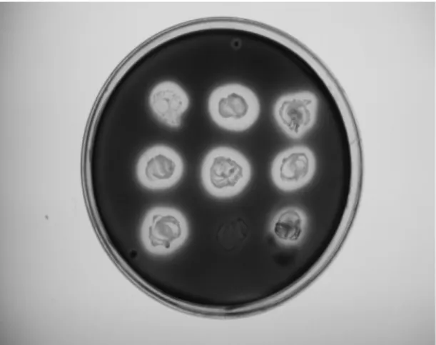
Beta-lactamase test: After inoculation of tested isolates on nutrient agar plates and overnight incubation at 37˚C, the
plates were overlaid with 1% molten agarose containing 0.2%
room temperature; iodine solution was poured onto the agar
plates. After 10 s, the residual iodine solution was damped out
and the plates were incubated at room temperature until a
discolouration zone appeared around -lactamase producing
colonies (35).
Plasmid profiles: Beta-lactamase producing strains were screened for their plasmid profile using alkaline lysis method
(2). Agarose gel electrophoresis was performed. The samples
were run on 0.8% agarose gel with Tris-Borate (TBE) buffer
and ethidium bromide (final concentration 0.5µg/ml). The gel
was observed under UV light and a picture was captured using
a digital camera.
Curing of -lactamase: Beta-lactamase producing strains were inoculated in Luria-Bertani (LB) broth containing
different concentrations of the curing agent, ethidium bromide
ranging from 0.25 to 128 µg/ml and incubated at 37˚C for
overnight. The cultures were diluted and subculture was done
on nutrient agar plates. The growths obtained were tested for
-lactamase production (7). The plasmid profile of the cured
derivatives was also analyzed.
Fluorometric assay: The accumulation of norfloxacin by S. aureus cells was determined using the modified fluorescence
method (9). Carbonyl cyanide m-chlorophenylhydrazone was
added to a parallel set of tubes to a final concentration of 100
µM. The concentration of norfloxacin accumulated was
determined on a Perkin-Elmer fluorescence spectrophotometer.
The norfloxacin excitation and emission wavelengths used
were 277 and 443 nm respectively.
Combination studies: Combinations of ciprofloxacin with oxacillin, cefepime, gentamicin and erythromycin were
investigated by the DAA with MRSA strains (32). For the
combination decimal mixtures, the mean zone size (
x
comb)was calculated from data obtained with combination mixtures,
95% confidence intervals (t distribution) were calculated for
this mean. Results were considered indicative of synergism if
x
comb >x
drug A andx
drug B and the 95% confidence intervalsfor
x
comb did not overlap those forx
drug A orx
drugB,antagonism if
x
comb <x
drug A andx
drug B and the 95%confidence intervals did not overlap and all other results were
considered additive.
RESULTS
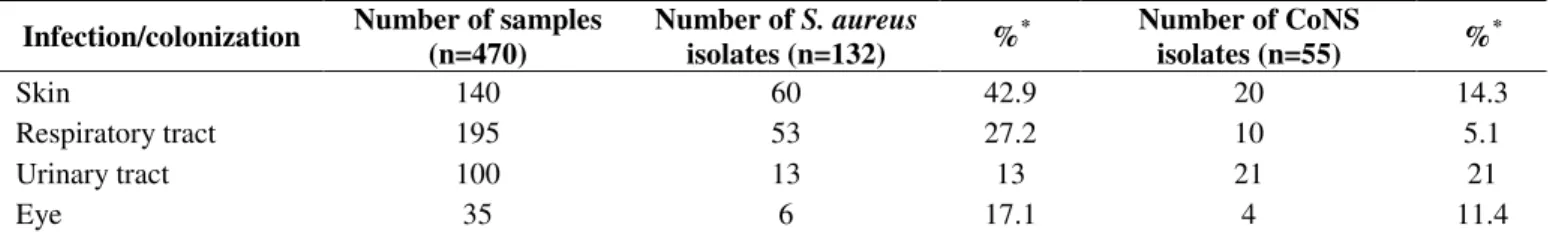
Incidence of staphylococcal species in clinical specimens Out of the 470 clinical specimens, 187 (39.8%)
staphylococcal strains were isolated and identified. Out of the
187 isolates, 132 were S. aureus and 55 were CoNS (70.6%
and 29.4% respectively). The incidence of staphylococcal
isolates was shown in Table 1.
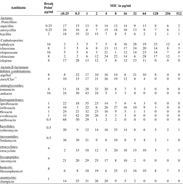
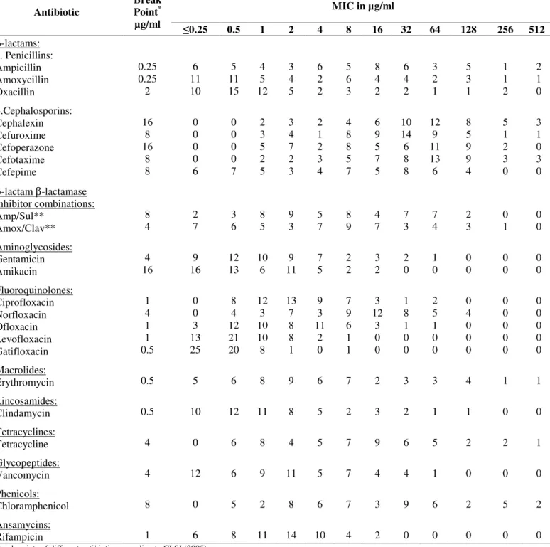
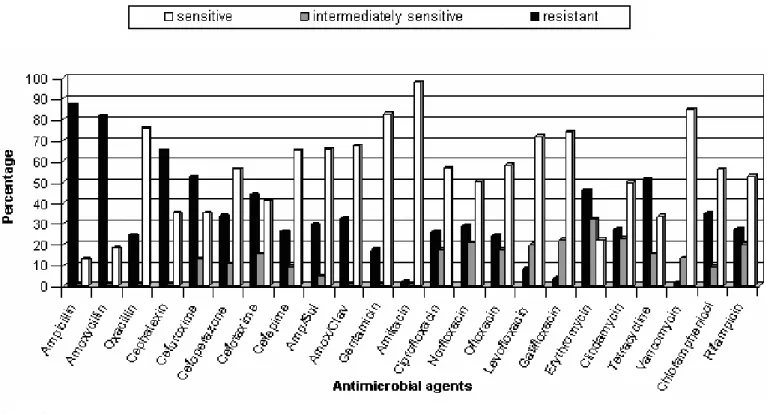
Antibiotic susceptibility and determination of MICs
Tables 2 and 3 show the respective MIC distributions of
different antibiotics for staphylococcal isolates. Figures 1 and 2
show whether the bacteria were sensitive, intermediately
sensitive or resistant to each antibiotic. The isolated S. aureus
strains showed lower resistance to vancomycin, amikacin and
gatifloxacin. Vancomycin was the most effective antibiotic
against CoNS.
Table 1. Incidence of staphylococcal species in clinical specimens
Infection/colonization Number of samples (n=470)
Number of S. aureus
isolates (n=132) %
* Number of CoNS
isolates (n=55) % *
Skin 140 60 42.9 20 14.3
Respiratory tract 195 53 27.2 10 5.1
Urinary tract 100 13 13 21 21
Eye 35 6 17.1 4 11.4
Table 2. Distribution of MICs of different antibiotics among S. aureus isolates (132 isolates)
MIC in µg/ml Antibiotic
Break Point* µg/ml
0.25 0.5 1 2 4 8 16 32 64 128 256 512
β-lactams: a. Penicillins: Ampicillin Amoxycillin Oxacillin b.Cephalosporins: Cephalexin Cefuroxime Cefoperazone Cefotaxime Cefepime
Table 3. Distribution of MICs of different antibiotics against CoNS isolates (55 isolates)
MIC in µg/ml Antibiotic
Break Point* µg/ml
0.25 0.5 1 2 4 8 16 32 64 128 256 512
β-lactams: a. Penicillins: Ampicillin Amoxycillin Oxacillin b.Cephalosporins: Cephalexin Cefuroxime Cefoperazone Cefotaxime Cefepime
Figure 1. Antibiotic susceptibility ofS. aureus isolates
Resistance through ββββ-lactamase production
It was found that 61.7% of ampicillin-resistant S. aureus
strains and 42.9% of ampicillinresistant CoNS strains were
-lactamase producers (Figure 3).
Figure 3. Beta-lactamase production by the isolated staphylococci
Plasmid-mediated antibiotic resistance
Some -lactamase producing strains carried at least one
plasmid and others don't (Figure 4). Results of curing revealed
that when these cells were exposed to curing agent ethidium
bromide, the production of the -lactamase was lost (Figure 5).
When the plasmid profile of the cured derivatives was
analyzed, none of the isolates harbored plasmids. This indicates
that -lactamase production by these strains were
plasmid-mediated. On the other hand, strains which were resistant to
ampicillin, -lactamase producers and did not show plasmid
bands were not cured and did not loose their enzyme activity.
This evidence that -lactamase production by these strains
might be chromasomally mediated.
Figure 4. Agarose gel electrophoresis of plasmid DNA of the selected ampicillin-resistant isolates.
Figure 5. Beta-lactamase detection after plasmid curing.
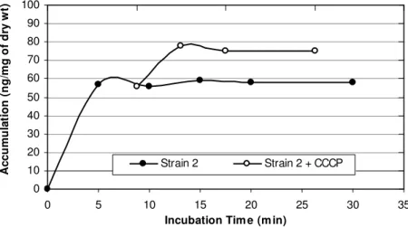
Resistance through the efflux system
A biphasic pattern of accumulation of norfloxacin was
observed with an initial rapid phase of accumulation seen
within the first 5 minutes. The rapid phase was followed by a
plateau phase. Norfloxacin-sensitive strain 1 took up higher
amount of norfloxacin than did any of the resistant strains
tested (2, 3, and 4) and the addition of CCCP did not affect the
accumulation (figure 6). In norfloxacin-resistant strains,
norfloxacin uptake was reduced when compared with that in
norfloxacin-sensitive strains, and the uptake of norfloxacin by
these strains increased rapidly after the addition of CCCP
(137%, 191% and 218% for 3 resistant strains) (Table 4). Thus
addition of uncoupler CCCP increased norfloxacin
accumulation in quinolone resistant S. aureus, suggesting
endogenous, energy-dependent efflux (Figure 6, 7, 8, and 9).
0 10 20 30 40 50 60 70 80 90 100
0 10 20 30 40
Incubation Tim e (m in)
A
c
c
u
m
u
la
ti
o
n
(
n
g
/m
g
o
f
d
ry
w
t)
Strain 1 Strain 1 + CCCP
0 10 20 30 40 50 60 70 80 90 100
0 5 10 15 20 25 30 35
Incubation Tim e (m in)
A
c
c
u
m
u
la
ti
o
n
(
n
g
/m
g
o
f
d
ry
w
t)
Strain 2 Strain 2 + CCCP
Figure 7. Norfloxacin accumulation in case of strain 2.
0 10 20 30 40 50 60 70 80 90 100
0 5 10 15 20 25 30 35
Incubation Time (min)
A
c
c
u
m
u
la
ti
o
n
(
n
g
/m
g
o
f
d
ry
w
t)
Strain 3 Strain 3 + CCCP
Figure 8. Norfloxacin accumulation in case of strain 3.
0 10 20 30 40 50 60 70 80 90 100
0 5 10 15 20 25 30 35
Incubation Time (min)
A
c
c
u
m
u
la
ti
o
n
(
n
g
/m
g
o
f
d
ry
w
t)
Strain 4 Strain 4 + CCCP
Table 4. Concentration of norfloxacin
±
100µM CCCP accumulated by staphylococcal strainsAccumulation (ng/mg dry cells)
Staphylococcus aureus strains
MIC (µg/mg)
Susceptibility to Norfloxacin
- CCCP + CCCP
Percentage of increase in accumulation
Strain 1 2 Sensitive 72 71 ---
Strain 2 16 Resistant 57 78 137%
Strain 3 128 Resistant 47 90 191%
Strain 4 128 Resistant 39 85 218%
Decimal assay for additivity method
For each pair of antibiotics tested, the selected target size
for zones of inhibition, the BEF, and the mean diameters of the
inhibition zones (with 95% confidence intervals) obtained are
provided in table 5. For each antibiotic the BEF was calculated
by using the linear regression equation for the standard
dose-response curve, similar to that illustrated in figure 10 for
ciprofloxacin.
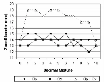
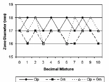
Interactions between ciprofloxacin and oxacillin,
cefepime, gentamicin and erythromycin were shown in figures
11, 12, 13 and 14.
Figure 10. Standard dose-response curve generated in disk diffusion assays with S. aureus strain and ciprofloxacin.
Figure 11. Synergy between ciprofloxacin and oxacillin.
Figure 13. Additivity between ciprofloxacin and gentamicin. Figure 14. Antagonism of ciprofloxacin by erythromycin.
Table 5. Interactions between pairs of antibiotics with MRSA strain
Antibiotics Target zone
diameter (mm) BEF (µg)
Mean diameter of zone of inhibition attained (mm)
95% CIa (mm)
Type of interaction Ciprofloxacin
Oxacillin
Ciprofloxacin+Oxacillin
14 14
25.06 309.03
14.3 13.3 18.2
13.8-14.8 12.6-14.0 17.4-19.0
Synergism
Ciprofloxacin Cefepime
Ciprofloxacin+Cefepime
13 13
21.53 918.33
14.3 12.9 13.7
13.8-14.8 12.4-13.4 13.1-14.3
Additive
Ciprofloxacin Gentamicin
Ciprofloxacin+Gentamicin
18 18
46.03 67.14
17.5 16.5 17.1
17.0-18.0 16.2-16.8 16.6-17.6
Additive
Ciprofloxacin Erythromycin
Ciprofloxacin+Erythromycin
18 18
46.03 74.64
17.4 17.1 14.1
17.1-17.7 16.6-17.6 13.2-15.0
Antagonism
a
Confidence interval.
DISCUSSION
Staphylococcal infections are a common and significant
clinical problem in medical practice (30). In the present work,
the incidence of staphylococci was studied among the clinical
specimens isolated in Minia. Out of the 470 clinical specimens,
187 (39.8%) staphylococcal strains were isolated and
identified. Out of the isolated staphylococci, 70.6% were S.
aureus and 29.4% were CoNS. Staphylococcus aureus was
more prevalent than CoNS in skin, respiratory and eye
infections, while CoNS were the most common isolates in
urinary tract infections.
Our study revealed high activity of vancomycin and
amikacin towards staphylococci. The effectiveness of
vancomycin against staphylococci is corroborated by data from
other groups (5,12,18).
In order to define the main mechanisms used by
production and for possess of plasmid and efflux-mediated
resistance. Liang et al. (21) reported that one of the major
mechanisms of resistance to -lactams was the expression of
ß-lactamases, such as penicillinase and cephalosporinase. We
observed high levels of -lactamase production in
staphylococcal isolates (61.7% in S. aureus and 42.9% in
CoNS). High levels of -lactamase production in staphylococci
had been reported elsewhere (4).
Other mechanisms of staphylococcal resistance to
-lactams were reported; such as intrinsic mechanism, which is
not due to drug inactivation, and accounts for
methicillin-resistance;and tolerance, in which there is a dissociation of the
inhibitoryand killing actions of beta-lactam antibiotics (31).
Beta-lactamase producing staphylococcal strains were
screened for their plasmid profile, results of the screening
revealed that some strains were carrying at least one plasmid
suggesting plasmid-mediated antibiotic resistance and others
did not. Plasmid-mediated antibiotic resistance was reported in
several studies (14,25). Results of -lactamase curing were
compatible with other studies (7,23). Also, Shakibaie et al. (33)
reported that the -lactamase-cured cells did not exhibit any
enzyme activity.
In this study the efflux mechanism in S. aureus strains was
measured by a modified fluorometric assay. Addition of
uncoupler CCCP increased norfloxacin accumulation in
quinolone resistant S. aureus strains. Numerous reports
involved fluorimetric method to study the efflux mechanisms
and the effect of CCCP on the accumulation (28,29). Kaatz et
al. (17) and Aeschlimann et al. (1) had shown increased
activity or accumulation of the hydrophilic quinolones,
norfloxacin and ciprofloxacin, in the presence of efflux
inhibitors. Takenouchi et al. (36) demonestrated an increase in
norfloxacin accumulated by S. aureus strains in the presence of
CCCP.
Our study revealed that, using DAA technique with
MRSA strain, synergistic interaction was observed between
ciprofloxacin and oxacillin, ciprofloxacin plus cefepime and
gentamicin appeared to be additive, while ciprofloxacin plus
erythromycin was antagonistic. Combinations of
fluoroquinolones with other antimicrobial agents against S.
aureus strains have been previously investigated (13,16). Also
the validity of DAA had been demonstrated (26,32).
CONCLUSION
This study examined the incidence of staphylococci, and
its susceptibility patterns to different antibiotics. Drug
resistance mediated by -lactamases, plasmid and efflux pumps
were important modes of cellular resistance to antimicrobial
agents in our study. It would be valuable to use efflux pump
inhibitors. Also, combination of antimicrobials which
potentiate the activity of the existing antibiotics may be useful
to decrease the resistance rate.
ACKNOWLEDGEMENT
The author gratefully acknowledges the referee, who made
useful suggestions and remarks which helped me improve the
paper.
REFERENCES
1. Aeschlimann, J.R.; Kaatz, G.W.; Rybak, M.J. (1999). The effects of NorA inhibition on the activities of levofloxacin, ciprofloxacin and norfloxacin against two genetically related strains of Staphylococcus aureus in an in-vitro infection model. J Antimicrob Chemother., 44(3), 343-349.
2. Alexander, S.T.; Strele, D.; Niles, M.J. (2004). Laboratory exercises in organismal and molecular microbiology. Mc. Grow Hill, USA.
3. Archer, G.L. (1998). Staphylococcus aureus: A well-armed pathogen. Clin Infect Dis, 26, 1179–1181.
4. Arslan, S.; Ozkarde, F. (2007). Slime production and antibiotic susceptibility in staphylococci isolated from clinical samples. Mem Inst Oswaldo Cruz., 102(1), 29-33.
5. Bashir, A.; Mujahid, T.Y.; Jehan, N. (2007). Antibiotic resistance profile: isolation and characterization of clinical isolates of staphylococci from patients with community-acquired skin infections. Pak J Pharm Sci., 20(4), 299-304.
6. Boneca, I.G.; Chiosis, G. (2003). Vancomycin resistance: occurrence, mechanisms and strategies to combat it. Expert Opin Ther Targets, 7, 311-328.
8. Casey, A.L.; Lambert, P.A.; Elliott, T.S.J. (2007). Staphylococci. Int J Antimicrob Agents, 29 (Suppl.3), 23–32.
9. Chapman, J.S.; Georgopapadakou, N.H. (1989). Fluorometric assay for fleroxacin uptake by bacterial cells. Antimicrob. Agents Chemother., 33(1), 27-29.
10. Clinical and Laboratory Standards Institute (CLSI). Performance standards for antimicrobial disk susceptibility tests, 9th ed. Approved. M2-A9 (2006). Clin. Lab. Stand. Inst., 26 (1), 1-172.
11. Collee, J.G.; Fraser, A.G.; Marmion, B.P.; Simmons, A. (1996). Tests for identification of bacteria. In: Mackie & McCartney. Practical medical microbiology. New York, USA.
12. Elouennass, M.; Sahnoun, I.; Zrara, A.; Bajjou, T.; Elhamzaoui, S. (2008). Epidemiology and susceptibility profile of blood culture isolates in an intensive care unit (2002-2005). Med Mal Infect, 38(1), 18-24. 13. Ferrara, A.; Dos-Santos, C.; Cimbro, M.; Gialdroni Grassi, G. (1997).
Effect of different combinations of sparfloxacin, oxacillin, and fosfomycin against methicillin-resistant staphylococci. Eur J Clin Microbiol Infect Dis., 16(7), 535-537.
14. Firth, N.; Apisiridej, S.; Berg, T.; O'Rourke, B.A.; Curnock, S.; Dyke, G.H.K.; Skurray, R.A. (2000). Replication of Staphylococcal Multiresistance Plasmids. J. Bacteriol., 182, 2170-2178.
15. Jain, A.; Agarwa, J.; Bansal, S. (2004). Prevalence of methicillin-resistant, coagulase-negative staphylococci in neonatal intensive care units: findings from a tertiary care hospital in India. J Med Microbiol, 53, 941–944.
16. Jenkins, S.G.; Lewis, J.W. (1995). Synergistic interaction between ofloxacin and cefotaxime against common clinical pathogens. Infection, 23(4): 245.
17. Kaatz, G.W.; Seo, S.M.; Ruble, C.A. (1991). Mechanisms of fluoroquinolone resistance in Staphylococcus aureus. Journal of infectious diseases., 163, 1080-1086.
18. Khosravi, A.D.; Mehdinejad, M.; Heidari, M. (2007). Bacteriological findings in patients with ocular infection and antibiotic susceptibility patterns of isolated pathogens. Singapore Med J., 48(8), 741-743. 19. Knauer, A.; Fladerer, P.; Strempfl, C. (2004). Effect of hospitalization
and antimicrobial therapy on bantimicrobial resistance of colonizing Staphylococcus epidermidis. Wien Klin Wochenschr, 116, 489–494. 20. Le Loir, Y.; Baron, F.; Gautier, M. (2003). Staphylococcus aureus and
food poisoning. Genet Mol Res., 2(1), 63-76
21. Liang, W.; Huang, H.; Lin, R.; Hou, W. (2003). Screening for natural inhibitors of penicillinase by copolymerization of hydrolyzed starch or glycogen in sodium dodecylsulfate polyacrylamide gels for detecting penicillinase activity. Bot. Bull. Acad. Sin., 44, 187-191.
22. Lynch, A.S. (2006). Efflux systems in bacterial pathogens: an opportunity for therapeutic intervention? An industry view. Biochem Pharmacol., 71(7): 949-956.
23. Mansouri, S. and Khalegi, M. (1997). Antibacterial resistance pattern and
frequency of methicillin resistant Staphylococcus aureus isolated from different sources in Southern Iran. Am. J. Med. Sci., 22, 89-93.
24. Marquez, B. (2005). Bacterial efflux systems and efflux pumps inhibitors. Biochimie., 87(12), 1137-1147.
25. Nayak, N.; Satpathy, G.; Vajpayee, R.B.; Mrudula, S. (2007). Phenotypic and plasmid pattern analysis of Staphylococcus epidermidis in bacterial keratitis. Indian J Ophthalmol., 55(1), 9-13.
26. Nworu, C.S.; Esimone, C.O. (2006). Comparative Evaluation of Three In Vitro Techniques in the Interaction of Ampicillin and Ciprofloxacin against Staphylococcus aureus and Escherichia coli. Tropical Journal of Pharmaceutical Research, 5(2), 605-611.
27. Palazzo, I.C.V.; Araujo, M.L.C.; Darini, A.L.C. (2005). First report of vancomycin-resistant Staphylococci isolated from healthy carriers in Brazil. J Clin Microbiol, 43, 179–185.
28. Piddock, L.J.; Jin, Y.F.; Griggs, D.J. (2001). Effect of hydrophobicity and molecular mass on the accumulation of fluoroquinolones by Staphylococcus aureus. J Antimicrob Chemother., 47(3), 261-270. 29. Piddock, L.J.; Jin, Y.F.; Webber, M.A.; Everett, M.J. (2002). Novel
ciprofloxacin-resistant, nalidixic acid-susceptible mutant of Staphylococcus aureus. Antimicrob Agents Chemother., 46(7), 2276-2278.
30. Rayner, C.; Munckhof, W.J. (2006). Antibiotics currently used in the treatment of infections caused by Staphylococcus aureus. Intern Med J., 36(2), 142-143.
31. Sabath, L.D. (1982). Mechanisms of resistance to beta-lactam antibiotics in strains of Staphylococcus aureus. Ann Intern Med, 97(3), 339-344. 32. Sanders, C.C.; Sanders, W.E.; Moland, E.S. (1993). Decimal assay for
additivity of drugs permits delineation of synergy and antagonism. Antimicrob Agents Chemother., 37(2), 260-264.
33. Shakibaie, M.R.; Mansouri, S.; Hakak, S. (2002). Plasmid pattern of antibiotic resistance in beta-lactamase producing Staphylococcus aureus strains isolated from hospitals in Kerman, Iran. Am. J. Med. Sci., 27, 80-83.
34. Skov, R.; Frimodt-Moller, N.; Espersen, F. (2003). Tentative interpretative zone diameters for fusidic Neosensitabs† on Mueller Hinton agar and three blood media. Int J Antimicrob Agents, 22, 502– 507.
35. Sykes, R.B. (1978). Methods for detecting -lactamases. In: Reeves, D.S.; Phillips, I.; Williams, J.D.; Wise, R. (eds). Laboratory methods in Antimicrobial Chemotherapy. Mulligan, Churchill Livingstone, p. 64-69. 36. Takenouchi, T.; Tabata, F.; Iwata, Y.; Hanzawa, H.; Sugawara, M.; Ohya,
S. (1996). Hydrophilicity of quinolones is not an exclusive factor for decreased activity in efflux-mediated resistant mutants of Staphylococcus aureus. Antimicrob. Agents Chemother., 40, 1835–1842.