doi: 10.1590/0037-8682-0227-2017
Short Communication
Corresponding author:Dr.Fereshteh Jabalameli
e-mail: [email protected]
Received 31 May 2017
Accepted 18 September 2017
Clonal relation and antimicrobial resistance pattern
of extended-spectrum and AmpC
β-lactamase-producing
Enterobacter
spp. isolated from different
clinical samples in Tehran, Iran
Roya Ghanavati
[1], Mohammad Emaneini
[1], Davood Kalantar-Neyestanaki
[2],
Azin Sattari Maraji
[1], Mosayyeb Dalvand
[1], Reza Beigverdi
[1]and Fereshteh Jabalameli
[1][1]. Department of Microbiology, School of Medicine, Tehran University of Medical Sciences, Tehran, Iran. [2]. Department of Microbiology, School of Medicine, Kerman University of Medical Sciences, Kerman, Iran.
Abstract
Introduction: Here, we determined the genes encoding antibiotic resistance enzymes and virulence factors and evaluated the genetic relationship between Enterobacter spp. isolated from different clinical samples. Methods: A total of 57 clinical isolates of Enterobacter spp. were tested for the production of extended-spectrum β-lactamases (ESBLs), carbapenemase, and AmpC
using phenotypic and genotypic methods. Results: The most common ESBLs and AmpC β-lactamases were blaTEM (63.3%) and blaEBC (57.7%), respectively. The most prevalent virulence gene was rpos (87.7%). The random amplified polymorphic DNA (RAPD) patterns of strains were genetically unrelated. Conclusions: RAPD polymerase chain reaction analysis revealed high
genetic diversity among isolates.
Keywords:Enterobacter. ESBL. AmpC. RAPD-PCR.
Enterobacter species may cause severe nosocomial infections, including bloodstream, respiratory tract, and central nervous system infections as well as endocarditis1,2.
Nosocomial infections caused by these microorganisms have
been associated with high rates of mortality and morbidity1.
Enterobacter cloacae and Enterobacter aerogenes are the most common species isolated from clinical samples3.
Several virulence genes are involved in the pathogenesis of these microorganisms4-7. Curli fimbria, encoded by csgBAC,
is an important factor for cell adhesion, aggregation, and
biofilm formation in many enterobacteria4. In addition, RpoS
regulation is known to play an important role in multiple stress conditions such as acid, heat, and oxidative stress, starvation, high osmolarity, and near UV exposure5. Another important
virulence factor is the type III secretion system encoded by FliI
that delivers a variety of effectors directly into the cytosol of host as well as aerobactin, encoded by the iutA, described as a
virulence factor related to iron acquisition from host-binding proteins6,7. β-lactam antibiotics, especially third-generation cephalosporins and carbapenems, are used to treat infections cause by several species of Enterobacter1-3. β-lactamase
enzymes, including extended-spectrum β-lactamases (ESBLs) and AmpC, are involved in the mechanism underlying resistance to β-lactam antibiotics in Enterobacter spp1-3. ESBLs are often
encoded by genes located on large plasmids that also carry genes for resistance to other antimicrobial agents such as
aminoglycosides and fluoroquinolones1. ESBLs are capable
of hydrolyzing penicillins, broad-spectrum cephalosporins, and aztreonam, but may not hydrolyze cephamycin, and are
inhibited by clavulanic acid. AmpC β-lactamases are usually
encoded on the bacterial chromosomeand in some cases on the
bacterial plasmid (plasmid-mediated AmpC)3. In Iran, ESBL
production was recently reported in 44.28% of E. cloacae
isolates1. Despite the high incidence of Enterobacter spp.
infection among Iranian patients, very little is known about the antibiotic resistance pattern, virulence factors, and molecular characteristics of Enterobacter spp. isolates. In the current study, the genes encoding antibiotic resistance enzymes and virulence factors were determined and the genetic relationship between
Bacterial isolates
A total of 57 isolates of Enterobacter spp. were obtained from different patients admitted to three teaching hospitals of the Tehran University of Medical Sciences between September 2013 and April 2014. The isolates were collected from various clinical samples, including urine, wounds, tracheal aspirate,
and blood. No duplicate isolates from the same patient and no
environmental strains were included in this study. All isolates of
Enterobacter spp. were identified by standard biochemical tests8.
Susceptibility testing
Antibiotic-containing discs (Mast, UK) were used to
deter-mine the susceptibility of Enterobacter spp. using the disc
diffusion method, as per the Clinical and Laboratory Standards Institute (CLSI) guidelines9. The antimicrobial agents used
were as follows: aztreonam-amikacin (30µg), amoxicillin-clavulanic acid (20/10µg), cefpodoxime (10µg), cefotaxime (30µg), ceftazidime (30µg), imipenem (10µg), cefepime (30µg), gatifloxacin (5 mg), cefoxitin (30µg), gentamicin (30µg), ciprofloxacin (30µg), levofloxacin (5µg), ertapenem (10µg), and meropenem (10µg). Isolates that showed resistance to at least three classes of antibiotics were defined as multi-drug resistant (MDR) strains1. ESBL-producing strains were detected
using the combined double-disc test1. In addition, organisms
were screened for carbapenemase production with the modified Hodge test (MHT)9. The minimum inhibitory concentration
(MIC) of imipenem was determined by the microbroth dilution method according to CLSI criteria9. AmpC overproduction was
confirmed according to the method of Kalantar-Neystanaki
et al10.
Detection of β-lactamases and virulence genes Genomic DNA was extracted by the boiling method2. The
genes encoding ESBLs (blaTEM, blaSHV, blaCTX-M, and blaPER), AmpC (blaACC, blaFOX, blaMOX, blaDHA, blaCIT, and blaEBC), and carbapenemase (blaIMP, blaVIM, blaNDM, blaKPC, blaGIM, and
blaOXA-48) were targeted by polymerase chain reaction (PCR) using specific primers10,11. The detection of seven different
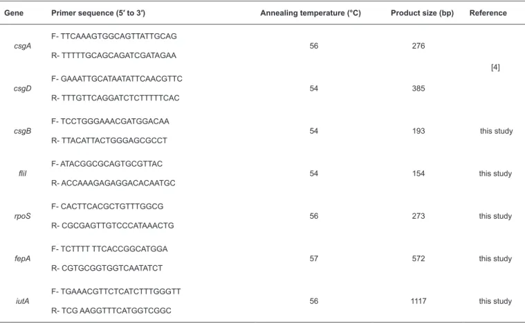
virulence genes (csgA, csgB, csgD, rpos, FliI, fepA, and iutA) was performed with PCR using the oligonucleotide primers
listed in Table 1.
Random amplified polymorphic DNA-PCR
For molecular analysis of isolates, random amplified
polymorphic DNA (RAPD)-PCR was performed as previously
described12. In brief, PCR protocol comprised a pre-denaturation
step at 95 °C for 5 min, followed by 30 cycles of 60 s at 95 °C, 60 s at 33 °C, and 60 s at 72 °C. A final extension step was performed at 72 °C for 10 min. PCR products were separated by electrophoresis on 1% agarose gels with 0.5× Tris-borate-ethylenediaminetetraacetic acid (EDTA) buffer (TBE buffer).
Gels were stained with ethidium bromide and the images were captured using a gel documentation system. Isolates that differed by more than two prominent bands were assigned to different types.
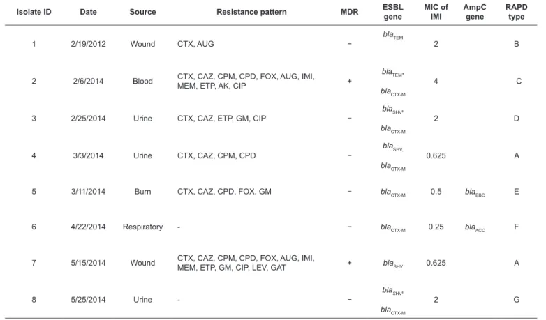
Of 57 isolates, 44 (77.1%) were E. cloacae and 13
(22.8%) were E. aerogenes. These were cultured from wounds
(n = 26), urine (n = 15), blood (n = 8), and other sources (n = 8). Resistance to cefoxitin (84.3%), cefotaxime (49.1%), cefpodoxime (36.8%), and ceftazidime (36.8%) was more prevalent, but only eight (14.1%), seven (12.3%), and six (10.5%) isolates were resistant to imipenem, levofloxacin, and gatifloxacin, respectively. Microbroth dilution method showed that 20 (35.1%) strains were resistant to imipenem. Ten (17.5%) isolates were defined as MDR. The phenotypic test for ESBL, AmpC β-lactamase, and carbapenemase production showed that 30 isolates (22 E. cloacae and 8 E. aerogenes) produced ESBL, 21 isolates (16 E. cloacae and 5 E. aerogenes) produced AmpC β-lactamases, and 8 isolates (6 E. cloacae and 2 E. aerogenes)
produced carbapenemases. The phenotypic and genotypic
characteristics of ESBL and AmpC-producing isolates of E. cloacae and E. aerogenes are shown in Table 2 and Table 3,
respectively. The genes encoding ESBL, blaTEM, blaCTX-M, and
blaSHV, were detected in 19 (63.3%), 19 (63.3%), and 8 (26.6%) isolates, respectively, making them the most prevalent ESBL
genes in these isolates. We failed to detect blaPER .
The gene for AmpC, blaEBC, was detected in only 17 (57%) isolates. Another common AmpC-associated gene, blaACC, was
detected in 5 (16.6%) isolates. The genes blaCIT and blaDHA were
detected in only 2 (6.6%) and 2 (6.6%) of E. cloacae isolates, respectively. The genes blaFOX and blaMOX were not detected. In addition, we failed to detect carbapenemase genes. The most prevalent genes were rpos and fliI reported in 50 (87.7%) isolates, followed by csgB, csgD, csgA, iutA, and fepA observed
in 40 (70.2%), 39 (68.4%), 34 (59.6%), 31 (54.4%), and 29 (50.9%) isolates, respectively. E. cloacae isolates were grouped
into 21 RAPD types, which were designated as type A (two isolates) to S (one isolate each) (Table 2). E. aerogenes isolates
were grouped into seven RAPD types, which were designated as type A (two isolates) to G (one isolate each) (Table 3). In
the present study, the most prevalent species was E. cloacae (77.1%) and its predominance was similar to that reported by
Khari et al. and Kanamori et al2,3. In recent years, E. cloacae is
the most common pathogen causing nosocomial infections1. In
this study, 84.3% of isolates were resistant to cefoxitin. High
level resistance to cefoxitin has been previously reported by other investigators2,3, suggesting that treatment with these drugs
should be avoided in Enterobacter infections.
Our study revealed that 35.1%, 12.3%, and 10.5% of isolates were resistant to imipenem, levofloxacin, and gatifloxacin, respectively. Previous reports from Iran have shown that
the resistance rate of Enterobacter isolates to imipenem and
gatifloxacin was 2% and 7%, respectively1. Our results indicated
the significant increase in the resistance to carbapenem and ciprofloxacin, which may be attributed to the inappropriate
and widespread use of antibiotics1. Of the 30 isolates that were
recognized as phenotypically positive for ESBL production in this study, 27 were positive for ESBL genotypes. In the study conducted by Kanamori et al. from Japan, 22 of 364 Enterobacter spp. were identified phenotypically positive for ESBL production, but only 11 isolates harbored ESBL genes; ESBL genes were undetected in the remaining 11 isolates2.
Discrepancy between disc tests and PCR detection results
TABLE 1: The oligonucleotide primers used in this study for the amplification of virulence genes.
Gene Primer sequence (5′ to 3′) Annealing temperature (°C) Product size (bp) Reference
csgA
F- TTCAAAGTGGCAGTTATTGCAG
R- TTTTTGCAGCAGATCGATAGAA
56 276
[4]
csgD
F- GAAATTGCATAATATTCAACGTTC
R- TTTGTTCAGGATCTCTTTTTCAC
54 385
csgB
F- TCCTGGGAAACGATGGACAA
R- TTACATTACTGGGAGCGCCT
54 193 this study
fliI F- ATACGGCGCAGTGCGTTAC R- ACCAAAGAGAGGACACAATGC
54 154 this study
rpoS
F- CACTTCACGCTGTTTGGCG
R- CGCGAGTTGTCCCATAAACTG
56 273 this study
fepA
F- TCTTTT TTCACCGGCATGGA
R- CGTGCGGTGGTCAATATCT
57 572 this study
iutA F- TGAAACGTTCTCATCTTTGGGTT R- TCG AAGGTTTCATGGTCGGC
56 1117 this study
TABLE 2: Characteristics of Enterobacter cloacae isolates.
Isolate ID Date Source Resistance pattern MDR ESBL gene
MIC
of IMI
AmpC gene
RAPD type
1 11/11/2013 Burn
CTX, CAZ, CPM, CPD, FOX, AUG, IMI, MEM, ETP, AK, GM, CIP, LEV,
GAT
+
blaTEM,
blaCTX-M
1 blaEBC D
2 11/11/2013 Burn CTX, CAZ, CPM, CPD, FOX, AUG,
AK, GM −
blaTEM,
blaCTX-M
2 blaEBC E
3 11/25/2013 Burn CTX, CAZ, CPM, CPD, FOX, AUG,
AK, GM, CIP +
blaTEM,
blaSHV
2 blaEBC F
4 12/21/2013 Eye CTX, CAZ, CPM, CPD, FOX, AUG,
AK, GM − blaTEM 4
blaACC,
blaEBC
G
5 12/29/2013 Respiratory CTX, CAZ, CPM, CPD, FOX, AUG, AK − blaTEM 4
blaACC,
blaDHA
H
6 12/28/2011 Urine CTX, CAZ, CPM, CPD, FOX, AUG, GM, CIP +
blaTEM,
blaCTX-M,
blaSHV
0.25 blaDHA,
blaEBC
B
7 12/28/2011 Wound CTX, FOX, AUG −
blaTEM,
blaCTX-M
2 blaACC,
blaEBC
C
CTX: cefotaxime; CAZ: ceftazidime; CPM: cefepime; CPD: cefpodoxime; FOX: cefoxitin; AUG: amoxicillin-clavulanate; IMI: imipenem; MEM: meropenem; ETP: ertapenem; AK: amikacin; GM: gentamicin; CIP: ciprofloxacin; LEV: levofloxacin; GAT: gatifloxacin; MDR: multi-drug resistant; ESBL: extended-spectrum β-lactamase; MIC: minimum inhibitory concentration; RAPD: random amplified polymorphic DNA.
8 1/12/2011 Wound CTX, FOX, AUG − blaTEM 2 blaEBC I
9 6/1/2012 Wound CTX, CAZ, CPM, CPD, FOX, AUG, GM −
blaTEM,
blaCTX-M
2 blaEBC C
10 12/13/2013 Wound CTX, CAZ, CPM, CPD, FOX, AUG,
AK, GM, CIP, LEV +
blaTEM,
blaCTX-M
4 blaEBC,
blaCIT
J
11 12/13/2013 Urine CTX, FOX, AUG −
blaTEM,
blaCTX-M
4 − K
12 2/25/2014 Urine CTX, CAZ, CPM, CPD, FOX, AUG,
AK, GM −
blaTEM,
blaCTX-M
1 blaACC,
blaEBC
L
13 3/11/2014 Burn
CTX, CAZ, CPM, CPD, FOX, AUG, IMI, MEM, ETP, AK, GM, CIP, LEV,
GAT
+
blaTEM,
blaCTX-M
64 blaEBC,
blaCIT
A
14 3/11/2014 Burn
CTX, CAZ, CPM, CPD, FOX, AUG, IMI, MEM, ETP, AK, GM, CIP, LEV,
GAT
+
blaTEM,
blaCTX-M
64 blaEBC A
15 4/22/2014 Urine CTX, FOX, AUG − blaCTX-M 4 blaEBC M
16 4/25/2014 Respiratory CTX, CAZ, CPM, CPD, AUG, GM − − 4 blaEBC B
17 4/29/2014 Respiratory CTX, CAZ, CPM, CPD, FOX, AUG, AK, GM −
blaTEM,
blaCTX-M
4 blaEBC N
18 5/5/2014 Blood CTX, CAZ, CPM, CPD, FOX, AUG, IMI, MEM, ETP, AK, GM, CIP, GAT + − 16 − O
19 5/6/2014 Blood CTX, CAZ, CPM, CPD, FOX, AUG,
IMI −
blaTEM,
blaCTX-M, blaSHV
16 blaEBC P
20 5/7/2014 Urine FOX, AUG − blaTEM 2 − Q
21 5/7/2014 Urine FOX, AUG − − 8 blaEBC R
22 5/15/2014 Wound CTX, CAZ, CPM, CPD, FOX, AUG,
IMI, MEM, ETP, GM, CIP, LEV, GAT + blaSHV 4 S
TABLE 2: Continuation.
for the detection of ESBLs in Enterobacter spp2. In the
present survey, 30 (52.6%) Enterobacter isolates were found
to be ESBL producers. Kanamori et al. also reported that 6% Enterobacter spp. were ESBL producers2. The high prevalence
of ESBL-positive isolates in our study may be associated with
the extensive use of third-generation cephalosporins for the treatment of Enterobacter infections. It should be noted that
10% (3/30) isolates were ESBL negative and eight isolates that
were recognized phenotypically positive for carbapenemase failed to show any carbapenemase-related genes, suggestive of the involvement of other resistance mechanisms. In our
study, 26.7% (8/30) of ESBL-positive isolates were MDR. Peymani et al. reported that all ESBL-positive Enterobacter isolates were MDR1. In our study, bla
TEM and blaCTX-M were the
most common ESBL resistance genes, which were frequently
reported in other countries2. In the present study, bla
TABLE 3: Characteristics of Enterobacter aerogenes isolates
CTX: cefotaxime; CAZ: ceftazidime; CPM: cefepime; CPD: cefpodoxime; FOX: cefoxitin; AUG: amoxicillin- clavulanate; IMI: imipenem; MEM: meropenem; ETP: ertapenem; AK: amikacin; GM: gentamicin; CIP: ciprofloxacin; LEV: levofloxacin; GAT: gatifloxacin; MDR: multi-drug resistant; ESBL: extended-spectrum β-lactamase; MIC: minimum inhibitory concentration; RAPD: random amplified polymorphic DNA.
Isolate ID Date Source Resistance pattern MDR ESBL gene MIC of IMI AmpC gene RAPD type
1 2/19/2012 Wound CTX, AUG −
blaTEM
2 B
2 2/6/2014 Blood CTX, CAZ, CPM, CPD, FOX, AUG, IMI, MEM, ETP, AK, CIP +
blaTEM,
blaCTX-M
4 C
3 2/25/2014 Urine CTX, CAZ, ETP, GM, CIP −
blaSHV,
blaCTX-M
2 D
4 3/3/2014 Urine CTX, CAZ, CPM, CPD −
blaSHV,
blaCTX-M
0.625 A
5 3/11/2014 Burn CTX, CAZ, CPD, FOX, GM − blaCTX-M 0.5 blaEBC E
6 4/22/2014 Respiratory - − blaCTX-M 0.25 blaACC F
7 5/15/2014 Wound CTX, CAZ, CPM, CPD, FOX, AUG, IMI,
MEM, ETP, GM, CIP, LEV, GAT + blaSHV 0.625 A
8 5/25/2014 Urine - −
blaSHV,
blaCTX-M
2 G
was the most common type of AmpC β-lactamase, followed
by blaACC (16.6%). Miró et al. reported that the CMY (78.3%) and DHA (19.5%) families were the most prevalent type of AmpC β-lactamase in 35 hospitals in Spain13. However, the
prevalence of ESBL and AmpC-producing Enterobacter spp. varied among different studies, which may be associated with the differences in the geographical area, type of infection, and
settings (hospital or community). Similar to previous reports, we observed the coexistence of ESBL-encoding genes in clinical
isolates1,2. Several virulence factors have been identified in the
pathogenesis of Enterobacter spp4-7. The majority of isolates
(87.7%) carried rpos and fliI. The high frequency of these genes may indicate that these genes are essential for the development
of disease. In contrast to the findings of our study, Krzyminska et al. observed that only 27% of isolates harbored fliI (TTSS gene)6. In the current study, the frequency of csgB, csgD,
and csgA was 70.2%, 68.4%, and 59.6%, respectively, which is
lower than that reported in the previous study by Akbari et al. These authors showed that csgD and csgA genes were present
in 100% and 77.75% of isolates, respectively14. The genes iutA
and fepA were found in 54.4% and 50.9% of isolates in our study. Mokracka et al. reported that 49% of E. cloacae strains produced aerobactin15. However, differences were observed in
the frequency of virulence genes reported in different studies;
this difference may be associated with the variation in the
geographical area, clinical samples, and other factors. RAPD-PCR analysis revealed the significant genetic heterogeneity. In addition, molecular analysis demonstrated that more than 90% (28/30) of ESBL-producing isolates were clonally unrelated,
indicating that the reported infections had no relation with clonal outbreak. In conclusion, blaTEM, blaCTX-M, and blaEBC are the most
common resistance gene types and more than 50% of isolates harbored virulence genes. RAPD-PCR analysis revealed high
genetic diversity among isolates.
Conflict of interests
The authors declare that there is no conflict of interest.
Financial support
This research has been supported by Tehran University of Medical Science of
Health Services grant 25744/93-02-30.
REFERENCES
1. Peymani A, Farivar TN, Sanikhani R, Javadi A, Najafipour R.
Emergence of TEM, SHV, and CTX-M-extended spectrum
isolates collected from hospitals of Tehran and Qazvin, Iran. Microb
Drug Resist. 2014;20(5):424-30.
2. Kanamori H, Yano H, Hirakata Y, Hirotani A, Arai K, Endo S, et al. Molecular characteristics of extended-spectrum beta-lactamases
and qnr determinants in Enterobacter species from Japan. PLoS One. 2012;7(6):e37967.
3. Mohd Khari FI, Karunakaran R, Rosli R, Tee Tay S. Genotypic
and phenotypic detection of ampC beta-lactamases in Enterobacter
spp. isolated from a Teaching Hospital in Malaysia. PLoS One. 2016;11(3):e0150643.
4. Kim SM, Lee HW, Choi YW, Kim SH, Lee JC, Lee YC, et
al. Involvement of curli fimbriae in the biofilm formation of
Enterobacter cloacae. J Microbiol. 2012;50(1):175-8.
5. Dong T, Schellhorn HE. Role of RpoS in virulence of pathogens.
Infect Immun. 2010;78(3):887-97.
6. Krzyminska S, Mokracka J, Koczura R, Kaznowski A. Cytotoxic
activity of Enterobacter cloacae human isolates. FEMS Immunol
Med Microbiol. 2009;56(3):248-52.
7. Johnson JR, Moseley SL, Roberts PL, Stamm WE. Aerobactin and other virulence factor genes among strains of Escherichia coli causing urosepsis: association with patient characteristics. Infect
Immun. 1988;56(2):405-12.
8. Mahon CR, Lehman DC, Manuselis G. Textbook of Diagnostic Microbiology. 5th edition. New York: Saunders; 2015. p: 429-30.
9. Clinical and Laboratory Standards Institute (CLSI). Performance Standards for Antimicrobial Susceptibility Testing. Document
M02-A12, M07-A10, and M11-A8. Wayne, PA: CLSI; 2017.
10. Neyestanaki DK, Mirsalehian A, Rezagholizadeh F, Jabalameli F,
Taherikalani M, Emaneini M. Determination of extended spectrum beta-lactamases, metallo-beta-lactamases and
AmpC-beta-lactamases among carbapenem resistant Pseudomonas aeruginosa
isolated from burn patients. Burns. 2014;40(8):1556-61.
11. Perez-Perez FJ, Hanson ND. Detection of plasmid-mediated AmpC
beta-lactamase genes in clinical isolates by using multiplex PCR. J Clin Microbiol. 2002;40(6):2153-62.
12. Mahenthiralingam E, Campbell ME, Foster J, Lam JS, Speert DP.
Random amplified polymorphic DNA typing of Pseudomonas aeruginosa isolates recovered from patients with cystic fibrosis.
J Clin Microbiol. 1996;34(5):1129-35.
13. Miro E, Aguero J, Larrosa MN, Fernandez A, Conejo MC, Bou G,
et al. Prevalence and molecular epidemiology of acquired AmpC beta-lactamases and carbapenemases in Enterobacteriaceae isolates from 35 hospitals in Spain. Eur J Clin Microbiol Infect Dis. 2013;32(2):253-9.
14. Akbari M, Bakhshi B, Najar Peerayeh S, Behmanesh M. Detection
of Curli Biogenesis Genes Among Enterobacter cloacae Isolated
From Blood Cultures. Int J Enteric Pathog. 2015;3(4):e28413.
15. Mokracka J, Koczura R, Kaznowski A. Yersiniabactin and other siderophores produced by clinical isolates of Enterobacter spp. and