www.nature.com/scientificreports
Deciphering
pyritization-kerogenization gradient for fish
soft-tissue preservation
Gabriel L.
Osés
1, setembrino petri
2, Cibele G. Voltani
3, Gustavo M. E. M.
prado
1, Douglas
Galante
4, Marcia A. Rizzutto
5, Isaac D. Rud
nitzki
6, Evandro P. da Silva
7, Fabio
Rodrigues
8,
Elidiane C.
Rangel
9, Paula A. Sucerquia
10& M. L. A. F.
pacheco
11Soft-tissue preservation provides palaeobiological information that is otherwise lost during
fossilization. In Brazil, the Early Cretaceous Santana Formation contains fish with integument, muscles, connective tissues, and eyes that are still preserved. Our study revealed that soft-tissues were pyritized or kerogenized in different microfacies, which yielded distinct preservation fidelities. Indeed, new data provided the first record of pyritized vertebrate muscles and eyes. We propose that the different taphonomic pathways were controlled by distinct sedimentation rates in two different microfacies. Through this process, carcasses deposited in each of these microfacies underwent different residence times in sulphate-reduction and methanogenesis zones, thus yielding pyritized or kerogenized soft-tissues, and a similar process has previously been suggested in studies of a late Ediacaran lagerstätte.
Exceptionally preserved fossils have palaeobiological novelties that are not often encountered elsewhere in the geological record. Housed in deposits known as Konservat-Lagerstätten1, investigation of fossil soft-tissues may
provide unique insights into ancient biology and palaeoenvironmental conditions. Among these deposits, the Mesozoic rocks from the Santana Formation (Araripe Basin, northeast Brazil) stand out for their often exquisitely preserved and diverse fossil content, including plants, vertebrates (e.g., fishes, pterosaurs, dinosaurs, turtles and lizards), and invertebrates (e.g., insects, arachnids, and crustaceans)2. A remarkable case is the recent description
of a fossilized heart and valves from fish, which provides insights into cardiac evolution3 and into the preservation
of soft-tissues in other vertebrates4, 5.
The Crato Member (Supplementary Figure 1) is one of the most diverse palaeontological deposits from the Early Cretaceous period. Among the fish fossils from this area, Dastible crandalli (Supplementary Note 1) is the most abundant and has been found throughout the entire unit. Even though previous studies have briefly assessed the taphonomic aspects of Crato Member fossils6, several issues regarding taphonomic processes remain
unresolved. Typically, the soft-tissues of these fossils have two distinct taphonomic modes: black carbonaceous compressions and orange iron oxyhydroxide, which occur in grey (GL) and beige (BL) limestones, respectively.
1Programa de Pós-graduação em Geoquímica e Geotectônica, instituto de Geociências, Universidade de São Paulo
- Rua do Lago 562, 05508080, Cidade Universitária, São Paulo-SP, Brazil. 2instituto de Geociências, Universidade
de São Paulo - Rua do Lago 562, 05508080, Cidade Universitária, São Paulo-SP, Brazil. 3instituto de Geociências e
Ciências Exatas, Universidade Estadual Paulista – Avenida 24A 1515, 13506900, Rio Claro-SP, Brazil. 4Laboratório
Nacional de Luz Síncrotron – Rua Giuseppe Maximo Scolfaro 10.000, 13083-970, Campinas-SP, Brazil. 5instituto
de Física, Universidade de São Paulo - Rua do Matão 1371, 05508090, Cidade Universitária, São Paulo-SP, Brazil.
6Departamento de Geologia, Universidade Federal de Ouro Preto – Morro do Cruzeiro s/n, 35400-000, Campus Morro
do Cruzeiro, Ouro Preto-MG, Brazil. 7Programa de Pós-Graduação em Química, instituto de Química, Universidade
de São Paulo – Avenida Prof. Lineu Prestes 748, 05508080, Cidade Universitária, São Paulo-SP, Brazil. 8Departamento
de Química Fundamental, Instituto de Química, Universidade de São Paulo – Avenida Prof. Lineu Prestes 748, 05508080, Cidade Universitária, São Paulo-SP, Brazil. 9Laboratório de Plasmas Tecnológicos, Universidade Estadual
Paulista – Avenida Três de Março 511, 18087-180, Sorocaba-SP, Brazil. 10Departamento de Geologia, Universidade
Federal de Pernambuco - Avenida Acadêmico Hélio Ramos s/n, 50740530, Cidade Universitária, Recife-PE, Brazil.
11Departamento de Biologia, Universidade Federal de São Carlos – Rodovia João Leme dos Santos, Km 110,
18052780, Sorocaba-SP, Brazil. Correspondence and requests for materials should be addressed to G.L.O. (email: [email protected])
Received: 21 July 2016 Accepted: 29 March 2017 Published: xx xx xxxx
Previous studies of insect preservation have interpreted this difference in preservation as a result of rock weath-ering2, but our evidence from fish tissues does not support this interpretation.
In this study, we hypothesized that fish have followed distinct preservation pathways according to palae-oenvironmental and/or microfacies variations. The results of our analyses confirmed that the fish were either pyritized or kerogenized in different sedimentary microfacies. We propose that the sedimentation rates varied in these microfacies, thus suggesting different carcass residence times either in sulphate-reduction (SR) or meth-anogenesis sedimentary microbial zones, which resulted in both different taphonomic pathways and preservation fidelities. Therefore, we suggest that the Crato Member fish fossilization occurred through a process similar to the kerogenization-pyritization gradient model for Neoproterozoic-Palaeozoic metazoan preservation7, 8.
Results
We compared soft-tissues from the fossil fish Dastilbe crandalli from the BL and GL microfacies. In Nova Olinda quarries, BL overlies GL9, 10, both differing mainly on siliciclastic influence. The BL are thinly laminated (ca.
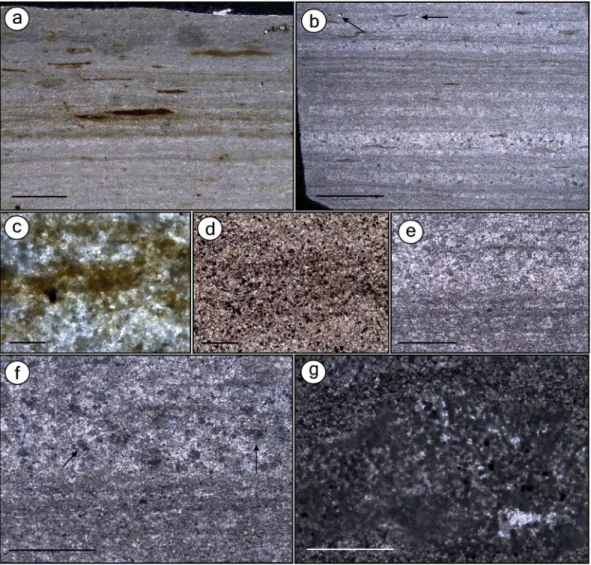
Figure 1. Thin sections of the beige limestone (BL) and the grey limestone (GL) microfacies. Thin sections GP/L 21 (a,d), GP/L 19 (b), GP/L 18 (g), GP/L 172 (c), and GP/L 16 (e,f). (a) BL is composed of thin laminasets of diffuse dark clay laminae (detail in c), interlaminated with pale pure microspar laminae. Elongated to round organic matter-rich dark lenses11 are indistinctly scattered. This microfacies is interpreted as the laminated
limestone (LL) Sm5 microfacies12. (b) GL is composed of dark-grey undulated laminasets formed by thin
laminae with fine blackish scattered material, likely clay/organic matter impurities10, 12 (detail in d). Laminasets
are interlaminated with paler microspar-dominated laminae. Scattered non-oriented detrital quartz is indicated by arrow. GL microfacies is interpreted as Sm1, a clay-carbonate rythmite (CCR)12. Besides GL having
significantly fewer dark lenses than BL, the clay/organic matter-rich laminasets are more frequent, regularly distributed, thicker and have more and closer-packed laminae. (c) Detail of BL dark laminae. (d) Detail of GL dark laminae, showing concentration of pyrite10. (e) Thin section of GL depicting microspar-dominated
www.nature.com/scientificreports/
0.5 mm) with dark clay interlaminated with pale pure microspar laminae (Fig. 1a,c) and have scattered dark, organic-rich lenses11. BL microfacies most probably corresponds to laminated limestones (LL)12. GL are
com-posed of 1–3-mm-thick layers12 and have microfaults (Supplementary Figure 2). Undulated dark-grey pyrite-rich
laminations10 exhibit fine black material (probably clay/organic impurities10, 12) and also peloids (Fig. 1b,d–g).
Such laminations are interlaminated with paler microspar-dominated layers, including neomorphic sparry anhe-dral crystals12. We interpret the GL microfacies to be Sm1 microfacies or clay-carbonate rhythmite (CCR)12,
which similarly to the GL, is composed of microfaults and millimetric plane-parallel and undulated laminae composed of intercalated clay and limestone.
Fish from the BL microfacies.
In the most complete BL specimen, we analysed three distinct regions: cau-dal fin base, dorsal fin, and anteroposterior axis (Supplementary Figures 3 and 4). In the caudal fin base, the fibres possess scarcely visible margins and are nearly indistinguishable from each other (Fig. 2a). The fibres are com-posed of sub-spherical to spherical grains of iron oxide/hydroxide larger than 1 µm that are locally merged and covered by a “fuzzy” coating of submicroscopic crystals (Fig. 2b,c). These grains replaced muscles of the entire specimen. Similar, but smaller grains (<1 µm) occur in pores among larger grains (Fig. 2b). Iron oxide/hydroxidewww.nature.com/scientificreports/
also may form a honeycomb-like texture characterized by subhedral to euhedral shaped hollows (Fig. 2c). We interpret that grains of up to 1 µm probably filled those spaces and were released after weathering oxidation13. The dorsal fin area exhibits muscles arranged in layers along the specimen’s depth, thus revealing how muscles are attached to the fin base and connected to muscles running along/around the column (Fig. 2d). Micrographs of the fish anteroposterior axis revealed muscular insertion into the vertebrae surface (including tendons attaching muscles to bones) and multiple stages of muscle decay around vertebra (Fig. 2e), and confirmed the fibre micro-fabric (Fig. 3a,b). These observations counter a previous analysis reporting rather indistinct fibres6. The eye area
is also preserved by iron oxide/hydroxide microfabrics (Fig. 3c,d).
Patches of orange amorphous iron oxide/hydroxide material are commonly seen in BL fossils. These orange regions have 3D muscle fibres, as has been reported elsewhere6. The fibres in our samples may be fragmented
and locally degraded, probably as a result of decay (Fig, 3e). Wide, elongated, flat, soft structures interpreted as sarcolemma (i.e., muscle cell membrane; Fig. 3e,f) occur between fibres. Despite being similar to putative EPS (extracellular polymeric substances) (below), the presence of these structures between fibres supports the hypoth-esis that they are sarcolemma, because a more random distribution would be present if they were EPS. Moreover, gaps between fibres also are present, which, along with occasional displacement of the sarcolemma in relation to such gaps14, may provide evidence for decay and dehydration/shrinkage of sarcolemma during fossilization. Some
samples have fibres subparallel to the vertebral column that are arranged in myomeres connected to the dorsal column area (Fig. 3g,h), and some fibres possess putative cell nuclei (Fig. 3h).
In a specimen, we identified a ribbon-like smooth structure with a pliable aspect that shapes itself according to the microfabric relief underneath and resembles bacterially secreted EPS (Fig. 3i). Indeed, it is richer in carbon content than microfabric (Supplementary Figure 5). Similar structures have been found in Crato insects15.
EDXRF analysis of fish from BL showed enhanced concentrations of phosphorus in bones from BL speci-mens compared with the rock matrix, whereas calcium is equally found in bones and in the carbonate matrix (Fig. 4a,b). The detection of a high abundance of phosphorus and calcium in soft-tissue areas is explained by the millimetric size of the X-ray beam of the system that was used, which measured bones scattered in these areas. We also measured lead, which correlated with the fossil and minor signals of sulphur and metals, such as iron, copper, and zinc, along soft-tissue regions (Fig. 4b).
EDS of one BL fish revealed original hydroxyapatite (Ca5(PO4)3(OH)) in bones, calcite-filled (CaCO3) bone cavities, and goethite (FeO(OH)) that replaced the soft-tissues surrounding vertebrae (Fig. 4c–e and Supplementary Figures 5 and 8). These results support the EDXRF data discussed above.
Fish from GL microfacies.
In carbonaceous fish, distinct cement types do occur (Supplementary Figure 2). Moreover, soft-tissues consist of a thick hard dark material that occurs consistently along the body. Almost every bone, in cross-section, is enveloped by dark opaque amorphous-to-sinuous fibrous material with alternating light/dark bands that are locally convoluted (Fig. 5a–c). Sometimes, black, beige, brown, and green bands alter-nate (Fig. 5d,e). We interpreted these features as preserved soft-tissue with muscle fibres (cells). In some regions,Figure 5. Thin section images and SEM of fish (Fig. 6a) with preserved carbonaceous soft-tissues. Thin sections GP/L 16 (a–c,f,g) and GP/L 17 (d,e,h–k). Dark opaque sinuous laminated, locally convolute muscle tissues (a–c), composed of alternating, differently coloured light/dark bands (d,e). Muscle tissue is composed of dark fibres (b,c; arrows). (f) Degraded soft-tissues and bones. (g) SEM micrograph of muscle fibre detail. (h,i)
www.nature.com/scientificreports/
both soft-tissues and bones present a degraded aspect (Fig. 5f). Carbonaceous fibres are poorly preserved and have no discernible microfabrics (Fig. 5g).
Some regions of the anterior-posterior axis (particularly near a vertebrae) possess a vesicular texture formed by bodies of various shapes (Fig. 5h,i). They are locally oriented in a curved fashion that yields apparently round
pockets. These bodies have been found to resemble muscle fibres in cross-section16. They are surrounded by a
light matrix that resembles endomysium (i.e., collagenous connective tissue). The pockets can be interpreted as fascicles (i.e., muscle bundles) that are separated by a structure resembling perimysium, which is also a connective tissue for muscle fibres. Locally, the outer fish margin is outlined by broken scales interlayered with calcite and labile-tissue that we interpreted as being carbonaceous skin (Fig. 5j,k) (Supplementary Note 2). These aspects have rarely been documented in the fossil record. A detailed description of well-preserved micro-morphologies may facilitate exploration of new avenues for physiological research (e.g., insights into muscular functions) with evolutionary and palaeoecological implications17.
The same bone composition from the BL was confirmed to be present in fish preserved in GL (Fig. 6a–e). In specimens from GL, iron and copper are more concentrated in soft-tissue regions (Fig. 6e,f), and zinc is more concentrated in bones (Fig. 6d,g). The soft-tissues from GL fish contain carbon (Fig. 6b,c) and sulphur (Supplementary Figure 9), thus revealing that they are mainly carbonaceous. Additionally, manganese is abun-dant in both the matrix and cement (Supplementary Figure 9).
Discussion
Pyritized Fish.
In the BL specimens, the concentration of phosphorus together with calcium in bones indi-cated that the original hydroxyapatite is probably unaltered. Iron dominance in soft-tissues reflects the presence of iron oxides/hydroxides concentrated in these regions. Thus, the presence of sulphur can be explained by sul-phate resulting from pyrite oxidation. Insects in BL are preserved as hematite/goethite replicas after pyrite15.Similarly, we interpret that fish soft-tissues in the BL were originally preserved by pyrite and later oxidized6, 18.
The microfabrics are mainly composed of iron oxide/hydroxide sub-spherical to spherical grains, which we interpret to be framboidal pyrite pseudomorphs19. This interpretation is enhanced by the occurrence of a
honeycomb-like texture that resembles framboids. Regarding the hollow spaces in this honeycomb-like texture, which was probably formerly filled with microcrystals <1 µm, their morphology, size, and organization are con-sistent, thus supporting the interpretation that the honeycomb-like texture was originally a pyrite framboid20, 21.
Soft-tissue pyritization is inhomogeneous in the Crato Member fish. In the same specimen, some regions have framboids that are not associated with soft-tissues, whereas other regions have pyritized 3D muscles, par-ticularly central trunk muscles surrounding the dorsal portion of the vertebral column, which is indeed the least decay-prone fish segment22. Myosepta (i.e., the collagen boundaries between myomeres) were not preserved in
our samples. Moreover, some fish have fibres organized in myomeres, whereas others have non-organized muscle fibres, thus revealing a preservation gradient. Taphonomic experiments on amphioxus, lampreys, and fish have shown that the ventral myomere portion is lost before the dorsal one, and gaps develop between myomeres as they shrink after 6 decay-days22 (Supplementary Figure 4). Interestingly, some specimens with good muscle fibre
preservation show evidence of integument rupture (Supplementary Figures 3 and 4; Supplementary Note 2), which should have supported sulphate-reducing bacteria (SRB) as well as sulphate and iron entry within car-casses, thereby promoting pyritization. Regarding eye preservation, the lens—which seems to be present in our specimen—is the least decay-prone eye structure, which may last more than 300 days under decay, as revealed by taphonomic experiments on lampreys22.
To our knowledge, despite of some reports of pyritized fish (fish from the Hunsrück Slate in Germany), this is the first description of pyritized vertebrate muscles and eyes in the fossil record. Surprisingly, Crato fish muscles are not phosphatized, as would be expected for vertebrate soft-tissue preservation (e.g., ref. 23). Because fish phosphatized soft-tissues are usually preserved by substrate microfabrics, phosphate crystals grow directly in decay sites, in a process requiring high availability of phosphate from external sources14, 24. Therefore, the lack
of phosphatized tissues in the Crato Member fish may be explained by three main factors. First, BL had low Corg and associated phosphate accumulation24, 25, which controls mineralization26. Consequently, there was
insuffi-cient phosphate accumulation to inhibit calcite formation24. Second, phosphatization is enhanced at Ca-depleted
continental basins27, which was not the case for the Crato palaeolake. Interestingly, Crato Member pterosaur
phosphatized soft-tissues4 suggest that this process was taxon-controlled and localized in certain tissues, although
a palaeoenvironmental influence cannot be ruled out. Future studies may test both hypotheses. Moreover, phos-phatization occurs more rapidly than pyritization after burial24. Nevertheless, the Crato palaeolake was a
eux-inic (H2S-rich) basin2, and, as such, pyritization might have started even before burial, which would have made pyritization the very first widespread28 taphonomic mineralization process available for fish muscle and eye
preservation.
Several conditions known to facilitate authigenic mineralization, such as anoxia29 and the lack of
burrow-ers30, were present in the Crato palaeolake, owing to water column stratification2. Anoxia facilitates
minerali-zation in the context of anaerobic respiration, and the absence of bioturbation prevents dilution of pore water electron acceptor concentrations, oxygenation of the substrate and the disruption of distinct geochemical gra-dients. Furthermore, microbial activity is thought to have influenced limestone deposition in the Crato palaeo-lake11. Therefore, the well-known role of microbial mats in controlling ion diffusion rates along the sediment30,
31 may also have influenced mineralization in the palaeolake. This ideal preservation scenario was ubiquitous
in Precambrian siliciclastic marine settings (ref. 30; references therein), even though these environments were quite different from the Crato Member in depositional context. Nevertheless, as shown above, some factors may have created ideal conditions for soft-tissue preservation long after the closing of the Precambrian taphonomic window.
Soft-tissue preservation primarily depends on early diagenetic authigenic mineralization during anaerobic decay25 when geochemical gradients (e.g., pH, Eh)26 are created and lead to C
www.nature.com/scientificreports/
zones (e.g., sulphate reduction, methanogenesis), and electron acceptors provide a higher free energy yield in shallower depths, thus resulting in mineral precipitation in geochemical zones (e.g., sulphidic or methanic)33.
However, corresponding metabolic and geochemical zones do not necessarily match horizontally, and there may be an overlap among the levels of each zone33.
Among the mineralization processes, pyritization is of particular interest. Sulphate reduction bacteria (SRB) decay organic matter (i.e., energy source) by using SO42− as an electron acceptor in anaerobic respiration and commonly drive Fe3+ reduction to Fe2+34, thus leading to the mineralization of organic matter28. The
precipita-tion of pyrite requires both reactive iron and sulphate environmental sources besides degradable organic matter28.
We predict that a very fine balance among these factors created optimal conditions for fish labile-tissue pyritiza-tion in the Crato palaeolake. These three factors may have been controlled primarily by the deposipyritiza-tional—e.g., the weathering-driven sources—and/or diagenetic processes discussed below. Indeed, the high productivity of the Crato palaeolake, which ultimately may reflect weathering nutrient contributions, has been proposed to have caused anoxia in the bottom waters2. The same argument has been used by Cui et al.35 to explain the conditions of
the interplay between pyritization and biomineralization in Ediacaran oceans.
The sediments of the Crato palaeolake, a euxinic basin2, were dominantly calcareous, which are commonly
iron-poor28. Therefore, pyrite genesis was iron-limited. The main source of sedimentary iron consists of Fe3+ oxy-hydroxides derived from weathering28. Therefore, the Crato palaeoenvironment would have had an appreciable
iron source, because its content was relatively significant compared to fossils, as supported by measurements of the matrix (Fig. 4b). Indeed, iron oxyhydroxides may have nourished the bottom waters of the palaeolake through pulses of freshwater11. Nevertheless, if iron quantities were still too low to yield carcass pyritization, this scenario
would have been balanced by the low Corg of the BL36, because few decay sites (i.e., low widespread organic matter) would allow for focused bacterial sulphate reduction, sulphide fixation by iron at decay sites, and thus pyritiza-tion25 in fish.
Notably, pyritization occurred during lacustrine carbonate deposition in the Crato palaeolake, because such freshwater environments are usually sulphate-poor24, and pyritization is sulphate-limited25, 28. Thus, pyritization
is commonly recorded in siliciclastic marine deposits18, 31, 37, 38 and in terrigenous lacustrine settings39, from oxic30,
37, 38 to dysoxic/anoxic39 water columns. However, considering the palaeolake water column O
2 stratification2, 11, 40, anoxic bottom waters yielded an either shallow or absent sedimentary oxic zone with prevailing aerobic decay30.
Therefore, bacterial iron/sulphate-reduction zones34 were shallow, thus facilitating iron/sulphate-enrichment32
and enhancing SR and pyritization20. Moreover, muscles and eyes (very labile tissues) probably provided intense
SR, thus compensating for low sulphate input to SRB. Indeed, pristine Corg yields high sulphate-reduction rates41. A total organic carbon (TOC) analysis of the Crato limestones revealed that the sediment was poor (<1%, although higher values have been recorded) in organic carbon36, possibly because of oxidation caused by bottom
pulses of freshwater11 or by water stratification36. This TOC value may account for pyritization in BL, because low
sedimentary Corg content widely explain pyritization37, 38. For carcasses being decomposed by SRB, hydrogen sul-phide (H2S) is formed and must be readily fixed by Fe2+ for pyrite production to be concentrated in carcasses20, 37,
38. In contrast, sediments rich in C
org yield widespread pyritization, thus decreasing the contributions of sulphate and iron to fossil pyritization. Therefore, pyritization requires the sedimentary iron concentrations to exceed that of the sulphide produced by SRB in carcasses20, 37, 38.
Beyond the major palaeoenvironmental factors controlling pyritization, the fish carcasses themselves influ-enced pyrite production in the Crato palaeolake. Iron monosulphides nucleate because of sulphide supersatura-tion favoured by organic matter19. Moreover, depending on its nature, concentration and adsorption properties,
organic matter can stabilize colloidal materials through particle aggregation (e.g., framboid formation) triggered by active Corg bridging, thus decreasing free energy19. Indeed, framboids have been widely associated with decay42,
43. Hence, putative EPS related to microfabrics (biofilm remains) may also have contributed to crystal binding of
organic matter. Researchers have demonstrated the role of biofilms in developing proper structural and geochem-ical conditions for framboid development44, 45. Framboidal pyrite also replaced the tissue of Crato Member insects
(interpreted as bacterially induced mineralization)15, and is also ubiquitous in other Konservat-Lagerstätten18, 37,
46. Moreover, the high abundance of copper, zinc, and lead (heavy metals commonly chelated by organic matter)
in BL fish labile-tissues in comparison to the host rock (Fig. 4b) suggests the presence of microbial activity47,
48. These conditions may also reflect the incorporation of these metals into pyrite, as is favoured by high pyrite
precipitation, especially in anoxic-sulphidic environments49 such as the Crato palaeolake. Indeed, framboids
con-tribute to the mobilization of heavy metals, which concentrate zinc and lead, as compared with the concentrations in the host rock50. Together, the evidence in this study provides geochemical and mineralogical data, including
putative EPS that suggest bacterially induced pyrite production.
There are some explanations for the distinct sizes of framboid pseudomorphs that replaced BL fish labile-tissues. The steps of framboidal pyrite precipitation19 involve (1) the nucleation and growth of iron
mono-sulphides (FeS; mackinawite) from the reaction of Fe2+ and HS−, (2) the transition of mackinawite to greigite, (3) the aggregation of greigite microcrystals, and (4) the conversion of greigite to pyrite, although greigite is not always required21. The framboid sphericity plus microcrystal size/morphology—and consequently
fram-boid size13—depend on the abovementioned steps. For example, in the process of monosulphide nucleation,
each step—sulphate/Fe3+ reduction, crystal nucleation and crystal growth—starts when the previous step has finished. Thus, equally sized microcrystals are formed in a single framboid19. Otherwise, the overlap between
nucleation and growth yields particles of different sizes, which lead to framboids of different sizes. Alternatively, the co-existence of distinct framboid sizes has been explained by the ‘particulation’ of original colloid drop-lets in a range of different final particle sizes51. It has also been argued that the variation of both framboid size
We observed framboids with distinct sizes in addition to EPS replacing decayed labile-tissues (Fig. 3i). This evidence indicates that distinct sizes of framboid pseudomorphs replacing BL fish labile-tissues were the result of the geochemical background of a decay microenvironment. This size variation requires variable pyrite nucleation rates and iron/sulphate diffusion balance over time7, 18, 21, thus suggesting that iron and sulphate concentrations
(besides pH values) vary over time, thus controlling framboid size (ref. 13; references therein). Whereas single pyrite crystals are formed under low Eh conditions, framboids precipitate under high Eh values, because the reac-tion of pyrite producreac-tion is thermodynamically stable, thus yielding sulphide supersaturareac-tion and fast nucleareac-tion rates21. Initially pristine labile-tissues (fish muscles and eyes) yield high decay rates because of their high
reac-tivity52, a key factor controlling pyrite production28. Consequently, sulphide supersaturation induces widespread
nucleation sites, thus favouring framboid precipitation21. This influence of the decay potential of labile-tissues to
pyrite habit (framboids or single euhedral crystals) has been demonstrated for Cambrian animals18 and is further
supported by our data. The rapid nucleation of minute framboids (Fig. 2b) requires that ion diffusion to nuclea-tion sites surpass the reacnuclea-tion speed21. In other words, iron and sulphate are used for creating more pyrite nuclei
instead of fueling crystal growth. This process diminishes highly the availability of reactive organic matter for SRB over time, thus decreasing framboidal pyrite nucleation rates, and consequently, with a continuous influx of reac-tants, sulphide fuels crystal growth7, as suggested for the Eocene London Clay plants53 and Cambrian animals18.
This ‘growth’ mechanism may account for the larger framboids that replaced the soft-tissues of the fish (Fig. 2c). Preserved area extension and preservation fidelity are usually higher in smaller specimens, whereas larger specimens have only very localized poor soft-tissue preservation. This pattern was expected because smaller car-casses are more readily pyritized, because the required iron and sulphate content is lower, and fixation of sulphide by iron20, 38 is faster because of lower H
2S production due to the already lower amount of decay-prone organic matter.
Kerogenized Fish.
We concluded that GL fish carbonaceous soft-tissues underwent kerogenization54, asevidenced by soft-tissue black colour, C composition18, lack of microfabrics, and isotropy at crossed nicols54.
Moreover, micro-Raman data revealed D and G bands of disordered and graphitic carbon (Supplementary Figure 8), which are considered to be kerogen because the attribution of the carbonaceous material to a well-known biogenic source and the low thermal imprint in the Crato unit40 made other processes of disordered
carbonaceous material genesis55—e.g., heating, metamorphism, and hydrothermalism—unlikely.
The two main mechanisms for resistant organic matter preservation56, degradation-recondensation and
selec-tive preservation, were not sufficient to explain the fish muscle preservation. However, muscle lipid-free aliphatic and ester-bound molecules can give rise to degradation-lasting polymeric components during diagenesis, which yields kerogen. Moreover, more labile-tissues undergo structural changes57, which might have accounted for
mus-cle kerogenization.
Cement-filled vertebrae and inter-bone gaps have manganese concentrations, similar to those of the matrix, thus revealing that cement composition is either derived from matrix dissolution-reprecipitation during diagen-esis or might reflect weathering-formed pyrolusite, as is frequently seen in Crato limestones2. Moreover, we
interpret zinc-bone enrichment as being a remnant of original bone composition. Indeed, extant fish retain zinc in their bones58, thus possibly reflecting dietary availability, which also influences physiological processes59.
Furthermore, iron and copper are concentrated in some soft-tissue regions in which high intensities of both elements are usually correlated (Fig. 6e,f). Because (1) evidence for mineralization has not been found in the kerogenized fish, and (2) zinc, iron and copper were highly restricted to either bones or muscles in our speci-mens, we considered whether these metals might have been incorporated during life, as seen in extant fish58, 60, 61.
Indeed, we did not observe a distributional gradient of these metals between soft t-issues and their host rock (as was the case of pyritized fish), and thus the migration of heavy metals from the surrounding sediment in the GL microfacies was ruled out.
Preservation Model.
In anaerobic bacterial respiration processes, particular electron acceptors are used in each respiration reaction (e.g., sulphate reduction and methane production), and the reactions ideally occur in a depth profile gradient within the sediment33 (Fig. 7). These respiration reactions yield geochemical products(e.g., sulphide and methane) that define sedimentary geochemical zones (e.g., sulphidic and methanic) that can overlap33 (Fig. 7). Schiffbauer et al.7 have proposed a model for predicting Neoproterozoic-Palaeozoic exceptional
preservation by using a kerogenization-pyritization gradient that has further been examined by Cui et al.35, who
have discussed the pyritization of Ediacaran fossils. This gradient is controlled by carcass residence time in dis-tinct sediment anaerobic respiration microbial zones, especially the sulphate reduction zone (SRZ) and the meth-anogenesis zone33, 62. Residence time is controlled by sedimentation rate; hence, slow rates increase residence time
in SRZ, yielding soft-tissue pyritization, whereas faster rates lead to shorter residence time in this zone, which favours kerogenization in the methanic zone7. We propose that this model can be extended to the Crato Member
to explain pyritization in the BL and kerogenization in the GL microfacies (Fig. 7).
The Crato palaeolake had suitable conditions—including bottom water anoxia and absence of bioturbation2—
for early diagenetic mineralization29, 30. The occurrence of neomorphic calcite in the limestone further supports
the conclusion that diagenetic anaerobic conditions prevailed after fossilization, given that alkalinity drives neo-morphism35. According to the rationale of Schiffbauer et al.7, the carcasses must have stayed in the SRZ for a
period sufficiently long for pervasive pyritization to occur. This process would have been achieved through low sedimentation rates, which is supported by low Corg accumulation in the BL microfacies36 (our thin sections revealed fewer organic matter/clay-rich laminae than GL). Low burial rates would have yielded a shallow SRZ, thus favouring iron/sulphate concentration and consequently pervasive pyritization.
www.nature.com/scientificreports/
Figure 7. Taphonomic proposal for pyritization and kerogenization in the Crato Member. The upper and lower diagrams show bacterial respiration processes (NR – Nitrate Reduction, MR – Manganese Reduction, IR – Iron Reduction, SR – Sulphate Reduction, M - Methanogenesis) on the left. The correspondent reactions are seen at figure bottom. Electron acceptor curves used in these respiration processes are depicted, showing electron acceptor depletion from right to left (indicated by arrow at figure top). Sediment depth is represented by vertical arrow. Either pyritized (upper diagram) or kerogenized (lower diagram) fossils are represented by an ellipsis, located in the correspondent simplified sediment geochemical zone (sulphidic or methanic). Curves of respiration geochemical products are depicted on the right, showing increase from left to right. We propose that BL (upper diagram) has been deposited at lower sedimentation rates than GL (lower diagram) microfacies, as evidenced by greater terrigenous clay/organic matter content and peloid levels in GL. Variable sedimentation rates are explained by transgressive-regressive climatic cycles12. As a consequence, carcasses in BL have
remained longer in the sulphidic zone, whereas carcasses in GL have both entered more rapidly and spent more time in the methanic zone, respectively yielding pyritized and kerogenized labile-tissues. In addition, variable cement contents in different carbonate microfacies40 plus clay in GL microfacies could have diminished sulphate
organic matter/clay content (compared with BL), owing to higher terrigenous influence, as also potentially evi-denced by its darker colour63. Moreover, round peloids do occur in specific layers, thus suggesting that they were
deposited during sedimentary pulses63. Alternatively, their round shape might suggest in situ formation by
micro-bial precipitation63. The more pronounced organic content in the host rock accounted for both pyrite scattered in
the carbonate—which probably was also favoured by high iron quantities provided by terrigenous input28 during
the GL deposition—and thus the lack of fish-pervasive pyritization (as explained in the above section). This rationale explains how carcasses passed through SRZ without being extensively pyritized7.
In addition to the presence of kerogen7, mineral paragenesis suggests the deposition of GL in the
methano-genesis zone. There iss evidence for the presence of barite (BaSO4), such as the correlation intensity of barium and sulphur in a GL thin section (Fig. 6c; Supplementary Figure 9). Presumably, biogenic barite was dissolved in the sulphate-poor methanogenesis zone and provided barium for diagenetic barite precipitation (cf. ref. 64).
In fact, the variable terrigenous input was controlled by climatic conditions (humid versus arid)11, 12. Even
considering that the clay contribution probably did not yield thick deposits at short intervals, the explanation above is sound. Indeed, it is known that slight depth changes (mm/cm-scale) are sufficient to change anaerobic zones in sediment33, 65. The faster sedimentation rates in GL may also have accounted for the lack of phosphatized
fish soft-tissues in the Crato Member, because the carcasses would have been placed in a zone unfavourable for phosphatization.
In addition to the differential sedimentation rate hypothesis, Crato carbonate microfacies have varied cement contents40, which, together with clay in GL microfacies, possibly may have decreased downwards sulphate
perco-lation and narrowed the SRZ7. The low microspar porosity might have enhanced this effect and also contributed
to narrow other microbial zones, thereby eliminating the need for very high sediment amounts to move carcasses through these zones. This scenario would have led to premature sulphate exhaustion by SR, thereby hastening methanogenesis onset62 and preventing sulphide migration downwards and, consequently, further pyrite
precip-itation in the methanogenesis zone33.
In conclusion, a sedimentological-controlled preservation/fidelity gradient does exist for Crato Member fish, because pyritization has recorded 3D muscle tissues, sarcolemma, putative cell nuclei, tendons, and eyes, whereas kerogenization has yielded connective tissues, integument, and compressed/distorted muscle fibres. There is clearly a general trend for the mineralization of labile-tissues and the kerogenization of more recalci-trant structures52. Moreover, the former process has yielded greater preservation fidelity than the latter, probably
because of the earlier onset of pyritization and to the higher degradability of labile-tissues, thereby leading to fast authigenic mineralization. Consequently, the types and the quality of preserved structures directly influence palaeobiological data retrieved from labile-tissues. Finally, we extended the taphonomic model of Schiffbauer et al.7—for Ediacaran-Cambrian early biomineralizing organisms from the Gaojiashan Formation (China)
silici-clastic marine beds—to early Cretaceous fish from the Crato Member plattenkalk. Therefore, the taphonomic model for fish preservation in the Crato Member can be considered an analogue for the preservation of Ediacaran fossils in the Gaojiashan Formation, thus potentially confirming the wide applicability of the Schiffbauer pyritization-kerogenization model beyond the terminal Ediacaran to wherever these mixed taphonomic modes are found.
Material and Methods
We have analyzed samples and thin sections (Supplementary Table) of the fossil fish Dastilbe crandalli. Although the exact provenance of this material was not specified after collection, it belongs to the Crato Member (Santana Formation, Araripe Basin, north-east Brazil) and is deposited in the Palaeontological Scientific Collection of the Institute of Geosciences of the University of São Paulo (USP).
Thin sections cross-cutting rock lamination and fossils were prepared. While 30 µm-thick thin sections with cover slips were used for rock and soft-tissue description, thin sections, thicker than 30 µm (ca. 50 µm) devoid of cover slips were employed in geochemical and scanning electron microscopy analyzes. The latter present more rock volume than usual 30 µm thin sections, which yields enhanced signal for mapping6. We took images from thin sections in transmitted light mode.
Energy dispersive X-ray fluorescence (EDXRF) analyzes were performed in the Institute of Physics of USP (IF-USP). We used a portable equipment setup that consists of a mini Amptek X-ray tube of Ag anode and a silicon drift detector (SDD), with 125 eV FWHM for the 5.9 keV line of Mn. The measurements were carried out with 30 kV voltage and 5 µA current and with an excitation/detection time of 100 s. Data were then processed in the software WinQXAS and Microsoft Excel
®
. The SR-µXRF measurements were made at the XRF beamline of the Brazilian Synchrotron Light Laboratory (LNLS/CNPEM)66 in micro-beam mode, using polychromaticexcitation, filtered with Fe and Al foils. A KB focusing system was employed to achieve a beam size of approx-imately 12 × 25 µm. Mappings were made with 20 µm steps. The accumulation time was of 0.2 s per point, and acquisition was made in fly-scan mode. Control spectra were collected in both the glass slide and the glue used for thin-section preparation, as well as in untreated fossils and their host rock to ensure that the fossils’ real data were being measured in thin sections. All data were treated using the PyMca software67 for the creation of elemental
concentration semi-quantitative maps.
www.nature.com/scientificreports/
Raman spectroscopy was performed both at the Research Unity in Astrobiology, Laboratory of Astrobiology (NAP/Astrobio-USP) and at the Laboratory of Molecular Spectroscopy (LEM) of the Institute of Chemistry of USP. Raman data were collected in micro mode with two different Renishaw inVia micro-Raman systems coupled to con-focal light microscopes, one with 532 nm excitation laser line, and another with 785 nm excitation line. Equipment was set to provide spectral resolutions of about 4 cm−1, being calibrated by the Si band at 520.7 cm−1. We used a 20X objective lens, exposure times of 10 s and 15 s, and laser powers of 0.05% and 5%. Point spectra were produced using both Renishaw WiRETM 4.1 and Origin
®
8 software. Data were interpreted using the RRUFF database.References
1. Seilacher, A., Reif, W.-E. & Westphal, F. Sedimentological, ecological and temporal patterns of fossil lagerstätten. Philos. Trans. R. Soc. Lond. B Biol. Sci. 311, 5–23, doi:10.1098/rstb.1985.0134 (1985).
2. Martill, D. M., Bechly, G. & Loveridge, R. The Crato Fossil Beds of Brazil: Window To An Ancient World (Cambridge University Press, New York, 2007).
3. Maldanis, L. et al. Heart fossilization is possible and informs the evolution of cardiac outflow tract in vertebrates. eLife 5, 1–12, doi:10.7554/eLife.14698 (2016).
4. Pinheiro, F. L., Horn, B. L. D., Schultz, C. L., de Andrade, J. A. F. G. & Sucerquia, P. A. Fossilized bacteria in a Cretaceous pterosaur headcrest. Lethaia 45, 495–499, doi:10.1111/j.1502-3931.2012.00309.x (2012).
5. Fielding, S., Martill, D. M. & Naish, D. Solnhofen-style soft-tissue preservation in a new species of turtle from the Crato Formation (early Cretaceous, Aptian) of north-east Brazil. Palaeontology 48, 1301–1310, doi:10.1111/j.1475-4983.2005.00508.x (2005). 6. Davis, S. P. & Martill, D. M. The gonorynchiform fish Dastilbe from the lower Cretaceous of Brazil. Palaeontology 42, 715–740,
doi:10.1111/pala.1999.42.issue-4 (1999).
7. Schiffbauer, J. D. et al. A unifying model for Neoproterozoic–Palaeozoic exceptional fossil preservation through pyritization and carbonaceous compression. Nat. Commun. 5, 5754, doi:10.1038/ncomms6754 (2014).
8. Cai, Y., Schiffbauer, J. D., Hong, H. & Xiao, S. Preservational modes in the Ediacaran Gaojiashan Lagerstatte: Pyritization, aluminosilicification, and carbonaceous compression. Palaeogeogr. Palaeoclimatol. Palaeoecol 326–328, 109–117, doi:10.1016/j. palaeo.2012.02.009 (2012).
9. Silva, A. L. Estratigrafia Física e Deformação do Sistema Lacustre Carbonático (Aptiano-Albiano) da Bacia do Araripe em Afloramentos Selecionados (Dissertação de Mestrado, Universidade Federal de Pernambuco, Recife, 2003).
10. Dias-Brito, D. & Tibana, P. Calcários Lacustres Crato, Laminados e Não Laminados: Bacia do Araripe, Aptiano Superior (Alagoas Superior) (IGCE/UNESP, UNESPetro Obra 1, Rio Claro, 2015).
11. Catto, B., Jahnert, R. J., Warren, L. V., Varejao, F. G., Assine, M. L. The microbial nature of laminated limestones: lessons from the Upper Aptian, Araripe Basin, Brazil. Sediment. Geol. doi:10.1016/j.sedgeo.2016.05.007 (2016).
12. Neumann, V. H. M. L. Estratigrafia, Sedimentologia, Geoquimica y Diagenesis de los Sistemas Lacustres Aptiense-Albienses de la Cuenca de Araripe (Noreste De Brasil) (Tesis de Doctorado, Universitat de Barcelona, Barcelona, 1999).
13. Sawlowicz, Z. Pyrite framboids and their development: a new conceptual mechanism. Geol. Rundsch. 82, 148–156, doi:10.1007/ BF00563277 (1993).
14. Wilby, P. R. & Briggs, D. E. G. Taxonomic trends in the resolution of detail preserved in fossil phosphatized soft tissues. Geobios 20, 493–502, doi:10.1016/S0016-6995(97)80056-3 (1997).
15. Delgado, A. & de, O. et al. Paleometry: a brand new area in Brazilian science. Mater. Res. 17, 1434–1441, doi: 10.1590/1516-1439.288514 (2014).
16. Chayen, N. E., Rowlerson, A. M. & Squire, J. M. Fish muscle structure: fibre types in flatfish mullet fin muscles using histochemistry and antimyosin antibody labelling. J. Muscle Res. Cell Motil. 14, 533–542, doi:10.1007/BF00297216 (1993).
17. Schweitzer, M. H. Soft Tissue preservation in terrestrial Mesozoic vertebrates. Annu. Rev. Earth Planet. Sci. 39, 187–216, doi:10.1146/ annurev-earth-040610-133502 (2011).
18. Gabbott, S. E., Xian-guang, H., Norry, M. J. & Siveter, D. J. Preservation of early Cambrian animals of the Chengjiang biota. Geology
32, 901–904, doi:10.1130/G20640.1 (2004).
19. Wilkin, R. T. & Barnes, H. L. Formation processes of framboidal pyrite. Geochim. Cosmochim. Acta 61, 323–339, doi: 10.1016/S0016-7037(96)00320-1 (1997).
20. Canfield, D. E. & Raiswell, R. Pyrite Formation and Fossil Preservation Ch 7 (Plenum Press, New York, 1991).
21. Butler, I. B. & Rickard, D. Framboidal pyrite formation via the oxidation of iron (II) monosulphide by hydrogen sulphide. Geochim. Cosmochim. Acta 64, 2665–2672, doi:10.1016/S0016-7037(00)00387-2 (2000).
22. Sansom, R. S., Gabbott, S. E. & Purnell, M. A. Atlas of vertebrate decay: a visual and taphonomic guide to fossil interpretation. Palaeontology 56, 457–474, doi:10.1111/pala.2013.56.issue-3 (2013).
23. Martill, D. M. Macromolecular resolution of fossilized muscle tissue from an elopomorph fish. Lett. Nature 346, 171–172, doi:10.1038/346171a0 (1990).
24. Briggs, D. The role of decay and mineralization in the preservation of soft-bodied fossils. Annu Rev. Earth Planet. Sci. 31, 275–301, doi:10.1146/annurev.earth.31.100901.144746 (2003).
25. Allison, P. A. Konservat-Lagerstatten: cause and classification. Paleobiology 14, 331–344, doi:10.1017/S0094837300012082 (1988). 26. Sagemann, J., Bale, S. J., Briggs, D. E. G. & Parkes, R. J. Controls on the formation of authigenic minerals in association with decaying
organic matter: an experimental approach. Geochim. Cosmochim. Acta 63, 1083–1095, doi:10.1016/S0016-7037(99)00087-3 (1999). 27. Martínez-Delclòs, X., Briggs, D. E. G. & Peñalver, E. Taphonomy of insects in carbonates and amber. Palaeogeogr. Palaeoclimatol.
Palaeoecol. 203, 19–64, doi:10.1016/S0031-0182(03)00643-6 (2004).
28. Berner, R. A. Sedimentary pyrite formation: An update. Geochim. Cosmochim. Acta. 48, 605–615, doi: 10.1016/0016-7037(84)90089-9 (1984).
29. Allison, P. L. The role of anoxia in the decay and mineralization of proteinaceous macro-fossils. Paleobiology 14(2), 139–154, doi:10.1017/S009483730001188X (1988).
30. Callow, R. H. T. & Brasier, M. D. Remarkable preservation of microbial mats in Neoproterozoic siliciclastic settings: implications for Ediacaran taphonomic models. Earth-Sci. Rev. 96, 207–219, doi:10.1016/j.earscirev.2009.07.002 (2009).
31. Gehling, J. G. Microbial mats in terminal Proterozoic siliciclastics: Ediacaran death masks. Palaios Res. Rep. 14, 40–57, doi:10.2307/3515360 (1999).
32. Konhauser, K. O. Introduction to Geomicrobiology (Blackwell Publishing, Padstow, 2007).
33. Canfield, D. E. & Thamdrup, B. Towards a consistent classification scheme for geochemical environments, or, why we wish the term ‘suboxic’ would go away. Geobiology 7, 385–392, doi:10.1111/j.1472-4669.2009.00214.x (2009).
34. Coleman, M. L., Hedrick, D. B., Lovley, D. R., White, D. C. & Pye, K. Reduction of Fe (III) in sediments by sulphate-reducing bacteria. Nature 361, 436–438, doi:10.1038/361436a0 (1993).
35. Cui, H. et al. Environmental context for the terminal Ediacaran biomineralization of animals. Geobiology. 14, 344–363, doi:10.1111/ gbi.2016.14.issue-4 (2016).
37. Briggs et al. Controls on pyritization of exceptionally preserved fossils: an analysis of the Lower Devonian Hunsrück Slate of Germany. Am. J. Sci. 296, 633–663, doi:10.2475/ajs.296.6.633 (1996).
38. Briggs, D. E. G., Bottrell, S. H. & Raiswell, R. Pyritization of soft-bodied fossils: Beecher’s Trilobite Bed Upper Ordovician, New York State. Geology 19, 1221–1224, doi:10.1130/0091-7613(1991)019<1221:POSBFB>2.3.CO;2 (1991).
39. Pan, Y., Sha, J. & Fürsich, F. T. A model for organic fossilization of the early Cretaceous Jehol lagerstätte based on the taphonomy of Ephemeropsis trisetalis. Palaios 29, 363–377, doi:10.2110/palo.2013.119 (2014).
40. Heimhofer, U. et al. Deciphering the depositional environment of the laminated Crato fossil beds (early Cretaceous, Araripe Basin, north-eastern Brazil). Sedimentology 57, 677–694, doi:10.1111/j.1365-3091.2009.01114.x (2010).
41. Westrich, J. T. & Berner, R. A. Organics and sulfate reduction. Limnol. Oceanogr. 29, 236–249, doi:10.4319/lo.1984.29.2.0236 (1984). 42. Szczepanik, P., Sawlowicz, Z. & Bąk, M. Pyrite framboids in pyritized skeletons (mid-Cretaceous of the Pieniny Klippen Belt,
Western Carpathians, Poland). Ann. Soci. Geol. Pol. 74, 35–41 (2004).
43. Peterson, J. E., Lenczewski, M. E. & Scherer, R. P. Influence of microbial biofilms on the preservation of primary soft tissue in fossil and extant archosaurs. PLoS ONE 5(10), e13334, doi:10.1371/journal.pone.0013334 (2010).
44. Wacey et al. Uncovering framboidal pyrite biogenicity using nano-scale Cnorg Mapping. Geology doi:10.1130/G36048.1 (2015). 45. MacLean, L. C. W. et al. A high-resolution chemical and structural study of framboidal pyrite formed within a low-temperature
bacterial biofilm. Geobiology 6, 471–480, doi:10.1111/j.1472-4669.2008.00174.x (2008).
46. Wang, B., Zhao, F., Zhang, H., Fang, Y. & Zheng, D. Widespread pyritization of insects in the early Cretaceous Jehol biota. Palaios 27, 707–711, doi:10.2110/palo.2012.p12-029r (2012).
47. Lambrez, M. et al. Formation of sphalerite (ZnS) deposits in natural biofilms of sulfate-reducing bacteria. Science 290, 1744–1777, doi:10.1126/science.290.5497.1744 (2000).
48. Kan, J. et al. Apatite and chitin amendments promote microbial activity and augment metal removal in marine sediments. Open. J. Met. 3, 51–61, doi:10.4236/ojmetal.2013.32A1007 (2013).
49. Huerta-Diaz, M. A. & Morse, J. A. Pyritization of trace metals in anoxic marine sediments. Geochim. Cosmochim. Acta. 56, 2681–2702, doi:10.1016/0016-7037(92)90353-K (1992).
50. Sugawara, H., Sakakibara, M., Belton, D. & Susuki, T. Quantitative micro-PIXE analysis of heavy-metal-rich framboidal pyrite. J. Mineral. Petrol. Sci. 103, 131–134, doi:10.2465/jmps.071019 (2008).
51. Kalliokoski, J. & Cathles, L. Morphology, mode of formation, and diagenetic changes in framboids. Bull. Geol. Soc. Finland 41, 125–133 (1969).
52. Butterfield, N. J. Secular distribution of Burgess-Shale-type preservation. Lethaia 28, 1–13, doi:10.1111/let.1995.28.issue-1 (1995). 53. Grimes, S. T. et al. Fossil plants from the Eocene London Clay: the use of pyrite textures to determine the mechanism of pyritization.
J. Geol. Soc. 159, 493–501, doi:10.1144/0016-764901-176 (2002).
54. Butterfield, N. J. Organic preservation of non-mineralizing organisms and the taphonomy of the Burgess Shale. Paleobiology 16, 272–286, doi:10.1017/S0094837300009994 (1990).
55. Pasteris, J. D. & Wopenka, B. Necessary, but not sufficient: Raman identification of disordered carbon as a signature of ancient life. Astrobiology 3, 727–738, doi:10.1089/153110703322736051 (2003).
56. Vandenbroucke, M. & Largeau, C. Kerogen origin, evolution and structure. Organ. Geochem. 38, 719–833, doi:10.1016/j. orggeochem.2007.01.001 (2007).
57. Stankiewicz, B. A. et al. Alternative origin of aliphatic polymer in kerogen. Geology 28, 559–562, doi:10.1130/0091-7613(2000)28< ;559:AOOAPI>2.0.CO;2 (2000).
58. Staniskiene, B., Matusevicius, P., Budreckiene, R. & Skibniewska, K. A. Distribution of heavy metals in tissues of freshwater fish in Lithuania. Polish. J. Environ. Stud. 15, 585–591 (2006).
59. Ramseyer, L., Garling, D., Hill, G. & Link, J. Effect of dietary zinc supplementation and phytase pre-treatment of soybean meal or corn gluten meal on growth, zinc status and zinc-related metabolism in rainbow trout, Oncorhynchus mykiss. Fish Physiol. Biochem.
20, 251–261, doi:10.1023/A:1007719722459 (1999).
60. Bergmann, U. et al. Archaeopteryx feathers and bone chemistry fully revealed via synchrotron imaging. Proc. Natl. Acad. Sci. USA.
107, 9060–9065, doi:10.1073/pnas.1001569107 (2010).
61. El-Moselhy, K. M., Othman, A. I., El-Azem, H. A. & El-Metwally, M. E. A. Bioaccumulation of heavy metals in some tissues of fish in the Red Sea, Egypt. Egypt. J. Bas. Appl. Sci. 11(2), 97–105, doi:10.1016/j.ejbas.2014.06.001 (2014).
62. Berner, R. A. A new geochemical classification of sedimentary environments. J. Sediment. Petrol. 51, 359–365 (1981). 63. Flügel, E. Microfacies of Carbonate Rocks: Analysis, Interpretation and Application (Springer-Verlag, Berlin, 2004).
64. Zabini, C., Schiffbauer, J. D., Xiao, S. & Kowalewski, M. Biomineralization, taphonomy, and diagenesis of Paleozoic lingulide brachiopod shells preserved in silicified mudstone concretions. Palaeogeogr. Palaeoclimatol. Palaeoecol 326–328, 118–127, doi:10.1016/j.palaeo.2012.02.010 (2012).
65. Pfeffer, C. et al. Filamentous bacteria transport electrons over centimetre distances. Nature 491, 218–221, doi:10.1038/nature11586
(2012).
66. Perez, C. A., Radtke, M., Sánchez, H. J., Tolentino, H. & Neuenshwander et al. Synchrotron radiation X-ray fluorescence at the LNLS: beamline instrumentation and experiments. X Ray Spectrom. 28, 320–326, doi:10.1002/(ISSN)1097-4539 (1999).
67. Sole, V. A., Papillon, E., Cotte, M., Walter, P. & Susini, J. A multiplatform code for the analysis of energy-dispersive X-ray fluorescence spectra. Spectrochim. Acta Part B At. Spectrosc. 62, 63–68, doi:10.1016/j.sab.2006.12.002 (2007).
68. Gaines, R. R. et al. Mechanism for Burgess Shale-type preservation. Proc. Natl. Acad. Sci. USA 109, 5180–5184, doi:10.1073/ pnas.1111784109 (2012).
Acknowledgements
www.nature.com/scientificreports/
Author Contributions
G.L.O., S.P., and M.L.A.F.P. conceived and designed the work. C.G.V. identified the fossils. D.G., P.A.S., G.L.O., and G.M.E.M.P. carried out the SR-µXRF analysis. M.A.R. and G.L.O. conducted the EDXRF experiments. I.D.R. and G.L.O. analyzed thin sections. E.P. da S., G.L.O., and F.R. performed micro-Raman measurements and interpreted obtained data. E.C.R., P.A.S., and G.L.O. did SEM/EDS analyzes. G.L.O., S.P., C.G.V., G.M.E.M.P., D.G., I.D.R., P.A.S., and M.L.A.F.P. wrote the manuscript.
Additional Information
Supplementary information accompanies this paper at doi:10.1038/s41598-017-01563-0
Competing Interests: The authors declare that they have no competing interests.
Publisher's note: Springer Nature remains neutral with regard to jurisdictional claims in published maps and institutional affiliations.
Open Access This article is licensed under a Creative Commons Attribution 4.0 International License, which permits use, sharing, adaptation, distribution and reproduction in any medium or format, as long as you give appropriate credit to the original author(s) and the source, provide a link to the Cre-ative Commons license, and indicate if changes were made. The images or other third party material in this article are included in the article’s Creative Commons license, unless indicated otherwise in a credit line to the material. If material is not included in the article’s Creative Commons license and your intended use is not per-mitted by statutory regulation or exceeds the perper-mitted use, you will need to obtain permission directly from the copyright holder. To view a copy of this license, visit http://creativecommons.org/licenses/by/4.0/.