Anoxia- and hypoxia-induced expression of LDH-A* in the Amazon Oscar,
Astronotus crassipinis
Vera Maria Fonseca Almeida-Val
1, Alice Reis Oliveira
1, Maria de Nazaré Paula da Silva
1,
Monica Stropa Ferreira-Nozawa
2, Roziete Mendes Araújo
1, Adalberto Luis Val
1and Sérgio Ricardo Nozawa
1,21
Laboratório de Ecofisiologia e Evolução Molecular, Instituto Nacional de Pesquisas da Amazônia,
Manaus, AM, Brazil.
2
Laboratório de Expressão Gênica, Centro Universitário Nilton Lins, Manaus, AM, Brazil.
Abstract
Adaptation or acclimation to hypoxia occurs via the modulation of physiologically relevant genes, such as erythropoi-etin, transferrin, vascular endothelial growth factor, phosphofructokinase and lactate dehydrogenase A. In the pres-ent study, we have cloned, sequenced and examined the modulation of theLDH-A gene after an Amazonian fish species,Astronotus crassipinis (the Oscar), was exposed to hypoxia and anoxia. In earlier studies, we have discov-ered that adults of this species are extremely tolerant to hypoxia and anoxia, while the juveniles are less tolerant. Ex-posure of juveniles to acute hypoxia and anoxia resulted in increasedLDH-A gene expression in skeletal and cardiac muscles. When exposed to graded hypoxia juveniles show decreasedLDH-A expression. In adults, the levels of LDH-A mRNA did not increase in hypoxic or anoxic conditions. Our results demonstrate that, when given time for ac-climation, fish at different life-stages are able to respond differently to survive hypoxic episodes.
Key words:Amazon, fishes, gene expression, RT-PCR, LDH-A, hypoxia, anoxia.
Received: December 23, 2009; Accepted: August 24, 2010.
Introduction
Lactate dehydrogenase (LDH, E.C. 1.1.1.37) is a key enzyme in the control of energy metabolism, catalyzing the interconversion of pyruvate to lactate and regulating the levels of these metabolites in accordance with oxygen availability. In vertebrates, the active form of the enzyme is a tetramer composed of four polypeptide subunits encoded
by two genes:LDH-AandLDH-B. Under anaerobic
condi-tions, the isoform LDH-A4 (isozyme A4) preferentially
converts pyruvate to lactate. This isoform is found predom-inantly in poorly vascularized tissues with low partial pres-sure of oxygen (pO2), such as skeletal muscle. On the other
hand, the isoform LDH-B4(isozyme B4) is more active in
aerobic conditions, converting lactate to pyruvate in well-oxygenated tissues, such as cardiac muscle. A third gene, LDH-C, is present in vertebrates, and its expression is re-stricted to a few tissues, such as primary spermatocytes in mammals or in the brain and retina of advanced teleosts (Markert, 1984).
The isoform (or isozyme) systems, especially LDH, have been the subject of several studies in Amazon fishes
(reviewed by Almeida-Valet al., 1995. 1999a,b, 2000).
Based upon the data from more than 50 species, two differ-ent models for LDH distribution in Amazon fish species have been proposed: (i) predominance of isozyme B4in the
heart, indicating the maintenance of low rates of aerobic metabolism during episodes of hypoxia; and (ii) low
ex-pression of theLDH-Bgene combined with a strong
ex-pression of theLDH-Agene in all tissues, suggesting acti-vation of anaerobic metabolism during hypoxia (reviewed by Almeida-Val and Val, 1993). These models are mutu-ally exclusive, and do not occur simultaneously in the ana-lyzed groups. Similar distribution,i.e., reduction inLDH-B gene expression in the heart, was first observed in wild flat-fish by Markert and Holmes (1969) and in stickleback by Rooney and Ferguson (1985) and was explained as a strat-egy to increase hypoxia tolerance.
Previous studies have shown that LDH tissue distri-bution in Amazon cichlids, includingAstronotus, is related to their ability to tolerate hypoxic environments and to ex-press some degree of phenotypic plasticity in the heart, thereby revealing their choice of habitats (Almeida-Valet al., 1995). One good example is the regulation of LDH
dis-tribution in the heart of Cichlassoma amazonarum.
De-pending on the availability of oxygen in their habitat, these animals' hearts exhibit the predominance of one of two
www.sbg.org.br
Send correspondence to Sergio Ricardo Nozawa. Laboratório de Ecofisiologia e Evolução Molecular, Instituto Nacional de Pes-quisas da Amazônia, Av. André Araújo 2936, 69060-001 Manaus, AM, Brazil. E-mail: [email protected].
isoforms: B4 or A4. When exposed to severe hypoxia
(±30 mmHg or±4 kPa) for 51 days, LDH-A4expression
increases in the heart and brain, while isozyme B4increases
in the liver and no longer appears in skeletal muscle (see Almeida-Valet al., 1995, for details). The most significant changes, however, were detected in the brain, which adopted muscle-type kinetics due to the new LDH distribu-tion (Almeida-Valet al., 1995; Valet al., 1998).
The plasticity in regulating the expression ofLDH-A and LDH-B in Amazon cichlids indicates the ability of these animals to base their metabolism on anaerobic
glycol-ysis through the increase ofLDH-Aexpression when
oxy-gen availability is low. Therefore, such plasticity allows these animals to visit environments with low oxygen con-centrations for feeding, breeding, etc. Cichlids are territo-rial fish with very aggressive behavior and strong parental care (Chellapaet al., 1999). Consequently, burst swimming is a common trait in their lives that depends upon anaerobic metabolism. The relative amount of red muscle fibers in Cichlids is reduced compared to other teleosts (Almeida-Valet al., 2006), and so are their heart somatic indexes (un-published data Almeida-Val). Both characteristics support the idea that anaerobic glycolysis takes place during peri-ods of limited oxygen availability and that heart work is sustained for short periods of time at the expense of
glu-cose. The LDH-A4isozyme will be more useful than
LDH-B4 in such situations, since lactate accumulation can be
avoided by a quick washout from the relatively small heart (unpublished results Maciel).
Oxygen concentrations in water bodies of the Ama-zon vary spatially, diurnally, and seasonally (Almeida-Val et al., 1999b). Due to low water oxygen capacitance, respi-ration of aquatic organisms and organic material decompo-sition, oxygen tension can reach very low levels, especially during the night when no photosynthesis occurs. This char-acteristic of Amazon aquatic ecosystems has been impor-tant for the appearance of several adaptive characteristics in the evolution of the Amazonian fishes, particularly some cichlid species (Almeida-Val et al., 1999a). The cichlid Astronotus ocellatusis extremely tolerant to low oxygen concentrations and anoxia, as is its sibling species Astronotus crassipinis(Muuszeet al., 1998; Almeida-Val et al., 2000; Chippari-Gomeset al., 2005). Increased hypo-xia tolerance was found in growingAstronotus ocellatus. Based on enzyme measurements, including LDH, we have described a size-dependent hypoxia tolerance and
anaero-bic/aerobic ratio in this species (Almeida-Val et al.,
1999b). Both species (Astronotus ocellatus and A.
crassipinis) show a decrease in metabolic rate when
ex-posed to hypoxia (Almeida-Val et al., 2000;
Chippari-Gomeset al., 2005). Based on experiments in the labora-tory we also showed that these species increase their toler-ance to hypoxia if dissolved oxygen is gradually decreased, compared to acute hypoxia exposure, when the animal is
suddenly transferred from normoxic to hypoxic environ-ments.
Adaptation to hypoxia occurs through the modulation of physiologically relevant genes. According to the
litera-ture (reviewed by Hochachka, 1997), low O2
concentra-tions induce the stabilization of the hypoxia-inducible factor (HIF-1), which induces the transcription of
hypo-xia-inducible genes like erythropoietin, transferrin,
vascular endothelial growth factor,phosphofructokinase
and lactate dehydrogenase A, among many others
(Wen-ger, 2000).
Considering the different responses to hypoxia between juvenile and adult Amazonian Oscar, and consid-ering that graded and acute hypoxia induce different accli-mation responses in these species, the present work was planned to study the contributions of LDH regulation to these adjustments, particularly LDH-A, which is one of the genes induced by HIF-1 under hypoxia. Thus, we have
cloned, sequenced and tested the modulation of LDH-A
gene expression during exposure of the juvenile and adult
Amazonian cichlid Astronotus crassipinis to acute and
graded hypoxia and anoxia.
Material and Methods
Experimental animals and exposure to hypoxia
Different-sized specimens ofAstronotus crassipinis were purchased from a fish farm (Amazonfish) located at 230 km from Manaus, on the highway AM-010 that links the cities of Manaus (59°59’14” N, 3°5’54” S) and Itacoa-tiara, and used for acute hypoxia experiments. Addi-tionally, specimens (most of them larger adults) were collected from Catalão Lake (59°54’29” N, 3°9’47” S) at the Negro River, near Manaus, Amazon, Brazil, and used for graded hypoxia experiments. All animals were first ac-climated to the laboratory in aerated water in 500-L indoor
tanks at room temperature (26.0±2.0 °C) and normoxic
water for 30 days at the Laboratory of Ecophysiology and Molecular Evolution, INPA, Manaus. The animals were fed ad libitum fish pellets every other day. The animals were exposed to two experimental conditions: (i) acute hypoxia and anoxia; and (ii) graded hypoxia and anoxia.
Acute hypoxia and anoxia exposure
For this experiment, two groups of animals were in-vestigated: juveniles weighing 38.25±6.71 g and
measur-ing 10.33± 0.73 cm of fork length, and adults weighing
animals was exposed to each of the following situations: normoxia, hypoxia, and anoxia.
To expose the fish to acute hypoxia, the dissolved
ox-ygen in the water (pO2) was reduced quickly to 8.5 kPa
(5 animals), 4.3 kPa (5 animals) and 0 kPa (5 animals) air saturation by reducing the aeration of the cell water over 1 h and 15 min. One group of animals was exposed to anoxia for 0 min, one for 30 min and one for 2 h. When the desired water pO2or time under anoxia was reached, fish were bled
and sacrificed with a sharp blow to their heads. Then, tis-sues were excised, immediately homogenized and pooled for RNA extraction. Samples from animals exposed to air-saturation water were used as a control group (nor-moxia). An Oxymeter YSI model 85 was used to monitor dissolved oxygen in water during the experiments.
In a second experiment mRNA from 18 muscle sam-ples of juveniles were extracted individually and amplified with specific LDH-A primers. This experiment was plan-ned to check homogeneity of responses among individual fishes, and only two experimental situations were tested: hypoxia (pO28.5 kPa), and anoxia (pO20.0 kPa) during one
hour. The control group was exposed to normoxia (pO2
16.9 kPa) (see Figure S3 for details).
Graded hypoxia and anoxia exposure
For this experiment, juveniles of Astronotus
crassipinisweighing between 43 and 67 g were acquired from a local hatchery or collected in the field and trans-ported to the Laboratory of Ecophysiology and Molecular Evolution (INPA) where they were held indoors in 500-L fiberglass static holding tanks for at least 20 days prior to the experiments (35-45 fish/tank). Water temperature and dissolved oxygen were monitored regularly and were
29±0.5 °C and 16 kPa, respectively, and 12L:12D cycle.
Tanks were also equipped with glass wool filters to remove particulate material from the water. 50% of the water was replaced every other day. Fish were fed commercial dry food pelletsad libitumdaily.
To assess the tolerance to graded hypoxia and to
eval-uate LDH-A gene expression, 70 specimens of A.
crassipiniswere individually placed into a 15-L metabolic chamber (flow cell) without access to the surface. The chamber was fitted with water inlet and outlet ports. Each chamber was immersed in a 100-L polystyrene box filled with water to maintain temperature and oxygen partial pressure levels. Water was re-circulated from the outer polystyrene chamber through the metabolic chamber with a submersible pump, providing a closed flow-through system in order to control the chamber and surrounding water properties (see Chippari-Gomeset al., 2005, for details). Fish were acclimated in normoxia inside the metabolic chamber (~16 kPa) for 12 h prior to experimentation. Dur-ing any of the experiments, the oxygen concentration was slowly decreased to 8.8 kPa, 4.3 kPa, 2.2 kPa, 1.7 kPa, 1.0 kPa, and 0.2 kPa (n = 10 for each oxygen level). Final
oxygen levels were obtained by bubbling N2 into the
polystyrene container (20% decrease per hour), and the temperature was kept constant at 29 °C±0.5 °C. Fish were held at each oxygen level for 8 h, and the loss of equilib-rium indicated the lowest survivable oxygen level. Dis-solved oxygen levels were continuously monitored using an oxygen meter, YSI model 85. Again, when the desired water pO2or time under anoxia was reached, fish were bled
and sacrificed with a sharp blow to their heads. Then, tis-sues were excised, immediately homogenized and pooled for RNA extraction.
Design of PCR primers, cloning and sequencing LDH-A gene
Total RNA was extracted from white muscle with Trizol (Invitrogen, Life Technologies) according to the manufacturer's instructions. Genomic DNA was eliminated from the samples using DNase according to the manufac-turer's instructions (Invitrogen, Life Technologies). The quality of the RNA was assessed with a UV/Vis Spectro-photometer, with a 260/280 nm absorbance ratio of 1.8-2.0. Agarose gel 1% (w/v) electrophoresis with ethidium bro-mide staining was performed to verify the integrity of the rRNAs. Both methods were used to check RNA purity.
LDH-A sequences were obtained from GenBank (NCBI) and were aligned using Bioedit bioinformatic tools (Hall, 1999). The degenerate primers used were: LDH-A13 (5’- AAI TTA AAG GGC GAG GCI ATG G - 3’) and LDH-A14 (5’- TTI GGT TGA GGI TTT GAA GAG - 3’),
which correspond to a conserved region of all theLDH-A
genes. Amplified PCR products of all actual cDNAs were ligated into pGEM-T easy vector (Promega) and amplified
in DH5'-alpha E. coli competent cells, after which the
plasmids were extracted. Plasmids containing a LDH-A
gene insert were sequenced to ensure that the correct mRNA sequences were semi-quantified. The fragments were sequenced with BigDye version 3.1 fluorescent dye (Applied Biosystems) and run on an ABI 310 automatic DNA sequencer (Applied Biosystems). The obtained se-quences were analyzed using a BLAST (Megablast)
pro-gram. The obtained sequence forLDH-A(A. crassipinis–
GenBank accession no. 154940275) was used as a probe in a northern blot.
Northern blot analysis
Total RNA (10-15mg) was fractionated by agarose
gel electrophoresis (1.2% (w/v), 2.2 M formaldehyde)/, transferred to Gene Screen Plus filters (New England Nu-clear) using 10x SSC (1.5 M NaCl, 0.15 M sodium citrate)
and hybridized toAstronotus crassipinis LDH-Aand
hu-man rRNA gene probes. The obtained fragment with the
above-mentioned primer was used as a probe and 32
for 18 h and the final wash was performed in 0.1x SSC and 0.2% SDS at 55 °C. Quantification of the hybridized RNA was performed using a densitometer (GS-700 model, BIORAD) and human 45S precursor rRNA was used to normalize the amount of RNA loaded in each sample.
To amplify a 480 bp DNA fragment from A.
cassiprinis,we used degenerate primers based on the
se-quence of knownLDH-A genes from a variety of fishes.
Cloning and DNA sequence analysis revealed that the am-plified fragment encoded a highly conserved nucleotide se-quence that was characteristic ofLDH-Agenes (Figure S1). BLAST results revealed an E-value of e = 0.0 with Oreochromis niloticus (GenBank accession no.
EU313200.1) and an E-value of 2e-175 with Chromis
xanthochira(GenBank accession no. AY289562.1). Anal-ysis of the deduced amino acid sequence (Supplementary material, Figure S2) in the NCBI databank (CDART: Con-served Domain Architecture Retrieval Tool) showed an E-value of 2e-63for the encoded protein, suggesting that this gene is a member of the family of NAD-dependent 2-hydroxycarboxylate dehydrogenases.
Statistical analysis
The results obtained through RT-PCR were scanned and relative concentration of each band was measured and presented as means±SEM (means and standard error of the means) in bar graphs as shown in Figure S3. One-way ANOVA was used for comparison of results (normoxia, hypoxia and anoxia). Different letters mean significant dif-ferences among them at 5% level.
Results
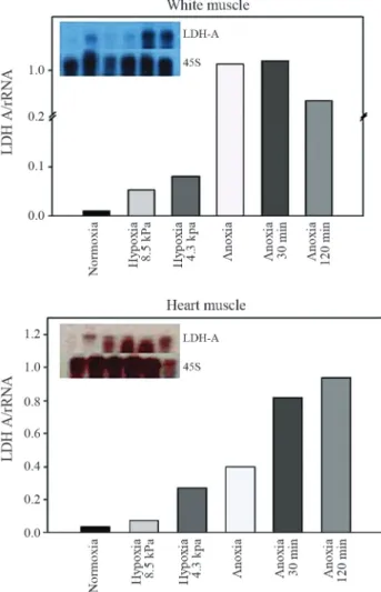
Acute hypoxia exposure
LDH-AmRNA expression was detected in skeletal and cardiac muscle in all studied groups. No expression of LDH-AmRNA was found in liver.LDH-AmRNA levels increased in white and heart muscles of juveniles exposed
to acute hypoxia (Figure 1). A 10-fold increase inLDH-A
expression was observed in the white muscle after 30 min of anoxia followed by a decrease after two hours. A three-fold increase inLDH-Atranscription was observed in car-diac muscle after deep hypoxia exposure (4.3 kPa). These higher levels of expression were retained for two hours in anoxic conditions, reaching a 10-fold increase at this time (Figure 1). No relationship between hypoxia and increasing
levels ofLDH-A mRNA expression was observed in the
adults, which expressed highLDH-Alevels in normoxia,
keeping the same LDH-A expression in anoxic animals
(Figure 2).
The semi-quantitative RT-PCR method was used withLDH-Aspecific primers forA.crassipinisand showed an increase in gene expression after anoxia (Figure S3), corroborating the results of pooled samples at the individ-ual level.
Graded hypoxia exposure
When juveniles were steadily exposed to hypoxia or
anoxia and kept in this condition for eight hours,LDH-A
expression increased in the skeletal muscle of the group
submitted to 1.7 kPa pO2. However, animals exposed to
0.9 kPa and 0.0 kPa pO2showed a decrease inLDH-A
mes-sage levels, that were similar to normoxic levels, indicating metabolic suppression (Figure 3). We observed no loss of equilibrium (behavior that precedes death) during the
whole experiment (see details in Chippari-Gomeset al.,
2005).
Discussion
The fish group has successfully radiated and is one of the most numerous groups among vertebrates, inhabiting all kinds of aquatic environments (Nelson, 2006). The evo-lution of the fish group occurred during the last 500 million
years, under low and unstable atmospheric O2 (Webster,
2003). Hypoxia-responsive elements date from 0.6 to
Figure 1- Autoradiography and estimated relative expression of RNA in white muscle (top) and in heart (bottom) of juvenileAstronotus crassipinis
0.4 billion years ago. Aquatic hypoxia was, probably, a strong regulator of gene expression and, as a consequence, of anaerobic metabolism (D'Angio and Finkelstein, 2000,
Prabhakar,2001; Semenza, 2001).
Fishes acquired the first coordinated hypoxia regula-tion system during the Silurian. The appearance of HIF-1a
(hypoxia induction factor) coincides with the development of multicellular systems and metabolic compartmentali-zation (Webster, 2003). LDH-A is one of the glycolytic en-zymes with a HIF-1asite. The HIF-1apathway is activated at oxygen saturation lower than 5% (1 kPa), and although pO2in the atmosphere is around 20 kPa, the normal pO2in
most tissues ranges between 6.7 and 9.3 kPa (Iyeret al., 1998; Hochachka, 1999; Semenza, 2001).
The sequence found forA. crassipinis LDH-Agene in the present work is extremely conservative, with very little difference from other fish species, particularly species be-longing to order Perciformes. The deduced amino acid se-quence for the protein suggests that this gene is a member of the NAD-dependent 2-hydroxycarboxylate dehydroge-nase family, of which LDH's enzyme class is a member.
Further studies ofLDH-Agene sequences in fish species
belonging to this family will be necessary to confirm the conservative character ofLDH-A.
Studies carried out in our laboratory indicate that LDH enzyme activities and hypoxia survivorship are di-rectly related toAstronotus ocellatussize (Almeida-Valet al., 2000). This increased ability to survive severe hypoxia in fishes with bigger sizes can be explained as an increase in their anaerobic power (represented by LDH absolute activi-ties in their tissues and organs) rather than a decrease in specific metabolic rates (Almeida-Valet al., 1999b, 2000). Furthermore, cichlid species of the genusAstronotusshow high tolerance to hypoxic and anoxic environments (Muus-zeet al., 1998; Chippari-Gomeset al., 2002). Both species ofAstronotus(A. crassipinis and A. ocellatus) have similar responses to hypoxia and present scaling metabolism ac-cordingly (Slomanet al.2008). In fact, in the present study, the results showed an increase in the amount of mRNA for LDH-A in adult fishes compared to juveniles in normoxia (Figures 1 and 2).
As mentioned above, increased expression ofLDH-A
along with a simultaneous reduction in the expression of LDH-Boccurs in the heart of several species of cichlids, and these characteristics are related to the degree of hypo-xia tolerance of each species (Almeida-Valet al., 1995).
The regulation ofLDH-Ain the heart is required to
com-pensate for lactate accumulation during hypoxic and anoxic episodes. The presence of the LDH-A4isoform in a highly
aerobic tissue such as the heart was observed in hypo-xia-tolerant species and represents the anaerobic
“prefer-ence” of the cardiac muscle (Almeida-Val et al., 1993,
1995).
The current analysis ofLDH-AmRNA expression in
A. crassipinisexposed to experimental hypoxia revealed higher transcription rates of this gene in white and heart muscles of juveniles. We observed a maximum increase in LDH-A expression after 30 min of anoxia in their white muscle, although the induction was more effective in car-diac muscle. After exposure to 20% air saturation, we ob-served an increase inLDH-Atranscription, and this higher level of expression was kept during the following 2 h of an-oxia. The different induction patterns observed in these two tissues are possibly due to differences in the ability of these tissues to cope with hypoxia. While white muscle endures hypoxia throughout metabolic suppression, cardiac muscle maintains lower but constant ATP generation at the ex-penses of anaerobic glycolysis. With regard to both LDH tissue distribution and the ability to deal with hypoxia, these responses are also observed in mammalian cells (Firthet al., 1994).
In adults, however, hypoxia did not affect the
expres-sion ofLDH-AmRNA. Therefore, we can infer that adult
animals have developed other mechanisms of tolerance and do not depend exclusively on gene regulation to survive hypoxic episodes. Previous studies have shown that hypo-xia tolerance in the sibling speciesA. ocellatusis directly related to the increase in size correlating to the increase in the routine metabolic rate, to a decrease in the mass-spe-Figure 2-LDH-Aexpression in white muscle and heart of juvenile and
adultAstronotus crassipinisunder normoxia and after 30 min of anoxia exposure.
cific metabolic rate and to an increase in anaerobic power in its tissues (Almeida-Valet al., 1999b, 2000). Based upon experiments with the rainbow trout gonad epithelial cell line, a further decrease in oxygen levels (down to 0.2 kPa) results in reduced accumulation of HIF-1a. Therefore, it is possible that a similar process occurs in other fish cell sys-tems and even in other species, given that these responses are very similar between mammalian and fish cells (Soi-tamoet al., 2001). As mentioned above,LDH-A,along with 100 other genes, is controlled by HIF-1aduring hypoxia.
Thus, we can conclude that the regulation ofLDH-Agene
expression in the present work is probably due to HIF-1a
induction.
Prolonging anoxia exposure for 120 min induced both an increase and a decrease inLDH-Agene expression in the heart and muscle of juveniles, respectively. A decrease in LDH-Aexpression in the muscles of juveniles allowed fish to acclimate for more than three hours in anoxia. Also, the responses of the adults seem to reflect another physiologi-cal status of both heart and skeletal muscles. In an extensive review, Nikinmaa (2002) suggested that the possible oxy-gen tensions leading to the stabilization of HIF-1aprotein are poorly studied, and that they may vary between species and cell types. If this is true, the stabilization or decrease in LDH-Aexpression can be explained as a result of this pro-cess. Furthermore, these properties also vary along animal development, as suggested by Guet al.(2000), who studied the different responses of genes of the “Per Arnt Sim” (PAS) superfamily in animals of different sizes and their roles in detecting and adapting to environmental changes.
In contrast with acute exposure, graded hypoxia ex-posure (animals exposed for 8 h to 8.5 kPa air saturation) resulted in the induction ofLDH-Aexpression. Further creases in dissolved oxygen concentrations resulted in a
de-crease ofLDH-Aexpression and can be explained as the
result of a generalized metabolic suppression induced by
the low levels of oxygen. Chippari-Gomes et al. (2005)
measured the metabolic responses in the same experiment and described an increase in blood glucose and lactate in these animals, concomitant with a decrease in glycolytic function (showed by PK, LDH and MDH activities) in the muscle of acclimated animals. Muscle LDH enzyme activi-ties dropped at low levels of hypoxia (approximately 5 kPa) and returned to control levels at higher hypoxic levels (1.7 kPa), accounting for the high levels of blood lactate de-tected. Due to LDH kinetic properties and its stability in the cell milieu, the first increase in mRNA levels can account for maintaining a continuous rate of anaerobic glycolysis in these animals, since this is a regular response in animals ex-posed to deep and prolonged hypoxia (Hochachka and Somero, 2002).
In vitrostudies of hypoxia modulation of LDH ex-pression using cultures of hepatocytes, myocytes and tu-mour cells as models have shown that the expression of LDH-AmRNA and/or protein is also induced by hypoxia
(Robinet al., 1984; Webster, 1987; Martiet al., 1994; Firth et al,.1994). As an example ofin vivostudy, the elegant ex-periment done by Graceyet al.(2001) has shown the profile of the hypoxia-induced genes by microarray analysis in the teleost fishGillichthys mirabilis. Among the many genes
identified,LDH-Awas induced in liver and white muscle,
but not in heart of that species. The induction ofLDH-Ain the heart observed inAstronotus crassipiniscould be an in-dication of its greater tolerance to hypoxia.
Studies withAstronotusspp. in our laboratory have shown that hypoxia tolerance in juveniles is accounted for
by activation of anaerobic metabolism (Muusze et al.,
1998; Chippari-Gomeset al., 2005), reduction of ATP turn-over, and suppression of the overall metabolism (Slomanet al., 2006; Richardet al., 2007) including decreases in pro-tein synthesis (Lewiset al., 2007) and partial arrest of gill ion fluxes (Wood et al., 2007) to avoid passive ion leak across the gills. Respiratory responses to progressive hypo-xia in Oscar showed that the oxygen transfer factor in-creases transiently in moderate hypoxia and dein-creases in deep hypoxia. Based on all these measurements, we con-cluded that the high tolerance of acidic anaerobic by-pro-ducts is more important to survive hypoxic conditions than a superior oxygen supply capacity (Scottet al., 2008).
In conclusion, the results presented in this study dem-onstrate that gene transcription responds immediately after hypoxia exposure, and that the intensity of this response is tissue-specific and varies according to animal size, period of acclimation and degree of hypoxia. Post-transcriptional facts, which change enzyme activities in the cells, is cer-tainly one of the regulatory mechanisms that occur in this species, since we could not find a direct relationship be-tween gene expression (present study) and absolute enzyme activities (Chippari-Gomeset al., 2005). As already stated in the literature, the kinetics of vertebrate LDH can be mod-ulated by substrate, enzyme, and co-factor concentration,
as well as by temperature, pH and pressure (Hochachkaet
al., 2002). Our results confirm previous observations on scaling hypoxia tolerance and on the higher anaerobic ca-pacity that adults ofAstronotus crassipinishave, compared to their juveniles. We also show that the activation of anaer-obic metabolism is regulated in different ways in different tissues according to the physiological characteristics of dif-ferent-sized animals. As expected, hypoxia tolerance in Astronotus crassipinisis based, at least in part, on gene reg-ulation that results from a very refined oxygen-sensing sys-tem developed and conserved throughout the evolution of vertebrates.
Acknowledgments
(#380521/00-8); ALV and VMFAV are the recipients of a Research fellowship from CNPq. The authors acknowledge Collin Brauner and Jeff Richards for useful comments on the first draft of this work. The results presented here are part of the data bank I of INCT – ADAPTA, funded by CNPq/MCT and FAPEAM.
References
Almeida-Val VMF and Val AL (1993) Evolutionary trends of LDH isozymes in fishes. Comp Biochem Physiol 105:21-28.
Almeida-Val VMF, Val AL and Hochachka PW (1993) Hypoxia tolerance in Amazon fishes: Status of an under-explored bi-ological “goldmine”. In: Hochachka PW, Lutz PL, Sick T, Rosenthal M and Van den Thillart G (eds) Surviving Hy-poxia: Mechanisms of Control and Adaptation. CRC Press, Boca Raton, pp 435-445.
Almeida-Val VMF, Farias IP, Silva MNP and Duncan WP (1995) Biochemical adjustments to hypoxia in Amazon Cichlids. Braz J Med Biol Res 28:1257-1263.
Almeida-Val VMF, Val AL and Walker I (1999a) Long- and short-term adaptation of amazon fishes to varying O2levels:
Intra-specific phenotypic plasticity and interspecific varia-tion. In: Val AL and Almeida-Val VMF (eds) Biology of Tropical Fishes. Editora do INPA, Manaus, pp 185-206. Almeida-Val VMF, Paula-Silva MN, Duncan WP, Lopes NP and
Val AL (1999b) Increase of anaerobic potential during growth of an Amazonian cichlid, Astronotus ocellatus. Survivorship and LDH regulation after hypoxia exposure. In: Val AL and Almeida-Val VMF (eds) Biology of Tropical Fishes. Editora do INPA, Manaus, pp 437-448.
Almeida-Val VMF, Val AL, Duncan WP, Souza FCA, Paula-Silva MN and Land S (2000) Scaling effects on hypoxia tol-erance in the Amazon fishAstronotus ocellatus (Percifor-mes, Cichlidae): Contribution of tissue enzyme levels. Comp Biochem Physiol 125B:219-126.
Almeida-Val VMF, Chipari-Gomes AR and Lopes NP (2006) Metabolic and physiological adjustments to low oxygen and high temperature in fishes of the Amazon. In: Val AL, Almeida-Val VMF and Randall DJ (eds) The Physiology of Tropical Fishes, v. 21. Series Fish Physiology. Elsevier, London, pp 443-500.
Chellapa S, Yamamoto ME and Cacho MSRF (1999) Reproduc-tive behavior and ecology of two species of cichlid fishes. In: Val AL and Almeida-Val VMF (eds) Biology of Tropical Fishes. Editora do INPA, Manaus, pp 113-126.
Chippari-Gomes AR, Gomes LC, Lopes NP, Val AL and Almei-da-Val VMF (2005) Metabolic adjustments in two Amazo-nian cichlids exposed to hypoxia and anoxia. Comp Bio-chem Physiol B BioBio-chem Mol Biol 141:347-355.
D’Angio CT and Finkelstein JN (2000) Oxygen regulation of gene expression: A study in opposites. Mol Genet Metab 71:371-380.
Firth JD, Ebert BL, Pugh CW and Ratcliffe PJ (1994) Oxy-gen-regulated control elements in the phosphoglycerate ki-nase 1 and lactate dehydrogeki-nase A genes: Similarities with the erythropoietin enhancer. Proc Natl Acad Sci USA 91:6496-6500.
Gracey AY, Troll JV and Somero GN (2001) Hypoxia-induced gene expression profiling in the euryoxic fishGallichthys mirabilis. Proc Natl Acad Sci USA 88:1993-1998. Gu YZ, Hogenesch JB and Bradfield CA (2000) The PAS
super-family: Sensors of environmental and developmental sig-nals. Annu Rev Pharmacol Toxicol 40:519-561.
Hall TA (1999) BioEdit: A user-friendly biological sequence alignment editor and analysis program for Windows 95/98/NT. Nucleic Acids Symp Ser 41:95-98.
Hochachka PW (1997) Oxygen - A key regulatory metabolite in metabolic defense against hypoxia. Am Zool 37:595-603. Hochachka PW (1999) Two research paths for probing the roles of
oxygen in metabolic regulation. Braz J Med Biol Res 32:661-672.
Hochachka PW and Somero GN (2002) Biochemical Adaptation: Mechanism and Process in Physiological Evolution. Oxford University Press, New York, 466 pp.
Hochachka PW, Beatty CL, Burelle Y, Trump ME, McKenzie DC and Matheson GO (2002) The lactate paradox in human high-altitude physiological performance. News Physiol Sci 17:122-126.
Iyer NV, Leung SW and Semenza GL (1998) The human hypo-xia-inducible factor 1alpha gene: HIF1A structure and evo-lutionary conservation. Genomics 52:159-165.
Lewis JM, Costa I, Val AL, Almeida-Val VMF, Gamperl AK and Driedzic WR (2007) Responses to hypoxia and recovery: Repayment of oxygen debt is not associated with compensa-tory protein synthesis in the Amazonian cichlid,Astronotus ocellatus. J Exp Biol 210:1935-1943.
Markert CL (1984) Lactate dehydrogenase. Biochemistry and function of lactate dehydrogenase. Cell Biochem Funct 2:131-134.
Markert CL and Holmes RS (1969) Lactate dehydrogenase isozy-mes of the flatfish, pleuronectiforisozy-mes: Kinetic, molecular and immunochemical analysis. J Exp Zool 171:85-104. Marti HH, Jung HH, Pfeilschifter J and Bauer C (1994) Hypoxia
and cobalt stimulate lactate dehydrogenase (LDH) activity in vascular smooth muscle cells. Pflugers Arch 429:216-222.
Muusze B, Marcon J, Van den Thillart G and Almeida-Val VMF (1998) Hypoxia tolerance of Amazon fish: Respirometry and energy metabolism of the cichlidAstronotus ocellatus. Comp Biochem Physiol 120A:151-156.
Nelson JS (2006) Fishes of the World. 4th edition. John Wiley, New York, 601 pp.
Nikinmaa M (2002) Oxygen-dependent cellular functions: Why fishes and their aquatic environment are a prime choice of study. Comp Biochem Physiol A Mol Integr Physiol 133:1-16.
Prabhakar NR (2001) Oxygen sensing during intermittent hypo-xia: Cellular and molecular mechanisms. J Appl Physiol 90:1986-1994.
Richard JG, Wang YS, Brauner CJ, Gonzalez RJ, Patrick ML, Schulte PM, Chippari-Gomes AR, Almeida-Val VMF and Val AL (2007) Metabolic and ionoregulatory responses of the Amazonian cichlid,Astronotus ocellatus, to severe hy-poxia. Comp Biochem Physiol B Biochem Mol Biol 177:361-374.
Rooney CH and Ferguson A (1985) Lactate dehydrogenase iso-zymes and alloiso-zymes of the nine-spined stickleback Pungitius pungitius (L.) (Osteichthyes, Gasterosteidae). Comp Biochem Physiol B 81:711-715.
Scott GR, Wood CM, Sloman KA, Iftikar FI, De Boeck G, Almeida-Val VMF and Val AL (2008) Respiratory respon-ses to progressive hypoxia in the Amazonian Oscar, Astronotus ocellatus. Resp Physiol Neurobiol 162:109-116. Semenza GL (2001) HIF-1, O(2), and the 3 PHDs: How animal
cells signal hypoxia to the nucleus. Cell 107:1-3.
Sloman KA, Wood CM, Scott GR, Wood S, Kajimura M, Johan-nsson OE, Almeida-Val VMF and Val AL (2006) Tribute to R. G. Boutilier: The effect of size on the physiological and behavioural responses of Oscar, Astronotus ocellatus, to hypoxia. J Exp Biol 209:1197-1205.
Soitamo AJ, Rabergh CM, Gassmann M, Sistonen L and Nikin-maa M (2001) Characterization of a hypoxia-inducible fac-tor (HIF-1alpha) from rainbow trout. Accumulation of pro-tein occurs at normal venous oxygen tension. J Biol Chem 276:19699-19705.
Val AL, Almeida-Val VMF and Paula-Silva MN (1998) Hypoxia adaptation in fish of the Amazon: A never-ending task. S Afr J Zool 33:107-114.
Webster KA (1987) Regulation of glycolytic enzyme RNA trans-criptional rates by oxygen availability in skeletal muscle cells. Mol Cell Biochem 77:19-28.
Webster KA (2003) Evolution of the coordinate regulation of glycolytic enzyme genes by hypoxia. J Exp Biol 206:2911-2922.
Wenger RH (2000) Mammalian oxygen sensing, signalling and gene regulation. J Exp Biol 203:1253-1263.
Wood CM, Kajimura M, Sloman KA, Scott GR, Walsh PJ, Almei-da-Val VMF and Val AL (2007) Rapid regulation of Na+ fluxes and ammonia excretion in response to acute environ-mental hypoxia in the Amazonian Oscar, Astronotus
ocellatus. Am J Physiol Regul Integr Comp Physiol 292:2048-2058.
Internet Resources
Chippari-Gomes AR, Val AL and Almeida-Val VMF (2002) Comparative responses of closely related cichlids to graded hypoxia. In: International Congress on the Biology of Fish, Vancouver. Responses of Fish to Aquatic Hypoxia, pp 9-13
(http://www-heb.pac.dfo-mpo.gc.ca/con-gress/2002/Hypoxia/Chippari-Gomes.pdf).
Supplementary Material
The following online material is available for this ar-ticle:
- Figure S1 - DNA sequence: Alignment of LDH-A
nucleotide sequences from Epinephelus coioides,
Lycodichthys dearborni, Notothenia angustata, Trematomus bernacchii , Pagothenia borchgrevinki, Dissostichus eleginoides, Coryphopterus nicholsi, Gillichthys seta, Gillichthys mirabilis and Astronotus crassipinnis.
- Figure S2 -LDH-A sequence from A. cassiprinis
used as a probe in Northern blotting experiments
- Figure S3 - RT-PCR expression of LDH-A in
Astronotus crassipinis juveniles exposed to normoxia, hypoxia and anoxia.
This material is available as part of the online article from http://www.scielo.br/gmb.
Associate Editor: Louis Bernard Klaczko
Lycodichthys dearborni GCCCTTGTTGACGTGATGGAGGAAAAGTTGAAGGGTGAGGTCATGGACCTGCAGCACGGCTCCCTCTTCCTCAAGACACACAAGATTGTAGCAGACAAAG Notothenia angustata GCCATGGTTGATGTGATGGAGGACAAGCTGAAGGGTGAGGTCATGGACCTGCAGCACGGATCCCTTTTCCTCAAGACA---AAGATTGTGGGAGACAAAG Trematomus bernacchii GCCATGGTTGATGTGATGGAGGACAAGCTGAAGGGTGAGGTCATGGACCTGCAGCACGGATCCCTCTTCCTCAAGACA---AAGATTGTGGGAGACAAAG Pagothenia borchgrevinki GCCATGGTTGATGTGATGGAGGACAAGCTGAAGGGTGAGGTCATGGACCTGCAGCACGGATCCCTCTTCCTCAAGACA---AAGATTGTGGGAGACAAAG Dissostichus eleginoides GCCATGGTTGATGTGATGGAGGACAAGCTGAAGGGGGAGGTCATGGACCTGCAGCACGGATCCCTCTTCCTCAAGACA---AAGATTGTGGGAGACAAAG Coryphopterus nicholsi GCCCTGGTTGATGTAATGGAGGACAAACTCAAGGGTGAGGTCATGGACTTGCAACATGGCTCCCTCTTCCTGAAGACCCACAAAATTGTTGGCGACAAAG Gillichthys seta GCCCTGGTTGATGTAATGGAGGACAAACTCAAGGGTGAGGTCATGGACTTGCAGCATGGCTCCCTCTTCCTGAAGACCCACAAAATTGTGGCTGACAAAG Gillichthys mirabilis GCCCTGGTTGATGTAATGGAGGACAAACTCAAGGGTGAGGTCATGGACTTGCAGCATGGCTCCCTCTTCCTGAAGACCCACAAAATTGTGGCTGACAAAG Astronotus crassipinnis ---AAGTTAAAGGGCGAGGCAATGGACCTGCAGCACGGATCCCTCTTCCTTAAGACGCACAAGATTGTAGCCGATAAAG
310 320 330 340 350 360 370 380 390 400 ....|....|....|....|....|....|....|....|....|....|....|....|....|....|....|....|....|....|....|....|
Epinephelus coioides ACTACAGTGTGACAGCCAACTCCAAAGTTGTGGTGGTGACGGCCGGTGCCCGCCAGCAGGAGGGCGAGAGCCGTCTTAACCTGGTCCAGCGCAACGTTAA Lycodichthys dearborni ACTACAGTGTGACAGCCAACTCCAAGGTGGTGGTGGTGACGGCCGGTGCCCGCCAGCAGGAAGGCGAGAGCCGCCTTAACCTGGTGCAGCGCAACGTCAA Notothenia angustata ACTACAGTGTGACAGCCAACTCCAAAGTGGTGGTGGTGACAGCTGGAGCCCGTCAGCAGGAGGGCGAGAGCCGTCTGAACCTTGTGCAGCGCAACGTCAA Trematomus bernacchii ACTACAGTGTGACAGCCAACTCCAAAGTGGTGGTGGTGACAGCTGGAGCCCGTCAGCAGGAGGGCGAGAGCCGTCTGAACCTTGTGCAGCGCAACGTCAA Pagothenia borchgrevinki ACTACAGTGTGACAGCCAACTCCAAAGTGGTGGTGGTGACAGCTGGAGCCCGTCAGCAGGAGGGCGAGAGCCGTCTGAACCTTGTGCAGCGCAACGTCAA Dissostichus eleginoides ACTACAGTGTGACAGCCAACTCCAAGGTGGTAGTGGTGACAGCTGGAGCCCGCCAGCAGGAGGGCGAGAGCCGTCTGAACCTTGTGCAGCGCAACGTCAA Coryphopterus nicholsi ACTACAGTGTCACAGCCAACTCCAGGGTGGTGGTGGTGACCGCCGGCGCCCGCCAGCAGGAGGGCGAGAGCCGTCTCAACCTGGTGCAGCGCAACGTCAA Gillichthys seta ACTACAGTGTCACAGCCAACTCCAGGGTGGTGGTGGTGACCGCCGGCGCCCGCCAGCAGGAGGGCGAGAGCCGTCTCAACCTGGTGCAGCGCAACGTCAA Gillichthys mirabilis ACTACAGTGTCACAGCCAACTCCAGGGTGGTGGTGGTGACCGCCGGCGCCCGCCAGCAGGAGGGCGAGAGCCGTCTCAACCTGGTGCAGCGCAACGTCAA Astronotus crassipinnis ACTACAGTGTGACAGCCAACTCCAAGGTAGTGGTGGTGACTGCAGGTGCCCGCCAGCAGGAGGGCNAAAGCCGTCTCAATNNGGTGCAACGCAACGTTAA
410 420 430 440 450 460 470 480 490 500 ....|....|....|....|....|....|....|....|....|....|....|....|....|....|....|....|....|....|....|....|
Epinephelus coioides CATCTTCAAGTTCATCATCCCGAACATCGTCAAGTACAGCCCCAACTGCATCCTGATGGTGGTTTCTAATCCAGTGGATATCCTGACCTACGTGGCCTGG Lycodichthys dearborni CATCTTCAAGTTCATCATCCCAAACATCGTCAAGTACAGCCCCAACTGCATCATCATGGTGGTTTCCAACCCAGTGGACATCCTGACCTACGTGGCCTGG Notothenia angustata CATCTTCAAGTTCATCATCCCAAACATCGTCAAGTACAGCCCCAACTGCATCCTGATGGTGGTTTCTAACCCAGTGGACATCCTGACCTATGTGGCCTGG Trematomus bernacchii CATCTTCAAGTTCATCATCCCAAACATCGTCAAGTACAGCCCCAACTGCATCTTGATGGTGGTGTCTAACCCAGTGGACATCCTGACCTATGTGGCCTGG Pagothenia borchgrevinki CATCTTCAAGTTCATCATCCCAAACATCGTCAAGTACAGCCCCAACTGCATCTTGATGGTGGTGTCTAACCCAGTGGACATCCTGACCTATGTGGCCTGG Dissostichus eleginoides CATCTTCAAGTTCATCATCCCAAACATCGTCAAGTACAGCCCCAACTGTATCCTGATGGTGGTTTCTAACCCAGTGGACATCCTGACCTATGTGGCCTGG Coryphopterus nicholsi CATCTTCAAGTTCATCATCCCCAACATCGTCAAGTACAGCCCCAACTGCATCCTGATGGTGGTCTCCAACCCAGTGGACATCCTGACCTACGTGGCCTGG Gillichthys seta CATCTTCAAGTTCATCATCCCCAACATCGTCAAGTACAGCCCCAACTGCATCCTGATGGTGGTCTCCAACCCAGTGGACATCCTGACCTACGTGGCCTGG Gillichthys mirabilis CATCTTCAAGTTCATCATCCCCAACATCGTCAAGTACAGCCCCAACTGCATCCTGATGGTGGTCTCCAACCCAGTGGACATCCTGACCTACGTGGCCTGG Astronotus crassipinnis CATCTTTAAGTTCATCATCCCCAACATCGTCAAGTACAGCCCCAACTGCATCCTGATGGTGGTCTCCAACCCAGTGGACATCCTGACCTACGTTGCCTGG
510 520 530 540 550 560 570 580 590 600 ....|....|....|....|....|....|....|....|....|....|....|....|....|....|....|....|....|....|....|....|
Epinephelus coioides AAGCTCAGCGGTTTCCCCCGTCACCGCGTCATCGGCTCCGGCACCAACCTGGACTCTGCCCGTTTCCGCCACCTCATGGGAGAGAAGCTCCACCTCCACC Lycodichthys dearborni AAGCTGAGTGGTTTCCCCCGCCACCGCGTCATTGGCTCCGGCACCAACCTGGACTCCGCCCGTTTCCGCCACCTCATGGGAGAGAAGCTCAACATCCACC Notothenia angustata AAGCTGAGTGGTTTCCCCCGTCACCGCGTCATTGGCTCTGGCACCAACCTGGACTCCGCCCGTTTCCGCCACCTCATTGGAGAGAAGCTCCACCTCCACC Trematomus bernacchii AAGCTGAGTGGTTTCCCCCGTCACCGCGTCATTGGCTCTGGCACCAACCTGGACTCCGCCCGTTTCCGCCACCTCATTGGAGAGAAGCTCCACCTCCACC Pagothenia borchgrevinki AAGCTGAGTGGTTTCCCCCGTCACCGCGTCATTGGCTCTGGCACCAACCTGGACTCCGCCCGTTTCCGCCACCTCATTGGAGAGAAGCTCCACCTCCACC Dissostichus eleginoides AAGCTGAGTGGTTTCCCCCGTAACCGCGTCATTGGCTCTGGCACCAACCTGGACTCCGCCCGTTTCCGCCACCTCATTGGAGAGAAGCTCCACCTCCACC Coryphopterus nicholsi AAGCTGAGCGGGTTCCCCCGCCACCGCGTCATCGGCTCTGGCACCAACCTGGACTCTGCCCGCTTCCGCCACATCATGGGAGAGAAGCTCCACCTCCACC Gillichthys seta AAGCTGAGCGGGTTCCCCCGCCACCGCGTCATCGGCTCTGGCACCAACCTGGACTCTGCCCGCTTCCGCCACATCATGGGAGAGAAGCTCCACCTCCACC Gillichthys mirabilis AAGCTGAGCGGGTTCCCCCGCCACCGCGTCATCGGCTCTGGCACCAACCTGGACTCTGCCCGCTTCCGCCACATCATGGGAGAGAAGCTCCACCTCCACC Astronotus crassipinnis AAACTGAGTGGCTTCCCCCGTCACCGTGTGGNTGGCTCCGGCACTAACCTCGACTCTGCTCGTTTCCGCCACATCATGGGAGAGAAGCTCCACCTGCATC
610 620 630 640 650 660 670 680 690 700 ....|....|....|....|....|....|....|....|....|....|....|....|....|....|....|....|....|....|....|....|
Epinephelus coioides CTTCCAGCTGCCATGGCTGGATCATCGGAGAGCATGGGGACTCCAGTGTGCCCGTATGGAGCGGTGTGAATGTTGCTGGAGTTTCTCTGCAGGCCCTCAA Lycodichthys dearborni CATCCAGCTGCCACGGCTGGATCGTCGGCGAGCATGGAGACTCCAGTGTGCCTGTGTGGAGCGGTGTGAATGTTGCTGGAGTTTCTCTGCAGGGCCTCAA Notothenia angustata CTTCCAGCTGCCACGCCTGGATCGTCGGAGAACATGGAGACTCCAGTGTGCCTGTCTGGAGCGGTGTGAATGTTGCTGGAGTGTCTCTGCAGGGTCTGAA Trematomus bernacchii CTTCCAGCTGCCACGCCTGGATCGTCGGAGAACATGGAGACTCCAGTGTGCCTGTTTGGAGCGGTGTGAATGTTGCTGGAGTTTCTCTGCAGGGTCTGAA Pagothenia borchgrevinki CTTCCAGCTGCCACGCCTGGATCGTCGGAGAACATGGAGACTCCAGTGTGCCTGTCTGGAGCGGTGTGAATGTTGCTGGAGTGTCTCTGCAGGGTCTGAA Dissostichus eleginoides CTTCCAGCTGCCACGCCTGGATCGTCGGAGAACATGGAGACTCCAGTGTGCCTGTCTGGAGCGGTGTGAATGTTGCTGGAGTGTCTCTGCAGGGTCTGAA Coryphopterus nicholsi CTTCCAGCTGCCACGGCTGGATCGTCGGAGAGCACGGAGACTCCAGTGTGCCCGTGTGGAGTGGAGTGAACGTTGCTGGAGTTTCTCTGCAGACCCTAAA Gillichthys seta CTTCCAGCTGCCACGGCTGGATCGTCGGAGAGCACGGAGACTCCAGTGTGCCCGTGTGGAGTGGAGTGAACGTTGCTGGAGTTTCTCTGCAGACCCTAAA Gillichthys mirabilis CTTCCAGCTGCCACGGCTGGATCGTCGGAGAGCACGGAGACTCCAGTGTGCCCGTGTGGAGTGGAGTGAACGTTGCTGGAGTTTCTCTGCAGACCCTAAA Astronotus crassipinnis CTTCTAGCTGCCATGGCTGGATCATCGGAGAGCATGGTGACTCCAGCGTGCCTGTGTGGAGTGGTGTGAATGTTGCTG-AGTTTCTCTTCAAAGCCTCAA
710 720 730 740 750 760 770 780 790 800 ....|....|....|....|....|....|....|....|....|....|....|....|....|....|....|....|....|....|....|....|
Epinephelus coioides CCCACAGATGGGTGCTGAGGGTGACGGTGAGAACTGGAAGGCAGTTCATAAGATGGTGGTTGATGGAGCCTATGAGGTCATCAAGCTGAAGGGCTACACT Lycodichthys dearborni CCCAAAGATGGGGGTCGAGGGTGACAGTGAGAACTGGAAGGCTGTGCATAAGCAGGTGGTTGATGGGGCCTACGAGGTTATCAGGCTGAAGGGCTACACT Notothenia angustata CCCACAGATGGGGACTGAGGGTGACGGTGAGAACTGGAAGGCTATTCACAAAGAGGTGGTTGATGGGGCCTATGAGGTTATTAAGCTGAAGGGCTACACC Trematomus bernacchii CCCACAGATGGGGACTGAGGGTGACGGTGAGAACTGGAAGGCTATTCACAAAGAGGTGGTTGATGGGGCCTATGAGGTTATTAAGCTGAAGGGCTACACC Pagothenia borchgrevinki CCCACAGATGGGGACTGAGGGTGACGGTGAGAACTGGAAGGCTATTCACAAAGAGGTGGTTGATGGGGCCTATGAGGTTATTAAGCTGAAGGGCTACACC Dissostichus eleginoides CCCACAGATGGGGACTGAGGGTGACGGTGAGAACTGGAAGGCTATTCACAAAGAGGTGGTTGATGGGGCCTATGAGGTTATTAAGCTGAAGGGCTACACC Coryphopterus nicholsi CCCAAAAATGGGGGCTGAGGGTGACAGCGAGAACTGGAAGGCGGTTCATAAGATGGTGGTTGATGGAGCCTACGAGGTTATCAAGCTGAAGGGCTACACT Gillichthys seta CCCAAAGATGGGGGCTGAGGGTGACAGCGAGAACTGGAAGGCGGTTCATAAGATGGTGGTTGATGGAGCCTACGAGGTTATCAAGCTGAAGGGCTACACT Gillichthys mirabilis CCCAAAAATGGGGGCTGAGGGTGACAGCGAGAACTGGAAGGCGGTTCATAAGATGGTGGTTGATGGAGCCTACGAGGTTATCAAGCTGAAGGGCTACACT Astronotus crassipinnis
+1fr: ·K··L··K··G··E··A··M··D··L··Q··H··G··S··L··F··L··K··T··H··K·
LDH: attgtagccgataaagactacagtgtgacagccaactccaaggtagtggtggtgactgca
+1fr: ·I··V··A··D··K··D··Y··S··V··T··A··N··S··K··V··V··V··V··T··A·
LDH: ggtgcccgccagcaggagggcnaaagccgtctcaatnnggtgcaacgcaacgttaacatc
+1fr: ·G··A··R··Q··Q··E··G··X··S··R··L··N··X··V··Q··R··N··V··N··I·
LDH: tttaagttcatcatccccaacatcgtcaagtacagccccaactgcatcctgatggtggtc
+1fr: ·F··K··F··I··I··P··N··I··V··K··Y··S··P··N··C··I··L··M··V··V·
LDH: tccaacccagtggacatcctgacctacgttgcctggaaactgagtggcttcccccgtcac
+1fr: ·S··N··P··V··D··I··L··T··Y··V··A··W··K··L··S··G··F··P··R··H·
LDH: cgtgtggntggctccggcactaacctcgactctgctcgtttccgccacatcatgggagag
+1fr: ·R··V··X··G··S··G··T··N··L··D··S··A··R··F··R··H··I··M··G··E·
LDH: aagctccacctgcatccttctagctgccatggctggatcatcggagagcatggtgactcc
+1fr: ·K··L··H··L··H··P··S··S··C··H··G··W··I··I··G··E··H··G··D··S·
LDH: agcgtgcctgtgtggagtggtgtgaatgttgctgagtttctcttcaaagcctcaacccaa
+1fr: ·S··V··P··V··W··S··G··V··N··V··A··E··F··L··F··K··A··S··T··Q·