839 839 839 839 839 Mem Inst Oswaldo Cruz, Rio de Janeiro, Vol. 102(7): 839-844, November 2007
Detection of diarrheagenic
Escherichia coli
from children with and
without diarrhea in Salvador, Bahia, Brazil
Vanessa Bueris, Marcelo Palma Sircili, Carla Romano Taddei, Maurilio Fernandes dos Santos,
Marcia Regina Franzolin/
+, Marina Baquerizo Martinez*, Suzana Ramos Ferrer**,
Mauricio Lima Barreto**, Luiz Rachid Trabulsi
Laboratório de Bacteriologia, Instituto Butantan, Av. Vital Brazil 1500, 05503-900 São Paulo, SP, Brasil *Faculdade de Ciências Farmacêuticas, Departamento de Análises Clínicas e Toxicológicas, Universidade de São Paulo, São Paulo, SP, Brasil **Instituto de
Saúde Coletiva, Universidade Federal da Bahia, Salvador, BA, Brasil
We identified different diarrheagenic (DEC) Escherichia coli pathotypes isolated from 1,207 children with and without acute endemic diarrhea in Salvador, Bahia, Brazil collected as part of a case-control study. Since the identification of DEC cannot be based on only biochemical and culture criteria, we used a multiplex poly-merase chain reaction developed by combining five specific primer pairs for Enteropathogenic Escherichia coli
(EPEC), Shiga toxin-producing E. coli/ Enterohaemorrhagic E. coli (STEC/EHEC), Enterotoxigenic E. coli (ETEC) and Enteroaggregative E. coli (EAEC) to detect these pathotypes simultaneously in a single-step reaction. In order to distinguish typical and atypical EPEC strains, these were tested for the presence of EAF plasmid. The prevalence of diarrheagenic E. coli in this sample of a global case-control study was 25.4% (259 patients) and 18.7% (35 patients) in the diarrhea group (1,020 patients) and the control group (187 patients), respectively. The most frequently isolated pathotype was EAEC (10.7%), followed by atypical EPEC (9.4%), ETEC (3.7%), and STEC (0.6%). Typical EPEC was detected only in one sample. The prevalence of the pathotypes studied in children with diarrhea was not significantly different from that in children without diarrhea.
Key words: diarrheagenic Escherichia coli - virulence - Multiplex PCR
Diarrhea continues to be one of the most common causes of morbidity and mortality among infants and chil-dren, especially in developing countries (WHO, 2002). The etiological agents of diarrhea include a wide range of viruses, bacteria and parasites. Among the bacterial pathogens, diarrheagenic Escherichia coli (DEC) is an important agent of endemic and epidemic diarrhea world-wide (Nataro & Kaper 1998).
DEC strains can be classified into six main pathotypes on the basis of their specific virulence properties, asso-ciation with some serotypes, and different epidemio-logical and clinical features.
Enteropathogenic E. coli (EPEC) cause a histopatho-logical lesion known as “attaching and effacing” (A/E). Strains of the A/E genotype, which do not possess the Enteropathogenic E. coli adherence factor (EAF) plas-mid, are classified as atypical EPEC (a-EPEC) (Trabulsi et al. 2002). The term Shiga toxin-producing E. coli
(STEC) is now used to define any E. coli strain that pro-duces Stx, and enterohemorrhagic E. coli (EHEC) is used to denote the Stx-positive strains that also contain locus of enterocyte effacement (LEE) (Kaper et al. 2004).
Enterotoxigenic E. coli (ETEC) are associated with wa-tery diarrhea among children in developing countries and are the main cause of traveler’s diarrhea in adults visit-ing endemic areas. Enteroaggregative E. coli (EAEC) are defined as E. coli strains that adhere in vitro to HEp-2 cells in a pattern known as autoaggregative. Enteroin-vasive E. coli (EIEC) are capable of invading the colonic epithelium and spreading from cell to cell, which may occasionally cause dysentery. Diffusely adherent E. coli
(DAEC) are defined by a characteristic diffuse pattern of adherence to HEp-2 cells (Nataro & Kaper 1998).
The identification of DEC cannot be based only on cultural and biochemical criteria, since they are indis-tinguishable from the non-pathogenic E. coli commonly found in human feces. Moreover, specific serotyping is not always correlated with pathogenicity. Since several virulence factors and DNA sequences of DEC have been identified, they can be determined by the presence of genes coding for specific virulence factors, which are absent in non-pathogenic strains. Polymerase chain re-action (PCR) is a commonly used method that gives rapid, reliable results, and shows high sensitivity and high speci-ficity. Several PCR methods, with both single and mul-tiple target genes, have been reported for detecting the different DEC pathotypes. In order to simplify the iden-tification, we developed a multiplex PCR combining five specific primer pairs for EPEC, STEC, ETEC, and EAEC virulence genes, to detect these pathotypes si-multaneously in a single-step reaction. Since the in-cidence of EIEC is very low and DAEC is not well defined as a distinct pathotype, their detection was not included in this study.
Financial support: Pronex (Contract 661086/1998-4) +Corresponding author: [email protected]
This work is dedicated to Luiz Rachid Trabulsi (in memoriam) who supervised this study and inspired us to continue his work. Received 21 May 2007
840 840 840 840
840 Diarrheagenic Escherichia coli in Salvador • Vanessa Bueris et al.
MATERIALS AND METHODS
Study population -From February, 2003 to Septem-ber, 2004, stool samples from 1,207 children (1 to 10 years of age) were collected as part of a case control study, with the aim of identifying risk factors for diar-rhea, involving 3,234 children (< 10 years of age) in the city of Salvador, which included 1,688 cases and 1,676 controls. Cases and controls were selected from patients of five outpatient clinics from various areas of Salvador, state of Bahia, Brazil. Cases were defined as children whose main complaint was acute diarrhea, characterized by the occurrence of three or more, loose, liquid, or watery stools in a 24 h period. Controls were children being seen at the same clinics for reasons other than di-arrhea, without any gastrointestinal symptoms for at least 21 days before inclusion in the study.Clinical and epi-demiological information were obtained through ques-tionnaires. The global case-control study sample con-sisted of 1,688 diarrhea cases and 390 controls (23% of the total 1,676 controls), and the present study in-cluded 1,020 diarrheic and 187 control children, repre-senting a partial analysis of the case-control study.
Pathogen identification - Stool samples collected in Cary-Blair transport medium were cultured on differ-ent solid media. MacConkey agar (Merck) was used to select E. coli isolates. As a rule 2-3 lactose-fermenting colonies, presumed to be E. coli by colony morphology, were selected and submitted to biochemical tests. E. coli
isolates were stored at -80°C in trypticase soy broth (TSB) supplemented with 20% glycerol for further pro-cedures. Bacterial testing for Salmonella, Shigella,
Yersinia, Aeromonas, and Campylobacter jejuni-coli
was conducted using standard procedures (Steele & McDermott 1984, Koneman et al. 1997). Adenovirus, Astrovirus, and Rotavirus (group A) were identified by enzyme immunoassay (Biomanguinhos, Oswaldo Cruz Foundation) and polyacrylamide gel electrophoresis
(PAGE). Parasitological examinations for helminthes and protozoa were conducted using the Kato-Katz (Katz et al. 1972) and sedimentation (Koneman et al. 1997) meth-ods. To test for Cryptosporidia sp, stool specimens were sieved and submitted to the formol-ether concentration method (Allen & Ridley 1970).
Multiplex PCR - DNA templates for PCR were ob-tained from overnight E. coli cultures that were pelleted, resuspended in 500 µl of sterile deionized water and boiled for 10 min. The multiplex PCR was developed by combining five specific primer pairs for EPEC, STEC/ EHEC, ETEC, and EAEC virulence genes, as described in Table I. Each multiplex reaction was performed in a 50 µl final volume containing 1 µl of the template DNA, 0.2 mM DNTPs, 10 mM Tris-HCl (pH8.8), 1.5 mM MgCl2, 50 mM KCl, 2 U Taq DNA polymerase (Invitrogen), and 10 pmol of each primer (Biosynthesis). The thermocycling conditions were as follows: 94°C for 5 min, 94°C for 45 s, 50°C for 1 min, and 2 min and 72°C for 35 cycles, with a final 9 min extension at 72°C. Amplified samples were evaluated by 1% agarose gel electrophoresis in Tris-bo-rate-EDTA buffer and ethidium bromide staining.
DEC detection - DNA templates from the stored E. coli isolates were submitted to multiplex PCR as de-scribed above. The eae-positive strains were tested by PCR for the presence of EAF plasmid, as described by Franke et al. (1994). In all experiments, the prototype EPEC E2348/69, EAEC 042, ETEC H10407 (LT), ETEC C275/15 (ST), and EHEC O157:H7 strains were used as positive control, while E. coli K12 DH5α was the nega-tive control.
Serotyping -The somatic (O) and flagellar (H) anti-gens of STEC (eae positive) strains and typical EPEC were determined by standard agglutination methods us-ing O1 to O172 and H1 to H51 (Orskov & Orskov 1984).
TABLE I
Sequence of polymerase chain reaction (PCR) primers, sizes of amplified DNA fragments and concentration of multiplex PCR
Product
Gene Sequence (5'- 3') size (bp) [pmol] Reference
eae IntFC – CCGGAATTCGGGATCGATTACCGTCAT 820 100 Batchelor et al. (1999)
(EPEC) IntRC – CCAAGCTTTTATTTATCAGCCTTAATCTC
LT LTFW – GGCGACAGATTATACCGTGC 696 20 Schultsz et al. (1994)
(ETEC) LTREV – CCGAATTCTGTTATATATGTC
ST STFW – TCCCTCAGGATGCTAAAC 240 20 Moseley et al. (1983)
(ETEC) STREV – GCAACAGGTACATACGTT
Stx StxUFW – GAACGAAATAATTTATATGT 500 75 Yamasaki et al. (1996)
(STEC) StxUREV – TTTGATTGTTACAGTCAT
AA pCVD432FW – CTGGCGAAAGACTGTATCAT 630 20 Schmidt et al. (1995)
(EAEC) pCVD432REV–CAATGTATAGAAATCCGCTGTT
841 841 841 841 841 Mem Inst Oswaldo Cruz, Rio de Janeiro, Vol. 102(7), November 2007
Statistical assessment was performed using Pearson’s chi-square test, where values < 0.05 were considered sta-tistically significant.
RESULTS
A total of 1,020 children with diarrhea and 187 con-trol children without diarrhea were studied. Diarrheagenic E. coli was isolated from 25.4% of the children with diarrhea (259 patients) compared with 18.7% of children without diarrhea (35 patients). The frequency of each DEC pathotype is shown in Table II. The most frequently isolated was EAEC (10.7%), fol-lowed by atypical EPEC (9.4%), ETEC (3.7%), and STEC (0.6%). Typical EPEC was detected only in one sample (0.1%), and it belonged to serotype O142:H34.
Thirty-two ETEC strains were positive for the LT gene and five for the ST gene, while seven strains possessed both genes. Only four patients displayed two pathotypes simulta-neously (a-EPEC and EAEC – 3 patients; EAEC and ETEC – 1 patient). E. coli strains containing the target genes were not identified in the stools from the other 152 (81.3%) patients without diarrhea and 761 (74.6%) pa-tients with diarrhea. The three STEC strains that had eae
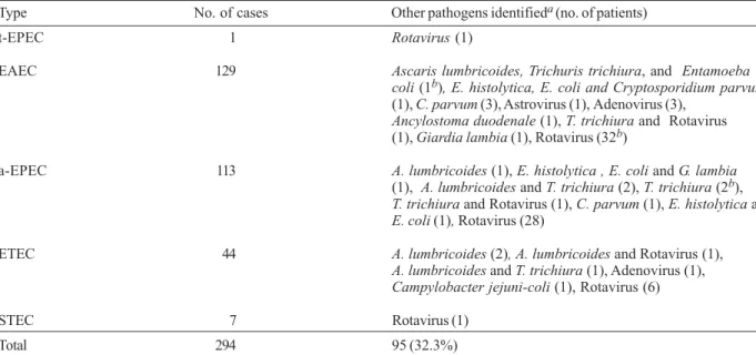
gene identified by PCR belonged to serotypes O157:H7, O26:H11 (control patients), and O111:H- (case patient). DEC was isolated in the stools as the sole enteric patho-gen apatho-gent in 199 (67.7%) of 294 patients, and in the re-maining 95 (32.3%) children, DEC was associated with some other pathogens (Table III).
TABLE II
Frequency of Escherichia coli pathotypes in children with and without diarrhea
E. coli pathotype Control patients (n = 187) (%) Case patients (n = 1020) (%) pa Total (n = 1207) (%)
EAEC 16 (8.6) 113 (11.1)b 0.30 129 (10.7)
a-EPECc 11 (5.9) 102 (10.0) 0.07 113 (9.4)
ETEC 6 (3.2)g 38 (3.7)d h 0.73 44 (3.7)i
STEC 2 (1.1)e 5 (0.5)f 7 (0.6)
t-EPEC 0 1 (0.1) 1 (0.1)
Total 35 (18.7) 259 (25.4) 0.05 294 (24.4)
a: chi-square test; b: three patients had Enteroaggregative E. coli (EAEC) strain and atypical Enteropathogenic E. coli (a-EPEC) strain; c: three strains had the LT gene; d: one patient had Enterotoxigenic E. coli (ETEC) strain (LT +) and Enteroaggregative E. coli (EAEC) strain; e: two strains had the eae gene, and belonged to serotypes O26:H11, and O157:H7; f: one strain had the eae gene (O111:H- serotype);g: LT gene - five strains; ST and LT genes - one strain; h: LT gene - 27 strains; ST and LT genes - six strains; ST gene - five strains; i: LT gene - 32 strains; ST gene - five strains; ST and LT - seven strains.
TABLE III
Pathogenic species identified in the fecal samples positive for diarrheagenic Escherichia coli
Type No. of cases Other pathogens identifieda (no. of patients)
t-EPEC 1 Rotavirus (1)
EAEC 129 Ascaris lumbricoides, Trichuris trichiura,and Entamoeba
coli (1b), E. histolytica, E. coli and Cryptosporidium parvum
(1), C. parvum (3), Astrovirus(1),Adenovirus(3), Ancylostoma duodenale (1), T. trichiura and Rotavirus (1), Giardia lambia (1),Rotavirus(32b)
a-EPEC 113 A. lumbricoides (1), E. histolytica , E. coli and G. lambia
(1), A. lumbricoides and T. trichiura (2), T. trichiura (2b),
T. trichiura andRotavirus (1), C. parvum (1), E. histolytica and E. coli (1), Rotavirus(28)
ETEC 44 A. lumbricoides (2), A. lumbricoides and Rotavirus(1),
A. lumbricoides and T. trichiura (1), Adenovirus(1), Campylobacter jejuni-coli (1), Rotavirus(6)
STEC 7 Rotavirus (1)
Total 294 95 (32.3%)
842 842 842 842
842 Diarrheagenic Escherichia coli in Salvador • Vanessa Bueris et al.
DISCUSSION
Phenotypic assays are routinely used in most labora-tories to characterize DEC strains. However, these meth-ods alone are not sufficient to identify all five pathotypes of DEC (Brandal et al. 2007). The genes encoding viru-lence factors are extensively studied and characterized, and several PCR methods have been developed to iden-tify the virulence genes of DEC (Kimata et al. 2005). Numerous multiplex PCR assays have been developed to identify DEC, and assays were recently described that are capable of distinguishing the five pathotypes of DEC (Aranda et al. 2004, Nguyen et al. 2005).
In this study, five distinct pathotypes and sub-pathotypes of diarrheagenic E. coli were detected in chil-dren with and without acute diarrhea in Salvador, Bahia. We were able to save time and effort involved in testing for various virulence factors, reducing the number of gene detection assays by developing a multiplex PCR method that has been shown to be highly sensitive and specific, using primers for the different DEC pathotypes in a single reaction.
EAEC was the most commonly isolated pathotype, followed by atypical EPEC, whereas the frequency of t-EPEC was very low. In addition, we identified seven STEC strains, and three of these strains harbored the eae gene, but two of them were from control patients. Interest-ingly, the only one O157:H7 STEC strain identified in this study was isolated from a child without diarrhea. The prevalence of the pathotypes studied in children with diarrhea was not significantly different from children without diarrhea.
ETEC and EAEC have been frequently detected in children with diarrhea in studies conducted in Brazil and in other countries (Gomes et al. 1991, 1996, Nataro & Kaper 1998, Scaletsky et al. 2002, Zamboni et al. 2004, Regua-Mangia et al. 2004), which is in agreement with the results reported in this study. EAEC is emerging as an enteric pathogen of great concern, responsible for acute and persistent diarrhea (≥ 14 days) and may cause malnutrition and growth defects in children. These strains have been associated with traveler’s diarrhea in both de-veloping and industrialized countries, and have been iso-lated in immunocompromised patients (Nataro 2005).
Previous studies were conducted in Salvador, Bahia to determine the presence of DEC in acute endemic di-arrhea. Tornieporth et al. (1995) analyzed stool samples from children with and without diarrhea by PCR. They only investigated virulence marker genes of ETEC, EPEC, and EIEC, identifying 16%, 8%, and 1% of the children with these pathotypes, respectively. Another epidemiological study conducted by our group examined children with acute endemic diarrhea in Salvador, by colony blot hybridization including other virulence fac-tors (Franzolin et al. 2005, Barreto et al. 2006). The most frequently isolated was atypical EPEC (10.1%) followed by ETEC (7.5%), and EAEC (4.2%). STEC eae-positive (serotype O26:H11), STEC and typical EPEC were each detected once.
In the present study we analyzed a greater number of patients and also included controls. The isolation of only
one typical EPEC strain confirms a recent trend that has been observed in São Paulo and in other Brazilian states (Gomes et al. 2004, Franzolin et al. 2005), showing a drastic decline in the frequency of typical EPEC with the frequency of atypical EPEC strains on the rise. Up to the 1990s, typical EPEC was the main cause of acute diarrhea in children younger than one year old, of low socio-economic status, in Brazil (Gomes et al. 1991, Trabulsi et al. 2002, Regua-Mangia et al. 2004), but such is currently very rare. This situation also occurred in developed countries, where up to the 1960s typical EPEC was frequently isolated as the cause of outbreaks and sporadic cases of diarrhea, but is currently very rare, while the frequency of atypical EPEC strains has in-creased (Afset et al. 2003). Outbreaks of adult gastro-enteritis due to atypical EPEC have been reported (Viljanen et al. 1990, Hedberg et al. 1997). t-EPEC have not been isolated from animals (Nataro & Kaper 1998), and thus humans appear to be the unique living reservoir for these organisms. It is likely that the frequency of t-EPEC has been influenced by the recently improved pub-lic health measures, such as more efficient control of hos-pital infections and implementation of sanitary conditions, more so than the frequency of a-EPEC and EHEC which are hosted by humans and animals (Trabulsi et al. 2002).
This is the second time that STEC/EHEC strains were isolated in Salvador, but to our knowledge, the O157:H7 serotype has been isolated in this city only in the present study. However, O157:H7 and O26:H11 serotypes were isolated from children belonging to the control group. No blood was reported in the feces of the children with STEC/EHEC. In Brazil, infections due to STEC have been identified in sporadic cases of non-bloody diarrhea, par-ticularly in young children (Gomes et al. 1996, Cantarelli et al. 2000, Guth et al. 2002, 2005, Bastos et al. 2006). Irino et al. (2002) isolated three O157:H7 strains from bloodydiarrhea, and one HUScase due to an O26:H11 strain has been recently described in São Paulo, Brazil (Guth et al. 2002).Among non-O157 STEC,serotypes O111:NM, O111:H8, and O26:H11 have been identified as agents of infantilediarrhea in São Paulo, since the late 1970s (Vaz et al. 2004). STEC/EHEC are frequently detected in food and in animal reservoirs in Brazil (Cerqueira et al. 1999, Leomil et al. 2003, Bastos et al. 2006), but only some of the serotypes identified in ani-mals have been recognized as causes of human illness. Bastos et al. (2006) studied eighteen strains of the O157:H7 serotype isolated from animals and from bloody diarrhea and HUS in humans during different surveys conducted in Brazil, suggesting that this serotype is emerging in this country. These findings point to the need for more ef-fective measures from our public health services.
843 843 843 843 843 Mem Inst Oswaldo Cruz, Rio de Janeiro, Vol. 102(7), November 2007
As the virulence determinants of atypical EPEC are relatively unknown, further studies are needed to inves-tigate the ecological, socio-economic and epidemio-logical basis of this emerging pathotype.
REFERENCES
Afset JE, Bergh K, Bevanger L 2003. High prevalence of atypi-cal enteropathogenic Escherichia coli (EPEC) in Norwe-gian children with diarrhoea. J Med Microbiol52: 1015-1019.
Allen AVD, Ridley DS 1970. Further observations on the formol-ether concentration technique for fecal parasites. J Clin Pathol23: 545.
Aranda KR, Fagundes-Neto U, Scaletsky IC 2004. Evaluation of multiplex PCRs for diagnosis of infection with diarrheagenic of Escherichia coli and Shigella spp. J Clin Microbiol42: 5849-5853.
Barreto ML, Milroy CA, Strina A, Prado MS, Leite JP, Ramos EA, Ribeiro H, Alcantara-Neves NM, Teixeira MD, Rodrigues LC, Ruf H, Guerreiro H, Trabulsi LR 2006. Com-munity-based monitoring of diarrhea in urban Brazilian chil-dren: incidence and associated pathogens. Trans R Soc Trop Med Hyg100: 234-242.
Bastos FC, Vaz TM, Irino K, Guth BE 2006. Phenotypic character-istics, virulence profile and genetic relatedness of O157 Shiga toxin-producing Escherichia coli isolated in Brazil and other Latin American countries. FEMS Microbiol Lett265: 89-97.
Batchelor M, Knutton S, Caprioli A, Huter V, Zanial M, Dougan G, Frankel G 1999. Development of a universal intimin anti-serum and PCR primers. J Clin Microbiol37: 3822-387.
Brandal LT, Lindstetd BA, Lena A, Stavnes TL, Lassen J, Kapperud G 2007. Octaplex PCR and fluorescence-based capillary electrophoresis for identification of human diarrheagenic Escherichia coli and Shigella ssp. J Microbiol Methods68: 331-342.
Cantarelli V, Nagayama K, Takahashi A, Honda T, Cauduro PF, Dias GAG, Mezzari A, Brodt T 2000. Isolation of Shiga toxin-producing Escherichia coli (STEC) serotype O91:H21 from a child with diarrhea in Porto Alegre city, RS, Brazil. Braz J Microbiol31: 266-270.
Cerqueira AMF, Guth BEC, Joaquim RM, Andrade JRC 1999. High occurrence of Shiga toxin-producing Escherichia coli (STEC) in healthy cattle at Rio de Janeiro State, Brazil. Vet Microbiol70: 111-121.
Franke J, Franke S, Schmidt H, Schwarzkopf A, Wieler LH, Baljer G, Beutin L, Karch H 1994. Nucleotide sequence analysis of enteropathogenic Escherichia coli (EPEC) adherence fac-tor probe and development of PCR for rapid detection of EPEC harboring virulence plasmids. J Clin Microbiol32: 2460-2463.
Franzolin MR, Alves RCB, Keller R, Gomes TAT, Beutin L, Barreto ML, Milroy CA, Strina A, Ribeiro H, Trabulsi LR 2005. Prevalence of diarrheagenic Escherichia coli in chil-dren with diarrhea in Salvador, Bahia, Brazil. Mem do Instituto Oswaldo Cruz100: 359-363.
Gomes TAT, Griffin PM, Ivey C, Trabulsi LR, Ramos SRTS 1996. EPEC infections in São Paulo. Rev Microbiol 27: 25-33.
Gomes TAT, Irino K, Girão DM, Girão VB, Guth BE, Vaz TM, Moreira FC, Chinarelli SH, Vieira MA 2004. Emerging en-teropathogenic Escherichia coli strains? Emerg Infect Dis 10: 1851-185.
Gomes TAT, Rassi V, MacDonald KL, Ramos SR, Trabulsi LR, Vieira MA 1991. Enteropathogens associated with acute di-arrheal disease in urban infants in São Paulo, Brazil. J Infect Dis164: 331-337.
Guth BEC, Lopes R, Vaz TMI, Irino K 2002. First Shiga toxin-producing Escherichia coli isolate from a patient with hemolytic uremic syndrome in Brazil. Emerg Infect Dis8: 535-536.
Guth BEC, Vaz TM, TA Gomes TA, Chinarelli SH, Rocha MM, de Castro AF, Irino K 2005. Re-emergence of O103:H2 Shiga toxin-producing Escherichia coli infections in São Paulo, Brazil. J Med Microbiol54: 805-806.
Hedberg C, Savarino S, Besser J, Paulus C, Thelen V, Myers L, Cameron DN, Barrett TJ, Kaper JB, Osterholm MT 1997. An outbreak of foodborne illness caused by Escherichia coli O39:NM, an agent not fitting into the existing scheme for classifying diarrheogenic E. coli. J Infect Dis176: 1625-1628.
Irino K, Vaz TMI, Kato MAMF, Naves ZVF, Lara RR, Marco MEC, Rocha MM, Moreira TP, Gomes TA, Guth BE 2002. O157:H7 Shiga toxin-producing Escherichia coli strains as-sociated with sporadic cases of diarrhea in São Paulo, Bra-zil. Emerg Infect Dis8: 446-447.
Kaper JB, Nataro JP, Mobley HLT 2004. Pathogenic Escheri-chia coli. Nat Rev Microbiol2: 123-40.
Katz N, Chaves A, Pellegrino J 1972. A simple device for quan-titative stool thick-smear technique in schistosomiasis mansoni. Rev Inst Med Trop São Paulo 14: 397-400.
Kimata K, Shima T, Shimizu M, Tanaka D, Isobe J, Gyobu Y, Watahiki M, Nagai Y 2005. Rapid Categorization of Patho-genic Escherichia coli by Multiplex PCR. Microbiol Immunol49: 485-492
Koneman EW, Allen SD, Janda WM, Schreckenberger PC, Winn Jr WC 1997. Diagnostic Microbiology. Color Atlas and Textbook, 5th ed., Lippincott. Philadelphia, Pennsylvania, 1395 pp.
Leomil L, Akdar-Ugrinovich L, Guth BE, Irino K, Vettorato MP, Onuma DL, de Castro AF 2003. Frequency of Shiga toxin-producing Escherichia coli (STEC) isolates among diarrheic and non-diarrheic calves in Brazil. Vet Microbiol97: 103-109.
Moseley SL, Hardy JW, Hug MI, Echeverria P, Falkow S 1983. Isolation and nucleotide sequence determination of a gene encoding a heat-stable enterotoxin of Escherichia coli. In-fect Immun 39: 1167-1174.
Nataro JP 2005. Enteroaggregative Escherichia coli pathogen-esis. Curr Opin Gastroenterol 21: 4-8.
Nataro JP, Kaper JB 1998. Diarrheagenic Escherichia coli. Clin Microbiol Rev 11: 142-201.
Nguyen TV, Le Van P, Le Huy C, Gia KN, Weintraub A 2005. Detection and characterization of diarrheagenic Escherichia coli from young children in Hanoi, Vietnam. J Clin Microbiol 43: 755-760.
Orskov F, Orskov I 1984. Serotyping of Escherichia coli. Meth-ods Microbiol14: 43-112.
844 844 844 844
844 Diarrheagenic Escherichia coli in Salvador • Vanessa Bueris et al.
Scaletsky IC, Fabbricotti SH, Silva SO, Morais MB, Fagundes-Neto U 2002. HEp-2-adherent Escherichia coli strains as-sociated with acute infantile diarrhea, São Paulo, Brazil. Emerg Infect Dis8: 855-858.
Schimidt H, Knop C, Franke S, Aleksic S, Heesemann J, Karch H 1995. Development of PCR for screening of enteroaggregative Escherichia coli. J Clin Microbiol33: 701-705.
Schultsz C, Pool GJ, van Ketel R, de Wever B, Speelman P, Dankert J 1994. Detection of enterotoxigenic Escherichia coli in stool samples by using nonradioactively labeled oligonucle-otide DNA probes and PCR. J Clin Microbiol32: 2393-2397.
Steele TW, McDermott SN 1984. The use of membrane filters applied directly to the surface of agar plates for the isolation of Campylobacter jejuni from feces. Pathol 16: 263-265.
Tornieporth NG, John J, Salgado K, de Jesus P, Latham E, Melo MC, Gunzburg ST, Riley LW 1995. Differentiation of patho-genic Escherichia coli strains in Brazilian children by PCR. J Clin Microbiol33: 1371-1374.
Trabulsi LR, Keller R, Gomes TAT 2002. Typical and atypical en-teropathogenic Escherichia coli. Emerg Infect Dis8: 508-513.
Vaz TM, Irino K, Kato MA, Dias AM, Gomes TA, Medeiros MI, Rocha MM, Guth BE 2004. Virulence properties and char-acteristics of Shiga toxin-producing Escherichia coli in São Paulo, Brazil, from 1976 through 1999. J Clin Microbiol42: 903-905.
Viljanen M, Peltola T, Junnila S, Olkkonen L, Jarvinen H, Kuistila M, Huovinen P 1990. Outbreak of diarrhoea due to Escheri-chia coli O111:B4 in school children and adults: association of Vi antigen-like reactivity. Lancet 336: 381-384.
WHO - World Health Organization 2002. Reducing Risks, Pro-moting Healthy Life. The World Health Report (http:// www.who.int/whr/en/).
Yamasaki S, Lin Z, Shirai H, Terai A, Oku Y, Ito H, Ohmura M, Karasawa T, Tsukamoto T, Kurazono H, Takeda Y 1996. Typing of verotoxins by DNA colony hybridization with poly-and oligonucleotide probes, a bead-enzyme-linked immunosorbent assay, and polymerase chain reaction. Microbiol Immunol40: 345-52.