Research Article
Experimental Autoimmune Encephalomyelitis Development Is
Aggravated by
Candida albicans
Infection
Thais F. C. Fraga-Silva,
1Luiza A. N. Mimura,
1Camila M. Marchetti,
2Fernanda Chiuso-Minicucci,
1Thais G. D. França,
1Sofia F. G. Zorzella-Pezavento,
1James Venturini,
2Maria S. P. Arruda,
2and Alexandrina Sartori
11Department of Microbiology and Immunology, Biosciences Institute, S˜ao Paulo State University (UNESP),
18618-000 Botucatu, SP, Brazil
2Department of Biological Sciences, School of Sciences, S˜ao Paulo State University (UNESP), 17033-360 Bauru, SP, Brazil
Correspondence should be addressed to Alexandrina Sartori; [email protected]
Received 18 September 2014; Revised 19 January 2015; Accepted 22 January 2015
Academic Editor: Oscar Bottasso
Copyright © 2015 hais F. C. Fraga-Silva et al. his is an open access article distributed under the Creative Commons Attribution License, which permits unrestricted use, distribution, and reproduction in any medium, provided the original work is properly cited.
Multiple sclerosis (MS) is an inlammatory/autoimmune disease of the central nervous system (CNS) mainly mediated by myelin speciic T cells. It is widely believed that environmental factors, including fungal infections, contribute to disease induction or
evolution. Even thoughCandidainfection among MS patients has been described, the participation of this fungus in this pathology
is not clear. he purpose of this work was to evaluate the efect of aCandida albicansinfection on experimental autoimmune
encephalomyelitis (EAE) that is a widely accepted model to study MS. Female C57BL/6 mice were infected withC. albicansand 3
days later, animals were submitted to EAE induction by immunization with myelin oligodendrocyte glycoprotein. Previous infection
increased the clinical score and also the body weight loss. EAE aggravation was associated with expansion of peripheral CD4+T
cells and production of high levels of TNF-�, IFN-�IL-6, and IL-17 by spleen and CNS cells. In addition to yeast and hyphae,
fungus speciic T cells were found in the CNS. hese indings suggest thatC. albicansinfection before EAE induction aggravates
EAE, and possibly MS, mainly by CNS dissemination and local induction of encephalitogenic cytokines. Peripheral production of encephalitogenic cytokines could also contribute to disease aggravation.
1. Introduction
Multiple sclerosis (MS) is an inlammatory/autoimmune and demyelinating disease of the central nervous system (CNS). It is considered one of the most common neurological disorders and causes of disability in young adults [1]. he estimated number of people with MS has increased from 2.1 million in 2008 to 2.3 million in 2013 [2]. Animal models, partic-ularly experimental autoimmune encephalomyelitis (EAE), have been essential to decipher the pathophysiology of MS [3–6]. MS and EAE are characterized by an autoimmune response against CNS proteins, mediated mainly by T cells, that culminates in inlammatory iniltrate, gliosis, damage of myelin sheath, and neuronal death [7–9].
dissemination to the CNS [12], they could contribute to local tissue destruction by their presence or, alternatively, by the induction of a local immune response.
Candidaspp. is one of these pathogens that could con-tribute to MS development.C. albicansis a pleomorphic fun-gus that colonizes the majority of healthy human individuals. his fungus can behave as a normal component of the micro-biota and also as an opportunistic pathogen that causes super-icial mucosal infections as well as disseminated disease [13, 14]. As the fourth most common cause of nosocomial infections,C. albicansis commonly isolated from immuno-compromised individuals, including those with HIV, those immunosuppressed due to cancer treatment, and premature babies [15]. A possible association between MS and Can-dida spp. has been suggested by serological evidences. A signiicantly higher level ofCandidaspeciic antibodies was detected in MS patients than in normal control individuals [16]. In addition, Candidaspp. antigens were also demon-strated in the cerebrospinal luid of some MS patients [17].
he possible contribution ofCandidaspp. to MS patho-genesis was initially attributed to cross-reactivity with human tissues, including brain structures [18]. More recently, it was proposed that Candida, sequestered in nonneuronal tissues, could release toxins that would destroy astrocytes and oligodendrocytes generating myelin debris that would then trigger a pathogenic immune response in the CNS [19]. Furthermore, the presence of yeast and hyphae in the brain recruits inlammatory cells and elicits expansion of microglia cells [20]. Considering that the possible contribution of C. albicansto MS needs to be investigated and that elucidation of this could afect the treatment of this disease, we evaluated the possible deleterious efect of a previousC. albicansinfection on EAE development.
2. Methods
2.1. Animals. Female C57BL/6 mice 9–11 weeks old were purchased from University of S˜ao Paulo (USP) (Ribeir˜ao Preto, SP, Brazil). he animals received sterilized food and waterad libitumand were manipulated in accordance with the local Ethics Committee for Animal Experimentation (CEEA), S˜ao Paulo State University (UNESP) (Botucatu, SP, Brazil; protocol number 351).
2.2. EAE Induction. MOG35–55 peptide (MEVGWYRSPF-SRVVHLYRNGK) was synthesized by Genemed Synthesis Inc. (San Antonio, Texas, USA). Mice were immunized subcutaneously with 100�g of MOG35–55 peptide emulsiied in 25�L of Complete Freund’s Adjuvant (CFA) containing 4 mg/mL ofMycobacterium tuberculosis. Mice also received 2 intraperitoneal doses, 0 and 48 hours ater immunization, of 200 ng ofBordetella pertussis toxin (Sigma-Aldrich Cor-poration, St. Louis, MO, USA). EAE clinical assessment was daily performed according to the following criteria: 0, no symptoms; 1, limp tail; 2, hind legs weakness; 3, partially paralyzed hind legs; 4, complete hind leg paralysis; and 5, complete paralysis/death. he % of weight loss and the maximum clinical score were calculated considering the highest body weight loss and the highest clinical score that
each animal reached during the experiment, independently of the period, and the result was expressed as the mean per experimental group.
2.3. Fungi. C. albicans strain FCF 14 (Genbank Accession EF591020) was originally obtained from the mycology collec-tion of the Faculdade de Odontologia de S˜ao Jos´e dos Cam-pos, UNESP, and maintained in our mycological collection on Sabouraud-dextrose agar (Difco Laboratories, Detroit, MI, USA). For mice infection,C. albicanswas cultured on solid media during 24 hours at 37∘C. he fungal concentration was adjusted to 5.0×10⁷/mL viable yeast cells in sterile saline solution (SSS). Fungus suspension was then inoculated into the lateral tail vein (0.1 mL/animal).
2.4. Fungal Load Determination. Samples from spleen, kid-ney, liver, brain, and spinal cord were weighted and mac-erated in 1.0 mL of SSS. Aterwards, 0.1 mL from each tis-sue homogenate was spread over culture plates containing Sabouraud-dextrose agar using a Drigalski T loop. he procedures were performed in duplicate. he plates were then sealed and incubated at 37∘C for 3 days. he number of colony forming units (CFU) was normalized per gram of tissue.
2.5. CNS-Mononuclear Cells Isolation. Fourteen days ater EAE induction, mice were anesthetized with ketamine/xyla-zine and perfused with 10 mL of SSS. Brain and spinal cord were collected, macerated, and digested with 2.5 mg/mL of collagenase D (Roche Applied Science, Indianapolis, IN, USA) in 4 mL of RPMI (Sigma) at 37∘C for 45 min. hen, suspensions were washed in RPMI and centrifuged at 450×g at 4∘C for 15 min. Cells were resuspended in Percoll (Sigma) 37% and gently laid over Percoll 70% in tubes of 15 mL. he tubes were centrifuged at 950×g for 20 min with centrifuge breaks turned of. Ater centrifugation the ring containing mononuclear cells was collected, washed in RPMI, and centrifuged at 450×g for 10 min. Cells were then resuspended in complete RPMI medium (RPMI supplemented with 10% of fetal bovine serum), counted, and analyzed.
2.6. Cell Culture Conditions and Cytokine Quantiication. Spleen and CNS-isolated cells were collected and adjusted to 5×10⁶cells/mL and 2×10⁵cells/mL, respectively, in complete RPMI medium. Spleen and CNS-isolated cells were plated and stimulated with MOG (20�g/mL and 50�g/mL, resp.) and withC. albicans (5 yeasts/1 cell). Cytokine levels were evaluated 48 h later by enzyme-linked immunosorbent assay (ELISA) in culture supernatants using IFN-� BD OptEIA Sets (Becton, Dickinson and Company, BD, Franklin, San Diego, CA, USA) and IL-2, IL-4, IL-6, IL-10, IL-17, and TNF-� Duosets (R&D Systems, Minneapolis, MN, USA). he assays were performed according to the manufacturer’s instructions.
0 1 2 3 4
3 7 14 21 30
a
a
b b
b
Days ater infection
L
og
10
(CFU/g)
(a)
0 1 2 3 4
3 7 14 21 30
b b
b a
a, b
Days ater infection
L
og
10
(CFU/g)
(b)
(c) (d)
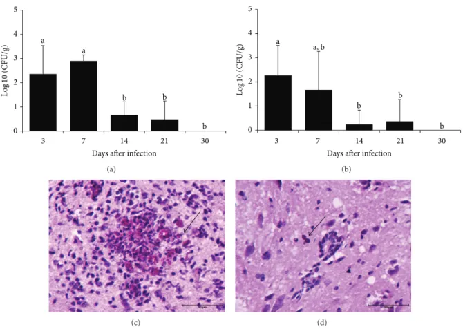
Figure 1: Dissemination ofC. albicansto the central nervous system. C57BL/6 mice were infected withC. albicansand fungal load was
evaluated 3, 7, 14, 21, and 30 days ater in the brain (a) and in the spinal cord (b). he results are expressed as mean±SEM (� = 5-6mice/group)
of the CFU (log10) per gram of tissue. ANOVA, Tukey’s test,� < 0.05. Diferent letters indicate statistical diference among the experimental
time points. Periodic acid-Schif revealed yeasts and hyphae in brain (c) and yeast in cervical spinal cord (d) sections.
48 h, cells were collected and stained. Spleen and CNS-extracted cells were blocked with rat serum 1% for 20 min to prevent nonspeciic binding via Fc receptor. Ater Fc blocking, cells were stained with 0.2�g of PerCP-conjugated anti-mouse CD3 and 0.25�g of FITC-conjugated anti-mouse CD4 for 20 min at 4∘C. Intracellular FoxP3 transcription factor analysis was performed only in spleen samples by using CD3-PercP, CD4-FITC plus 0.13�g of APC-conjugated anti-mouse CD25 and 0.2�g of PE-conjugated anti-mouse FoxP3 and staining set (eBiosciences, San Diego, CA, USA) according to manufacturer’s instructions. Ater staining, the cells were washed, resuspended in FACS bufer, and ixed in paraformaldehyde 1%. Analysis was performed using a FACSCanto II (BD) from Bioscience Institute (Botucatu, SP, Brazil) and the data were analyzed with FlowJo sotware (TreeStar, Ashland, OR, USA).
2.8. Histopathology of the CNS. Ater euthanasia, brain and lumbar spinal cord samples were removed and ixed in 10% neutral bufered formalin. Parain slides with 4�m were stained with hematoxylin and eosin (H&E) to evaluate the inlammatory process. A semiquantitative analysis of CNS inlammation was performed according to the fol-lowing criteria: (0) inlammatory iniltration absent; (+/++) mild/moderate inlammatory iniltration; (+++) intense
inlammatory iniltration. Sections were also stained with periodic acid-Schif to visualize fungal structures.
2.9. Statistical Analysis. Results were expressed as mean± standard deviation or with median and interquartile (25– 75%) ranges. To test for the normality of data, results were analyzed by Shapiro-Wilk’s test. Comparisons between two samples were made by �-test and more than three samples were made by one way ANOVA followed by Tukey’s test for parametric variables and by Kruskal-Wallis followed by Dunn’s test for nonparametric variables. Fisher’s test was performed to estimate the frequency ofC. albicans-positive tissues and to compare the semiquantitative analysis of CNS tissue inlammation. he data were analyzed using SigmaPlot statistical package for Windows version 2.0 (1995, Jandel Corporation, CA, USA) and values of� < 0.05were con-sidered statistically signiicant.
3. Results
TNF
-�
(pg/mL)
Days ater infection
3 7 14 21 30
CTL
600
450
300
150
0
∗∗ ∗
(a)
Days ater infection
3 7 14 21 30
IL
-6
(pg/mL)
CTL
100
80
60
40
20
0
∗
(b)
IFN-𝛾
(pg/mL)
Days ater infection
3 7 14 21 30
CTL
3000
2400
1800
1200
600
0
∗∗ ∗∗
(c)
Days ater infection
3 7 14 21 30
IL
-17
(pg/mL)
CTL
10
8
6
4
2
0
∗
(d)
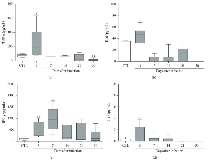
Figure 2: Kinetics of cytokine production by spleen cells from mice infected withC. albicans. C57BL/6 mice were inoculated withC. albicans
and the spontaneous production of cytokines by spleen cells was evaluated 3, 7, 14, 21, and 30 days ater fungal inoculation. he results are
expressed as median, 25–75% (box), and minimum-maximum (error bars) of 5-6 mice/group. Mann-Whitney test,∗� < 0.05and∗∗� < 0.01
indicate statistical diference between each experimental time point and the control group (uninfected).
also reached the CNS. As observed in Table 1, the viable fungi were recovered from all evaluated organs, including the brain and the spinal cord. Ater 30 days all organs, except the spleen, exhibited fungal clearance. he kinetics of fungal load, during 30 days, is showed in Figures 1(a) and1(b)for brain and spinal cord, respectively, and indicates that the fungus load is more accentuated in the irst week of infection. he presence of yeasts and hyphae in the brain and yeast in the spinal cord is illustrated in Figures1(c)and1(d), respectively.
3.2. Production of Potentially Encephalitogenic Cytokines dur-ing C. albicans Infection. As many of the most encephalito-genic cytokines are also involved in the defense againstC. albicansand other fungi, we tested their production during the time periods when the fungus was being detected. Spleen cell cultures from infected mice produced elevated levels of TNF-�, IL-6, IFN-�, and IL-17 (Figure 2). Cytokine levels were especially elevated in the 3rd day ater infection.
Table 1: Frequency ofC. albicans-positive tissues.
Period Tissue
Spleen Kidney Liver Brain Spinal cord
3 days 6/6 6/6 5/6 5/6 5/6
7 days 5/5 5/5 3/5 5/5 3/5
14 days 4/6 2/6 0/6 4/6 1/6
21 days 4/6 1/6 0/6 2/6 1/6
30 days 2/5 0/6 0/6 0/6 0/5
�value 0.0606 0.0022 0.0152 0.0152 0.0152
Data were expressed as number ofC. albicans-positive animals/total number of animals per group.
0 1 2 3
0 1 2 3 4 5 6 7 8 9 10 11 12 13 14 15 16 17 18 19 20 21
Clinical s
co
re
Days
−3 −2 −1
∗ ∗
∗
∗
EAE EAE+Ca
(a)
0 1 2 3 4 5
EAE EAE+Ca
M
axim
um s
co
re
∗
(b)
EAE+Ca 0
EAE
W
eig
h
t loss (%)
−10
−20
−30
−40
∗
(c)
Figure 3: Efect ofC. albicanson EAE development. C57BL/6 mice were infected withC. albicans3 days before EAE induction. Disease
development was followed during 21 days. Clinical scores (a) were checked every day and are expressed as mean; maximum clinical score (b)
and % of body weight loss (c) were calculated as described in Methods section. he results (a and b) are expressed as mean±SD (� = 6–8
mice/group). Unpaired�test,∗� < 0.05indicates diference between EAE and EAE+Ca groups.
severe form of encephalomyelitis. As shown in Figure3(a), these animals already showed paralysis signs at the 9th day ater EAE induction whereas the EAE control group pre-sented paralysis only 2 days later. his higher disease severity was detected during the whole acute disease phase. he average maximum clinical score, as depicted in Figure3(b), conirmed this worst clinical evolution. Weight loss was also more accentuated in this experimental group as can be observed in Figure3(c).
3.4. Peripheral Immunological Alterations during EAE Aggra-vation by C. albicans Infection. To evaluate if peripheral immunological parameters could explain this detrimental fungal efect on EAE, we tested the % of CD3+CD4+ and CD3+CD4+CD25+FoxP3+T-cell subsets. he cytokine pro-duction by spleen cells restimulated with MOG or with heat-killedC. albicansyeasts was also determined. Normal mice and mice only infected were also analyzed. A higher percentage of CD3+CD4+T cells were found in EAE+Ca and EAE groups in comparison to normal and infected groups. In addition, the % of this T-cell subset was signiicantly higher in the group that was previously infected with the fungus
(EAE+Ca) in comparison to the EAE group (Figure4(a)). he % of the FoxP3+ T cells was signiicantly higher in the EAE, but not in the Ca and EAE+Ca groups, in com-parison to the control group, as illustrated in Figure4(b). Concerning cytokines induced by MOG, the EAE+Ca group presented a signiicant production of TNF-�(Figure4(d)), IL-6 (Figure4(e)), and IL-17 (Figure4(f)) in comparison to all other experimental groups. IL-2 (Figure4(h)) and IL-4 (Figure4(i)) were similarly elevated in EAE and EAE+Ca groups. hese two groups also produced low and similar amounts of IL-10 (Figure4(c)). Comparison of EAE+Ca and EAE cytokine production induced by heat-killedC. albicans clearly showed that IL-10, IL-6, IL-17, IFN-�, IL-2, and IL-4 were signiicantly higher in the previously infected group.
0 10 20 30 40 50 c c b a CD 3 + CD 4 + (%) (a) 0 5 10 15 20 25
b a, b
a, b a CD 25 + Fox P 3 + (%) (b) 0 400 800 1200 1600 2000 b a b b a a b a, b
MOG C. albicans
IL -10 (pg/mL) (c) 0 500 1000 1500 2000 2500
b b b
a
a a
b b
MOG C. albicans
TNF -� (pg/mL) (d) 0 300 600 900 1200 1500 a b c b, c a b b b
MOG C. albicans
IL -6 (pg/mL) (e) 0 700 1400 2100 2800 3500 a b b b a b b b
MOG C. albicans
IL -17 (pg/mL) (f) 0 600 1200 1800 2400 3000 a c a b
MOG C. albicans
IFN-𝛾 (pg/mL) EAE Ca CTL EAE+Ca (g) 0 100 200 300 400 500 a b b b b b a a
MOG C. albicans
IL -2 (pg/mL) EAE Ca CTL EAE+Ca (h)
0 50 100 150 200
IL
-4 (pg/mL)
MOG C. albicans
b b
b b
a
a a, b
a, b
EAE Ca
CTL
EAE+Ca
(i)
Figure 4: Modulation of MOG-induced cytokine production by previous infection withC. albicans. C57BL/6 mice were infected withC.
albicansand 3 days later they were submitted to EAE induction. Fourteen days ater EAE induction, some immunological parameters were
evaluated in the spleen. he percentage of CD3+CD4+(a) and CD3+CD4+CD25+FoxP3+(b) was performed by cytometric analysis in 100.000
acquired events. IL-10 (c), TNF-�(d), IL-6 (e), IL-17 (f), IFN-�(g), IL-2 (h), and IL-4 (i) levels were measured in spleen cell cultures stimulated
with MOG or heat-killedC. albicans. he results are expressed as mean±SD (� = 6–8mice/group). ANOVA, Tukey’s test, and� < 0.05.
Diferent letters indicate statistical diference among the groups (a and b) or among the groups under the samein vitrostimulation (c, d, e, f,
g, h, and i).
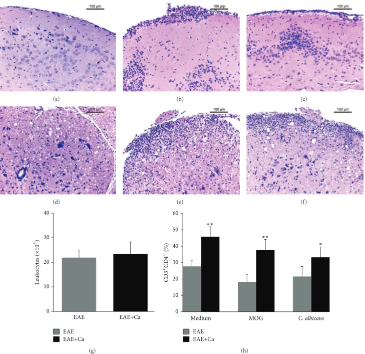
no inlammatory iniltrates were present in normal mice (Figures 5(a) and 5(d)). he amount of total leukocytes eluted from the CNS from both experimental groups was also similar as depicted in Figure5(g). he percentage of CD3+CD4+T cells was always higher in the EAE+Ca group, independently of their previous stimulation with MOG or heat-killedC. albicansyeasts (Figure5(h)). Cells eluted from the CNS of both groups respond in a similar way to in vitrostimulation with MOG, that is, they produced similar amounts of TNF-�, IL-17, IFN-�, IL-2, and IL-10 (Figure6). However, cells eluted from mice previously infected with C. albicans(EAE+Ca group) produced much more TNF-�, IL-6, IL-17, IFN-�, and IL-10 in response toC. albicans in vitro restimulation (Figure6).
4. Discussion
Multiple sclerosis (MS) is one of the world’s most common neurological disorders [2]. he disease develops as a result of interactions between the environment and the immune system in genetically susceptible individuals and it has long been recognized that infections may serve as environmental triggers for MS [11]. Even though viral agents have been more usually suspected as aggravating or triggering agents of this disease, fungi, especially their toxins, were recently incriminated as relevant underlying causes of MS and thus may ofer an approach towards a more efective adjunct treatment [19]. C. albicans is the most common fungal pathogen of humans and its spreading to the brain has been described during acute infections [21,22]. Interestingly, ity percent of patients with disseminated candidiasis underwent CNS fungal invasion [23]. Even thoughC. albicansis usually more prevalent in immunocompromised individuals, it has
also been reported to cause meningoencephalitis in healthy individuals [24]. Considering these aspects and the fact that a possible relationship betweenCandidaspp. and MS patients [16,17,19] was recently described, we evaluated the efect of an experimental infection with this fungus on the development of EAE, which is a largely accepted model to study the pathophysiological mechanisms of MS [25].
We initially evaluated the characteristics of C. albicans infection in C57BL/6 mice, which is one of the strains that develop encephalomyelitis upon immunization with antigens from the CNS [26]. his strain developed a widespread infection characterized by involvement of the majority of the organs, including the brain and the spinal cord. his difuse infection was, however, very well controlled by the immune system since almost no fungi were recovered ater 30 days of infection. his dissemination ofC. albicansto the brain was already demonstrated not only in C57BL/6 mice [20] but also in other mouse strains as BALB/c [27] and Swiss [28]. However, this is the irst report that indicates spreading of this fungus to the spinal cord portion of the CNS in mice.
(a) (b) (c)
(d) (e) (f)
0 10 20 30 40
L
euk
o
cyt
es
(×
10
5 )
EAE EAE+Ca
EAE EAE+Ca
(g)
0 10 20 30 40 50 60
Medium MOG C. albicans
CD
3
+ CD
4
+ (%)
∗ ∗∗
∗∗
EAE EAE+Ca
(h)
Figure 5: Previous infection withC. albicansincreases the amount of CD4+T cells in the CNS. C57BL/6 mice were infected withC. albicans
and 3 days later they were submitted to EAE induction. Fourteen days ater EAE induction, inlammation and % of CD4+T cells were evaluated
in the CNS. Inlammatory iniltrates detected by H&E staining are shown in brain samples from EAE (b) and from EAE+Ca (c) groups and in spinal cord samples from EAE (e) and from EAE+Ca (f) groups. A brain and spinal cord samples from a normal mouse is shown in (a) and
(d), respectively. Total leukocyte number (g) and percentage of CD3+CD4+T-cell subset (h) (analysis performed in 50.000 acquired events).
he results are expressed as mean±SD (� = 6-7mice/group). Unpaired�test,∗� < 0.05and∗∗� < 0.01indicate diference between EAE
and EAE+Ca groups under the samein vitrostimulation.
in addition, developed a more severe disease. Higher severity was characterized by both a higher body weight loss and a more accentuated degree of paralysis. To the best of our knowledge, this is the irst demonstration that a previous experimental infection with C. albicans triggered EAE exacerbation. hese indings are relevant because a direct contribution ofC. albicansto this neurological disease has not been deeply investigated. However, a series of indirect and epidemiological indings supports this possibility. For
MOG C. albicans
400
300
200
100
0
TNF
-�
(pg/mL)
∗
Medium
(a)
MOG C. albicans
∗
∗
∗
IL
-6
(pg/mL)
2400
1800
1200
600
0
Medium
(b)
IL
-17
(pg/mL)
1000
800
600
400
200
0
MOG C. albicans
∗
Medium
(c)
MOG C. albicans
∗∗
IFN-𝛾
(pg/mL)
2000
1500
1000
500
0
Medium
(d)
Medium MOG C. albicans
IL
-2
(pg/mL)
300
200
100
0
EAE EAE+Ca
(e)
MOG C. albicans
EAE EAE+Ca
IL
-10
(pg/mL)
500
400
300
200
100
0
∗
Medium
(f)
Figure 6:Candidaspeciic T cells contribute to elevated production of encephalitogenic cytokines in the CNS. CNS eluted cells were
restimulated with MOG or heat-killedC. albicansand TNF-�(a), IL-6 (b), IL-17 (c), IFN-�(d), IL-2 (e), and IL-10 (f) levels were measured
by ELISA. he results are expressed as median, 25–75% (box), and minimum-maximum (error bars) of 6 to 7 mice/group. Mann-Whitney
test,∗� < 0.05and∗∗� < 0.01indicate statistical diference between EAE and EAE+Ca groups under the samein vitrostimulation.
Candida spp. could be associated with increased odds of MS [16]. In addition to speciic antibodies, fungal macromolecules such as proteins, polysaccharides, and DNA were also detected in blood samples from MS patients [33]. Besides these serologic evidences, antibodies against
Candidaspp. [33] and fungal DNA [17] were also detected in the cerebrospinal luid of MS patients.
C. albicansinfected (Ca), and normal (CTL) experimental groups. Even though T regulatory (Treg) mediated responses remain poorly understood inCandidainfection, data indicate increased proportion of this subset during candidiasis [34, 35]. As FoxP3+ T cells are mostly responsible for EAE recovery in C57BL/6 mice [36,37], we initially hypothesized that Treg expansion could theoretically downregulate EAE development. his assumption was based on the fact that Treg cells induced during infectious diseases can regulate EAE in an apparently nonspeciic manner [38]. To test this possibility we evaluated the efect of the C. albicans infection on the percentage of this T-cell subset. he expected increase in the percentage of FoxP3+T cells was found in the spleen of the EAE group. However, the proportion of this T-cell subset was not modiied in Ca and in EAE+Ca groups. his inding can be attributed, at least partially, to the complex relationship, including cell plasticity, between Treg and h17 responses duringC. albicansinfection [35]. In addition to Treg cells we also evaluated the percentage of CD4+ T cells and cytokine production. Previous fungal infection increased CD4+T-cell subset in spleen of EAE-mice (EAE+Ca group) and clearly upmodulated the production of many encephalitogenic cytokines by spleen cells stimulated with MOG or heat-killed C. albicans. Even though the efect of EAE on fungal load was not the focus of this investigation, fungi recovery was usually signiicantly lower in the infected animals that had also EAE (not shown). his inding suggests that the immune response against MOG, or maybe the presence of the CFA, is increasing fungicidal activity of the immune system. he higher pro-duction of encephalitogenic cytokines by both stimuli, MOG andC. albicans, was interpreted as a possible cause of EAE increased severity as cytokines can easily cross the blood-brain barrier and directly afect CNS functions [39,40].
As the histopathology analysis from brain and spinal cord sections suggested similar degrees of inlammation, we compared the amounts of leukocytes and CD3+CD4+T cells eluted from the CNS. Conirming the H&E analysis, this comparison revealed the presence of similar numbers of total cells in EAE and EAE+Ca groups, demonstrating therefore that the higher disease severity was not due to a higher degree of inlammatory iniltration. Nevertheless, the immunophe-notyping analysis showed a higher proportion of CD3+CD4+ T-cell population in the EAE+Ca group. Culture of the cells eluted from the CNS showed, as expected, that they pro-duced proinlammatory cytokines in the presence of MOG. Interestingly, they also produced signiicant amounts of proinlammatory cytokines when stimulated withC. albicans. Together, these results are suggesting that both peripheral and local fungus efects are contributing to a more severe disease development. he translation of these indings to human patients certainly requires much more investigation in this area. However, we believe that these indings add more evidence thatC. albicansis one of the fungi that can afect this type of neurological pathology.
Conflict of Interests
he authors declare that they have no conlict of interests.
Acknowledgment
Special thanks are due to S˜ao Paulo Research Foundation (FAPESP), Grant no. 2013/14353-2 and Grant no. 2012/12540-7.
References
[1] R. Hohlfeld, “Multiple sclerosis: human model for EAE?”
Euro-pean Journal of Immunology, vol. 39, no. 8, pp. 2036–2039, 2009.
[2] Multiple Sclerosis International Federation—MSIF,he Atlas of
Multiple Sclerosis 2013, Multiple Sclerosis International Federa-tion, London, UK, 2013.
[3] E. Lavi and C. S. Constantinescu, Experimental Models of
Multiple Sclerosis, Springer, New York, NY, USA, 2005. [4] M. Sospedra and R. Martin, “Immunology of multiple sclerosis,”
Annual Review of Immunology, vol. 23, pp. 683–747, 2005. [5] A. G. Baxter, “he origin and application of experimental
auto-immune encephalomyelitis,”Nature Reviews Immunology, vol.
7, no. 11, pp. 904–912, 2007.
[6] C. Cassan and R. S. Liblau, “Immune tolerance and control of CNS autoimmunity: from animal models to MS patients,”
Journal of Neurochemistry, vol. 100, no. 4, pp. 883–892, 2007. [7] H. Neumann, “Molecular mechanisms of axonal damage in
inlammatory central nervous system diseases,”Current
Opin-ion in Neurology, vol. 16, no. 3, pp. 267–273, 2003.
[8] N. M. Rebenko-Moll, L. Liu, A. Cardona, and R. M. Ransohof, “Chemokines, mononuclear cells and the nervous system:
heaven (or hell) is in the details,”Current Opinion in
Immunol-ogy, vol. 18, no. 6, pp. 683–689, 2006.
[9] M. Rodriguez, “Efectors of demyelination and remyelination in
the CNS: implications for multiple sclerosis,”Brain Pathology,
vol. 17, no. 2, pp. 219–229, 2007.
[10] J. R. Oksenberg, “Decoding multiple sclerosis: an update on
genomics and future directions,”Expert Review of
Neurother-apeutics, vol. 13, no. 12, pp. 11–19, 2013.
[11] A. Venkatesan and R. T. Johnson, “Infections and multiple
sclerosis,”Handbook of Clinical Neurology, vol. 122, pp. 151–171,
2014.
[12] J. M. K. Murthy and C. Sundaram, “Fungal infections of the
central nervous system,”Handbook of Clinical Neurology, vol.
121, pp. 1383–1401, 2014.
[13] S. K. Fridkin and W. R. Jarvis, “Epidemiology of nosocomial
fungal infections,”Clinical Microbiology Reviews, vol. 9, no. 4,
pp. 499–511, 1996.
[14] F. L. van de Veerdonk, M. G. Netea, L. A. Joosten, J. W. M. van der Meer, and B. J. Kullberg, “Novel strategies for the
preventionand treatment ofCandidainfections: the potential of
immunotherapy,”FEMS Microbiology Reviews, vol. 34, no. 6, pp.
1063–1075, 2010.
[15] H. Wisplinghof, T. Bischof, S. M. Tallent, H. Seifert, R. P. Wen-zel, and M. B. Edmond, “Nosocomial bloodstream infections in US hospitals: analysis of 24, 179 cases from a prospective
nationwide surveillance study,”Clinical Infectious Diseases, vol.
39, p. 309, 2004.
[16] J. Benito-Le´on, D. Pisa, R. Alonso, P. Calleja, M. D´ıaz-S´anchez, and L. Carrasco, “Association between multiple sclerosis and
“Fungal infection in cerebrospinal luid from some patients with
multiple sclerosis,”European Journal of Clinical Microbiology
and Infectious Diseases, vol. 32, no. 6, pp. 795–801, 2013. [18] A. Vojdani, P. Rahimian, H. Kalhor, and E. Mordechai,
“Immunological cross reactivity betweenCandida albicansand
human tissue,”Journal of clinical & laboratory immunology, vol.
48, no. 1, pp. 1–15, 1996.
[19] C. B. Purzycki and D. H. Shain, “Fungal toxins and multiple
sclerosis: a compelling connection,”Brain Research Bulletin, vol.
82, no. 1-2, pp. 4–6, 2010.
[20] M. S. Lionakis, J. K. Lim, C.-C. R. Lee, and P. M. Murphy, “Organ-speciic innate immune responses in a mouse model of
invasive candidiasis,”Journal of Innate Immunity, vol. 3, no. 2,
pp. 180–199, 2011.
[21] P. A. Davis and P. T. Rudd,Neonatal Meningitis, McKeith Press,
London, UK, 1994.
[22] O. H. Del Brutto, “Central nervous mycotic infections,”Revue
Neurologique, vol. 30, pp. 447–459, 2000.
[23] J. S´anchez-Portocarrero, E. P´erez-Cecilia, O. Corral, J. Romero-Vivas, and J. J. Picazo, “he central nervous system and infection
by Candida species,” Diagnostic Microbiology and Infectious
Disease, vol. 37, no. 3, pp. 169–179, 2000.
[24] A. Borha, J.-J. Parienti, E. Emery, O. Coskun, S. Khouri, and
J.-M. Derlon, “Candida Albicans cerebral granuloma in an
immunocompetent patient. A case report,”Neurochirurgie, vol.
55, no. 1, pp. 57–62, 2009.
[25] A. Ben-Nun, N. Kaushansky, N. Kawakami et al., “From classic to spontaneous and humanized models of multiple sclerosis: impact on understanding pathogenesis and drug development,”
Journal of Autoimmunity, vol. 54, pp. 33–50, 2014.
[26] C. C. A. Bernard, T. G. Johns, A. Slavin et al., “Myelin oligodendrocyte glycoprotein: a novel candidate autoantigen in
multiple sclerosis,”Journal of Molecular Medicine, vol. 75, no. 2,
pp. 77–88, 1997.
[27] D. H. M. L. P. Navarathna, J. Munasinghe, M. J. Lizak, D. Nayak, D. B. Mcgavern, and D. D. Roberts, “MRI conirms loss of blood-brain barrier integrity in a mouse model of disseminated
candidiasis,”NMR in Biomedicine, vol. 26, no. 9, pp. 1125–1134,
2013.
[28] T. F. C. Fraga-Silva, J. Venturini, and M. S. P. de Arruda, “Traicking of phagocytic peritoneal cells in
hypoinsulinemic-hyperglycemic mice with systemic candidiasis,”BMC Infectious
Diseases, vol. 13, article 147, 2013.
[29] W. E. F. Klinkert, K. Kojima, W. Lesslauer, W. Rinner, H. Lassmann, and H. Wekerle, “TNF-alpha receptor fusion protein prevents experimental auto-immune encephalomyelitis and
demyelination in Lewis rats: an overview,”Journal of
Neuroim-munology, vol. 72, no. 2, pp. 163–168, 1997.
[30] C. Lock, G. Hermans, R. Pedotti et al., “Gene-microarray analysis of multiple sclerosis lesions yields new targets validated
in autoimmune encephalomyelitis,”Nature Medicine, vol. 8, no.
5, pp. 500–508, 2002.
[31] J. M. Fletcher, S. J. Lalor, C. M. Sweeney, N. Tubridy, and K. H. G. Mills, “T cells in multiple sclerosis and
experimen-tal autoimmune encephalomyelitis,”Clinical and Experimental
Immunology, vol. 162, no. 1, pp. 1–11, 2010.
[32] D. W. Luchtman, E. Ellwardt, C. Larochelle, and F. Zipp, “IL-17 and related cytokines involved in the pathology and immunotherapy of multiple sclerosis: current and future
devel-opments,”Cytokine & Growth Factor Reviews, vol. 25, no. 4, pp.
403–413, 2014.
patient with multiple sclerosis,” European Journal of Clinical
Microbiology and Infectious Diseases, vol. 30, no. 10, pp. 1173– 1180, 2011.
[34] P. Bonifazi, T. Zelante, C. D’Angelo et al., “Balancing
inlam-mation and tolerancein vivo through dendritic cells by the
commensalCandida albicans,”Mucosal Immunology, vol. 2, no.
4, pp. 362–374, 2009.
[35] N. Whibley, D. M. Maccallum, M. A. Vickers et al., “Expansion
of Foxp3+ T-cell populations by Candida albicans enhances
both h17-cell responses and fungal dissemination ater
intra-venous challenge,”European Journal of Immunology, vol. 44, no.
4, pp. 1069–1083, 2014.
[36] A. P. Kohm, P. A. Carpentier, H. A. Anger, and S. D. Miller,
“Cutting edge: CD4+CD25+regulatory T cells suppress
antigen-speciic autoreactive immune responses and central nervous system inlammation during active experimental autoimmune
encephalomyelitis,”he Journal of Immunology, vol. 169, no. 9,
pp. 4712–4716, 2002.
[37] S. F. G. Zorzella-Pezavento, F. Chiuso-Minicucci, T. G. D. Franc¸a et al., “Persistent inlammation in the CNS during chronic
EAE despite local absence of IL-17 production,”Mediators of
Inlammation, vol. 2013, Article ID 519627, 10 pages, 2013. [38] A. S. Farias, R. L. Talaisys, Y. C. Blanco et al., “Regulatory T cell
induction duringPlasmodium chabaudiinfection modiies the
clinical course of experimental autoimmune
encephalomyeli-tis,”PLoS ONE, vol. 6, no. 3, Article ID e17849, 2011.
[39] W. A. Banks, A. J. Kastin, and R. D. Broadwell, “Passage of
cytokines across the blood-brain barrier,”
Neuroimmunomodu-lation, vol. 2, no. 4, pp. 241–248, 1995.
[40] W. A. Banks, “Blood-brain barrier transport of cytokines:
a mechanism for neuropathology,” Current Pharmaceutical
Submit your manuscripts at
http://www.hindawi.com
Stem Cells
International
Hindawi Publishing Corporation
http://www.hindawi.com Volume 2014
Hindawi Publishing Corporation
http://www.hindawi.com Volume 2014
INFLAMMATION
Hindawi Publishing Corporation
http://www.hindawi.com Volume 2014
Behavioural
Neurology
Endocrinology
International Journal ofHindawi Publishing Corporation
http://www.hindawi.com Volume 2014
Hindawi Publishing Corporation
http://www.hindawi.com Volume 2014
Disease Markers
Hindawi Publishing Corporation
http://www.hindawi.com Volume 2014
BioMed
Research International
Oncology
Journal of Hindawi Publishing Corporationhttp://www.hindawi.com Volume 2014
Hindawi Publishing Corporation
http://www.hindawi.com Volume 2014
Oxidative Medicine and Cellular Longevity Hindawi Publishing Corporation
http://www.hindawi.com Volume 2014
PPAR Research
The Scientiic
World Journal
Hindawi Publishing Corporation
http://www.hindawi.com Volume 2014
Immunology Research
Hindawi Publishing Corporation
http://www.hindawi.com Volume 2014
Journal of
Obesity
Journal ofHindawi Publishing Corporation
http://www.hindawi.com Volume 2014
Hindawi Publishing Corporation
http://www.hindawi.com Volume 2014
Computational and Mathematical Methods in Medicine
Ophthalmology
Journal ofHindawi Publishing Corporation
http://www.hindawi.com Volume 2014
Diabetes Research
Journal ofHindawi Publishing Corporation
http://www.hindawi.com Volume 2014
Hindawi Publishing Corporation
http://www.hindawi.com Volume 2014
Research and Treatment
AIDS
Hindawi Publishing Corporation
http://www.hindawi.com Volume 2014
Gastroenterology Research and Practice
Hindawi Publishing Corporation
http://www.hindawi.com Volume 2014
Parkinson’s
Disease
Evidence-Based Complementary and Alternative Medicine
Volume 2014