Gene Expression in Mouse Skeletal Muscle
Kristen A. Baltgalvis1*, Sarah M. Greising2, Gordon L. Warren3, Dawn A. Lowe2
1Department of Biochemistry, Molecular Biology, and Biophysics, Medical School, University of Minnesota, Minneapolis, Minnesota, United States of America,
2Department of Physical Medicine and Rehabilitation, Medical School, University of Minnesota, Minneapolis, Minnesota, United States of America,3Division of Physical Therapy, Georgia State University, Atlanta, Georgia, United States of America
Abstract
Background:Estrogens are associated with the loss of skeletal muscle strength in women with age. Ovarian hormone removal by ovariectomy in mice leads to a loss of muscle strength, which is reversed with 17b-estradiol replacement. Aging is also associated with an increase in antioxidant stress, and estrogens can improve antioxidant status via their interaction with estrogen receptors (ER) to regulate antioxidant gene expression. The purpose of this study was to determine if ER and antioxidant gene expression in skeletal muscle are responsive to changes in circulating estradiol, and if ERs regulate antioxidant gene expression in this tissue.
Methodology/Principal Findings:Adult C57BL/6 mice underwent ovariectomies or sham surgeries to remove circulating estrogens. These mice were implanted with placebo or 17b-estradiol pellets acutely or chronically. A separate experiment examined mice that received weekly injections of Faslodex to chronically block ERs. Skeletal muscles were analyzed for expression of ER genes and proteins and antioxidant genes.ERawas the most abundant, followed byGperandERbin both soleus and EDL muscles. The loss of estrogens through ovariectomy inducedERagene and protein expression in the soleus, EDL, and TA muscles at both the acute and chronic time points.Gpx3mRNA was also induced both acutely and chronically in all 3 muscles in mice receiving 17b-estradiol. When ERs were blocked using Faslodex,Gpx3mRNA was downregulated in the soleus muscle, but not the EDL and TA muscles.
Conclusions/Significance:These data suggest thatGpx3andERagene expression are sensitive to circulating estrogens in skeletal muscle. ERs may regulateGpx3gene expression in the soleus muscle, but skeletal muscle regulation ofGpx3via ERs is dependent upon muscle type. Further work is needed to determine the indirect effects of estrogen andERaonGpx3 expression in skeletal muscle, and their importance in the aging process.
Citation:Baltgalvis KA, Greising SM, Warren GL, Lowe DA (2010) Estrogen Regulates Estrogen Receptors and Antioxidant Gene Expression in Mouse Skeletal Muscle. PLoS ONE 5(4): e10164. doi:10.1371/journal.pone.0010164
Editor:Maurizio C. Capogrossi, Istituto Dermopatico dell’Immacolata, Italy
ReceivedNovember 11, 2009;AcceptedMarch 22, 2010;PublishedApril 13, 2010
Copyright:ß2010 Baltgalvis et al. This is an open-access article distributed under the terms of the Creative Commons Attribution License, which permits unrestricted use, distribution, and reproduction in any medium, provided the original author and source are credited.
Funding:This work was funded by a grant awarded to Dawn A. Lowe from the NIH (R01AG031743). The funders had no role in study design, data collection and analysis, decision to publish, or preparation of the manuscript.
Competing Interests:The authors have declared that no competing interests exist. * E-mail: [email protected]
Introduction
There has been debate as to whether or not estrogen affects the force-generating capacity of skeletal muscle. A recent meta-analysis was conducted by our lab examining 23 studies in which postmenopausal women who were and were not taking estrogen hormone replacement were subjected to tests of muscle strength [1]. Our findings demonstrated a significant effect of approximately 5% greater strength in women who were taking estrogen replacement therapy. These findings are corroborated by a recent study that examined muscle strength in twins, in which one twin took estrogen replacement, and the other did not [2]. This paper showed that the hormone replacement therapy users walked at a maximal speed faster than non-users, and they had greater muscle power. We have extended the findings in humans using a mouse ovariectomy model. We showed that muscle and myosin functions were reduced,20% in ovariectomized mice [3], and that those losses in force generation at both the whole muscle and molecular levels were completely restored when mice were administered 17b-estradiol [4].
Most of the work that has been done to elucidate a role for the different ERs in muscle has been conducted in cell culture. Both ERa and ERb have been carefully characterized for their localization in C2C12 cells [13,14]. Human skeletal muscle cells treated with estrogen increase steroid receptor coactivator (SRC) and decrease silencing mediator for retinoid and thyroid hormone receptors (SMRT) mRNAs, suggesting transcriptional activity of the ER in response to estradiol [15]. Many potential ER-dependent mechanisms in myoblasts have been investigated, including their role in stimulating the PI3K/Akt pathway [16], Glut-4 expression [15], muscle differentiation via upregulation of myogenin and myosin heavy chain [17], MyoD activity [18], and prevention of apoptosis [19,20]. While studying the effects of estrogen and ERs in culture warrants merit and has yielded important information, intact skeletal muscle is composed of fused myotubes, is multi-nucleated, and innervated. A physiological consequence of estrogen deprivation in this complex environment is a reduction in the force-generating capacity of muscle in both women and rodents. Therefore, investigating the role of ER-dependent mechanisms in vivo is necessary to elucidate the mechanisms by which myosin is affected, and ultimately to understand the contractile dysfunction that occurs in estrogen-deficient, aged women.
A link between estradiol-induced changes in ER expression and muscle function is likely complicated. While the downstream targets of the ER are many, estrogen-responsive genes that regulate oxidative stress are interesting to consider. Ovariecto-mized mice have lower levels of many antioxidant enzymes in the heart, including glutathione peroxidase, catalase, and superoxide dismutase [21,22]. Mitochondria from the liver and brain of ovariectomized mice also produce more H2O2 [23]. However, these decrements are ablated when estrogen is replaced. These studies strongly indicate that estradiol plays an important role in balancing oxidative stress in non-skeletal muscle tissues. In skeletal muscle, balancing oxidative stress is crucial for myosin function [24] and overall muscle contractility, particularly during aging [25]. Elucidating a role for ERs in skeletal muscle most likely involves linking the ER with both estrogenic and aging effects, making antioxidant pathways worth investigating.
In summary, the literature illustrates that ERs exist in skeletal muscle, but whether or not they display typical steroid receptor responsiveness to their ligand, i.e., estradiol, has not been studied in intact skeletal muscle. The primary purpose of this study was to test the hypothesis thatERa,ERb, andGperin skeletal muscle are responsive to changes in circulating estradiol. We also began work to try to understand how estrogen and/or ERs confer a benefit to skeletal muscle function. Thus, a secondary purpose of this study was to test the hypothesis that antioxidant gene expression in skeletal muscle is responsive to changes in circulating estradiol. In order to probe for a link between ERs and antioxidant function, we systemically blocked ERs using the ER antagonist, Faslodex. We hypothesized that blocking ERs would have a detrimental effect on antioxidant gene expression, providing a ER-dependent mechanism for estrogen function in skeletal muscle.
Methods
Mice and estrogen manipulations
Four-month-old female C57BL/6 mice were acquired from Jackson Laboratories (Bar Harbor, ME). Mice were group housed and had access to phytoestrogen-free food (Harlan-Teklad; #2019) and water ad libitum. The room was maintained on a 12:12 light:dark cycle. The ovariectomy procedure [3] and 17b -estradiol pellet implantation procedure [4] were performed as
previously described. Three experiments were performed: (1) ovariectomy and acute replacement of 17b-estradiol for 48 hours, (2) ovariectomy and chronic replacement of 17b-estradiol for 3 weeks, and (3) inhibition of ERs for 1 month with Faslodex.
In the acute study, after 7 days of estrogen withdrawal, mice received a placebo pellet (OVX + Placebo; n= 6) or a 17b -estradiol pellet (OVX + E2; n= 6) containing 0.18 mg of 17b -estradiol in a matrix that is designed to release the hormone (or placebo) over a 60-day period (Innovative Research of America, Sarasota, FL). Sham operations were performed (Sham;n= 4) on an additional group of mice. All mice were returned to individual cages following the subcutaneous pellet implantation and remained there for 48 hours. In the chronic study, ovariectomies were performed and mice were implanted with placebo or 17b -estradiol pellets (n= 6 per group) for a total duration of 3 weeks. This time point was chosen since we previously reported that skeletal muscle contractile dysfunction occurs between 3 and 4 weeks following ovariectomy [3,4,26].
In the third study, mice were injected weekly with the ER antagonist ICI 182,780 (FaslodexH, AstraZeneca) at a dose of 10 mg/kg body mass, or a similar volume of mineral oil (n= 5 per group) for a duration of 1 month. A daily dose of approximately 0.1 to 3 mg/kg BM has previously been shown to reduce uterine mass in mice and rats [27–29]. Mice were placed in voluntary activity wheels for 3 days before the start of injections, as well as 2 weeks following treatment, as an indicator of physical activity. The total amount of voluntary physical activity was averaged over the 3 days at each time point. Food intake and body mass was also monitored on a weekly basis.
At each study’s end, mice were anesthetized with sodium pentobarbital (100 mg/kg body mass). Blood was collected by facial vein bleeds and plasma was stored at280uC for the acute study. Uterine masses were recorded for the chronic study and the Faslodex study to ensure the validity of the ovariectomies and effective blocking of ERs. The soleus and extensor digitorum longus (EDL) muscles were dissected and immediately processed for RNA isolation. Tibialis anterior (TA) muscles were also dissected, snap frozen in liquid nitrogen, and stored at280uC. All animal procedures were approved by the University of Minneso-ta’s Institutional Animal Care and Use Committee.
Circulating estradiol
An ELISA was used to measure 17b-estradiol in the plasma according to the manufacturers’ specifications (KA0234, Abnova Corporation, Taiwan). The sensitivity of the assay was 10 pg/mL. All samples were run in duplicate. Standards were graphed and fit using a 4-parameter logistic curve fit. Plasma estradiol levels in sham-operated mice averaged 20620 pg/ml. Circulating estradiol levels were below the level of detection (10 pg/ml) in all OVX+
Placebo mice, while OVX + E2 mice had an average of 246668 pg/mL.
RNA isolation and DNase treatment
precipitation with 500mL of isopropanol. Samples were incubated for 10 min at room temperature and centrifuged at 12,000 g for 8 min at 4uC. The supernatant was discarded, the pellet was washed in 75% EtOH, and the tubes were centrifuged at 7,500 g for 5 min at 4uC. The pellet was briefly allowed to air dry and was resuspended in 100mL of RNase-free H2O, and heated at 55uC for 10 minutes to help solubilize the RNA. RNA was purified and concentrated using the RNeasyHMinEluteHCleanup Kit (Qiagen, Valencia, CA) according to the manufacturers’ instructions. The RNA quantity was determined by reading 1mL of RNA using a NanoDrop ND-1000 spectrophotometer (Thermo Fisher Scientif-ic, Wilmington, DE) at wavelengths 260 and 280. All 260/280 ratios were$2.0. One or 2.5mg of RNA was diluted to a total volume of 8mL and was treated with 1mL of RQ1 RNase-free DNase (Promega, Madison, WI) and 1mL of DNase buffer at 37uC in a thermal cycler (Techne, Model TC-312, UK) for 30 min. The reaction was terminated using 1mL of stop solution and incubating the samples at 65uC for 15 minutes.
cDNA synthesis and PCR Array
cDNA was made using a RT2 First Strand cDNA kit from SABiosciences (Frederick, MD). Briefly, 1mg of DNase-treated RNA in a volume of 10mL was added to 4mL of RT buffer, 1mL of Primer and External Control Mix, 2mL of RT Enzyme Mix, and 3mL of RNase-free H20. This 20mL cocktail was incubated for 15 minutes at 42uC in a thermal cycler. The reaction was terminated by incubating the samples at 95uC for 5 minutes. The tubes were then diluted with 91mL of sterile ddH2O to have a final volume of 111mL of diluted cDNA, and stored at 220uC until the PCR arrays were run.
Oxidative stress and antioxidant defense PCR arrays (PAMM-065) from SABiosciences were prepared for OVX+Placebo (n = 6) and OVX+E2(n = 6) for both soleus and EDL muscles. One PCR array was used per sample. Each well on the plate was coated with primers specific for each gene. The PCR cocktail contained 1275mL of 2x SA Biosciences RT2master mix (containing SYBRH green dye), 102mL of diluted cDNA, and 1173 sterile ddH2O for a total volume of 2550mL; 25mL of this cocktail was added to each well on plate. Plates were run on a Stratagene Mx3000P quantitative PCR system. The parameters on the machine included 1 cycle of 10 minutes at 95uC to activate the HotStart DNA polymerase. This was followed by a 2-step cycling program consisting of 40 cycles of a denaturing step of 15 sec at 95uC and an annealing step of 1 minute at 60uC. SYBRHgreen fluorescence was detected during the last 15 s of the annealing step. The cycle threshold (CT) was calculated automatically. The 2‘(-DDCT) method was used for detecting changes in gene expression [30]. TheDCTwas calculated by subtracting the average CTfor all 5 housekeeping genes (Gusb, Hprt1, Hsp90ab1, Gapdh, and Actb) from the gene of interest. Melting curves were run and no primer dimers were detected. Any wells having a CTgreater than 35 were considered undetectable. Control wells were also used for genomic DNA contamination, efficiency for reverse transcription, and a positive PCR control to ensure consistency between the individual plates. All of these values were within the range suggested by the manufacturer.
cDNA synthesis and real-time PCR
Onemg of DNase-treated RNA was used to synthesize cDNA using the High Capacity cDNA Reverse Transcription kit from Applied Biosystems (Foster City, CA). Briefly, 11mL of DNase-treated RNA was added to a 9mL cocktail containing 2mL 10x RT buffer, 0.8mL 25x DNTPs, 2mL 10x random primers, 1mL RT, 1mL RNase-inhibitor, and 2.2mL of RNase-free H2O. The
mixture was incubated in a thermal cycler (Techne TC-312, UK) for 10 min at 25uC, 120 min at 37uC, 5 s at 85uC, and then held at 4uC overnight. Samples were diluted 1:10 and 1:100 in sterile ddH2O and stored at220uC until real-time PCR analysis.
The TaqmanHGene Expression Assays for mouse-specificERa
(Mm00433149_m1), ERb (Mm00599819_m1), Gper (Mm01194814_g1), Gpx1 (Mm00656767_g1), Gpx3 (Mm00492427_m1), Nox4 (Mm00479246_m1), Txnip (Mm00452393_m1), MyoD (Mm01203489_g1), and Glut-4 (Mm00436615_m1) were all purchased from Applied Biosystems (Foster City, CA). Eukaryotic 18S rRNA (4333760F) andb-actin (4352341E) were used as housekeeping genes. Each PCR reaction contained 9mL of diluted cDNA,10mL of 2x Taqman Master mix, 1mL of 20x primer, for a total volume of 20mL in each reaction. Samples were run in duplicate on ABI 7500 Fast Real-Time PCR System. The cycling conditions consisted of an initial step of 50uC for 2 minutes, followed by an initial denaturing step of 95uC for 10 minutes. The thermal cycler then followed a 2-step cycling program of denaturing at 95uC for 15 s and annealing at 60uC for 1 minute for a total of 40 cycles. FAM fluorescence was measured during last 15 s of the annealing step. The 2‘(-DDCT) method was used for detecting changes in gene expression [30]. The DCT was calculated by subtracting the average of the 18S rRNA andb-actin CTvalues from the gene of interest. It should be noted that the housekeeping genes did not differ between treatment groups.
Western blotting
Frozen TA muscle was homogenized in 1 ml of ice-cold RIPA buffer (50 mM Tris, pH 7.4, 150 mM NaCl, 1 mM EDTA, pH 8.0, 0.1% Triton-X, 0.1% SDS, 0.5% sodium deoxycholate, 5mg/mL protease inhibitors (Sigma; P8340), and 10mg/ml phosphatase inhibitors (Sigma; P5726). Homogenates were centrifuged at 10,000 g at 4uC for 10 min and the supernatants were stored at280uC. ERaprotein was measured by SDS-PAGE and Western blotting using an anti-mouse ERa(Clone MC-20, sc-542, Santa Cruz Biotechnology, Santa Cruz, CA) at a 1:250 in Odyssey Blocking Buffer overnight at 4uC. Visualization of the antigen-antibody interactions were detected with the secondary antibody (Goat Anti-Rabbit IRdyeH 680, LI-COR Biosciences, Lincoln, NE) at a dilution of 1:500 in Odyssey Blocking Buffer. The membranes were then scanned and quantified using the OdysseyHInfrared Imaging System (LI-COR Biosciences).
Data analysis
All gene and protein expression data were analyzed using student t-tests or one-way ANOVAs. Two-way ANOVAs were used to determine the effect of ER isoforms within muscle type, antioxidant gene expression within muscle type using the PCR array, and changes in body mass and wheel running activity over time. If a significant main effect or interaction existed, Tukey post-hoc tests were performed. Significance was set at P,0.05.
Results
relative to soleus muscle. However, the soleus muscle had 74% greater expression of Gper (P = 0.003) compared to the EDL muscle.
ER gene and protein expression are sensitive to ovariectomy and acute 17b-estradiol replacement
ERa,ERb, andGpermRNA levels were measured in the soleus and EDL muscles from sham-operated, OVX + Placebo, and OVX+E2mice. There was a significant effect of estradiol status onERaexpression in both the soleus (P = 0.002) and EDL muscles (P,0.001). Ovariectomy resulted in muscle ERa mRNA being ,70% greater compared to that in sham mice (Figure 2A). When 17b-estradiol was administered to ovariectomized mice, ERa
mRNA levels came back down to sham levels in both muscles. ERb mRNA levels were also altered in the EDL muscle (P = 0.038), with OVX+E2mice having lower amounts of ERb
compared to sham mice in the EDL muscle. There were no effects onGperexpression in skeletal muscle in response to ovariectomy in either the soleus or EDL muscles (Figure 2C). Collectively, these data show that onlyERaresponds to acute changes in circulating 17b-estradiol in skeletal muscle.
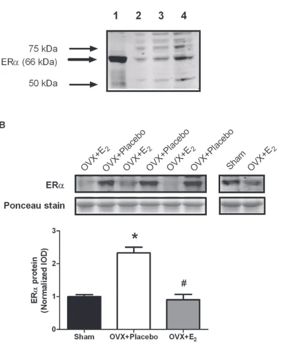
To determine if the changes in gene expression were representative of changes in protein expression, we examined ERaprotein levels. To confirm that we were measuring ERa, we first qualitatively examined ERa protein levels in uterine tissue and skeletal muscle tissue, and also performed a dose-response curve in skeletal muscle. Figure 3A shows the relative large amount of ERa protein in uterine tissue compared to skeletal muscle, and also verifies that we are able to measure ERaprotein in skeletal muscle. As shown in Figure 3B, ERaprotein levels in the TA muscle responded in the same manner as did mRNA levels (P,0.001). Muscle from OVX+ Placebo mice had about 2-fold more ERaprotein than did those from OVX+E2mice. Muscle from estradiol–replaced mice had similar ERa protein levels compared to those from sham mice.
Acute 17b-estradiol replacement in ovariectomized mice induces antioxidant gene expression in skeletal muscle
One of the mechanisms by which estrogens may exert a beneficial effect on skeletal muscle is through its role as an antioxidant, or by activating ERs to regulate genes related to
oxidative stress. We used PCR arrays to screen 84 genes related to the antioxidant defense system to determine if estradiol status was associated with regulation of these genes in skeletal muscle. We used a two-way ANOVA to compare the effect of treatment (placebo vs. 17b-estradiol) and muscle type (soleus vs. EDL) to determine global changes in skeletal muscle gene expression with estrogen. Our results show that 5 out of the 84 genes measured were greater in OVX+ E2 mice compared to OVX + Placebo mice in both soleus and EDL muscles (Table 1; P#0.027). These genes included Gpx3, Gpx2, Nox4, Txnip, and Gpx1. These data demonstrate that estrogen status can alter antioxidant gene expression in skeletal muscle.
Chronic estradiol deprivation alters both ER expression and antioxidant gene expression
For ERs and antioxidant genes to have an impact on skeletal muscle function, these effects must be chronic in nature. Our next study was designed to examined ER and antioxidant gene expression in skeletal muscle following 3 weeks of ovariectomy with or without 17b-estradiol replacement. First,ERa, ERb, and Gpergene expression were measured in the soleus, EDL, and TA muscles from estrogen-deficient and estrogen-replete mice. As shown in Figure 4A,ERaexpression in OVX+E2mice was 57%, 75%, and 69% of OVX+Placebo mice in the soleus, EDL, and TA muscles, respectively, similar to results from the previous acute study (P#0.038).ERbandGperexpression in skeletal muscle from this chronic study also mimicked the results from the acute study. Ovariectomized mice expressed similar amounts of ERband Gper in all muscles tested relative to those from mice replaced with 17b -estradiol. These data show that long-term deprivation of estradiol altersERagene expression in skeletal muscle.
Antioxidant gene expression was also quantified in these mice. Instead of screening 84 genes using the PCR arrays, individual PCR reactions were run for 4 out of the 5 genes that we found to be estradiol-responsive at the 48-hour time point: Gpx3, Gpx1, Nox4, and Txnip. Gpx2 was not analyzed because it is classically defined as a gastrointestinal glutathione peroxidase [31]. Only Gpx3 was responsive in all 3 muscle types following 3 weeks of ovariectomy and 17b-estradiol replacement (Figure 4B).Gpx3gene expression in 17b-estradiol-treated mice was 3.9-, 3.1-, and 3.4-fold greater, respectively, than that in ovariectomized mice (P#0.001 for all muscles). There were also modest effects of Figure 1. ER gene expression in skeletal muscles of 4-mo-old female wild-type mice.Data normalized toERbin the soleus. Values are means6SEM. *Signifies different fromERawithin a muscle type.#
Signifies different fromERbwithin a muscle type.$Signifies different from soleus muscle.
Gpx1 mRNA (P = 0.035) and Txnip mRNA (P = 0.003) in 17b -estradiol-treated mice, but this effect only occurred in the EDL muscle. These data suggest that Gpx3 is positively regulated by 17b-estradiol in skeletal muscle.
Inhibition of ER by Faslodex alters antioxidant gene expression
Our next study was designed to determine if ERs regulateGpx3 expression in skeletal muscle. We treated mice for 1 month with Faslodex, an ER antagonist. There were no differences between oil and Faslodex-treated mice regarding body mass before or at the study’s end, but both groups gained weight over the course of the study at the same rate (Figure 5A; P,0.001). Faslodex did not alter food intake by the mice (P$0.651). There were no differences in
acute wheel running activity between the 2 groups before the treatment, but there was a trend after treatment for Faslodex-treated mice to run 40% less than oil-injected mice (Figure 5B; P = 0.093). At the end of the study, uterine wet mass was 81% less in Faslodex-treated mice compared to mineral oil-injected control mice (Figure 5C; P = 0.015), validating that Faslodex did indeed block ERs. Although ERa gene expression in the uterus was unaltered with Faslodex,Gpx1was downregulated 58% (P = 0.046) andTxnipmRNA was upregulated 3-fold (Figure 5D; P = 0.016). Collectively these data validate that the Faslodex treatment was effective at blocking ERs to decrease uterine mass and alter antioxidant gene expression in that tissue.
Skeletal muscle mass, ERa, and Gpx3 gene expression were measured in the soleus, EDL, and TA muscles after Faslodex Figure 2. ER gene expression in skeletal muscle following ovariectomy and 48 hours of 17b-estradiol replacement.A.ERagene expression. B.ERbgene expression. C.Gpergene expression. Data are normalized to sham mice within each muscle. Values are means6SEM. *Signifies different from sham.#Signifies different from OVX
+Placebo.
treatment. Muscle mass was unaltered in all 3 muscles (P$0.208; Figure 5E) as was ERa gene expression (P$0.176; Figure 5F). There was a 50% decrease inGpx3gene expression in the soleus muscle in response to Faslodex treatment (P = 0.019), but Gpx3 levels remained the same in the EDL and TA muscles (Figure 5G).
Estrogens minimally alterMyoDandGlut-4mRNAin vivo Markers of muscle differentiation and hypertrophy, as well as glucose metabolism were also measured, since estrogen has robust effects on these markers in cell culture. We found that MyoD mRNA levels were reduced 45% after 48 hours (P = 0.046) in the EDL muscle with estrogen, but were unchanged in the soleus muscle (Figure 6A) compared to ovariectomized mice. Similar results were found after 3 weeks of replacement where
estrogen-treated mice hadMyoDmRNA levels that were,40% less in the EDL and TA muscles (P#0.019), butMyoDwas not affected in the soleus (Figure 6B). Glut-4mRNA levels were not altered in the soleus, TA, or EDL muscles by ovariectomy at any time point measured (Figures 6A & 6B).
Discussion
The main findings from this study were thatERa,Gper, andERb
are all expressed in skeletal muscle, but that onlyERais responsive to both acute and chronic changes in circulating estradiol. Acute and chronic changes in circulating estradiol also caused changes in Gpx3gene expression.Gpx3expression appeared to be regulated by ERs, but this was a muscle-specific response. These results are Figure 3. ERaprotein expression in the TA muscle following ovariectomy and 48 hours of 17b-estradiol replacement.A. Preliminary work examining ERaexpression in uterine tissue and skeletal muscle. Lane 1 = 10mg of uterine homogenate. Lanes 2-4 = 10, 20, and 40mg of skeletal
muscle homogenate. B. ERaprotein expression in muscle from sham, OVX+Placebo, and OVX+E2mice. Data are normalized to sham mice. Values
are means6SEM. *Signifies different from sham.#
Signifies different from OVX+Placebo.
vital first steps in discovering estrogen-mediated mechanisms that influence skeletal muscle contractility. On the whole, this is an important topic because the decline in circulating estrogens with age has been associated with muscle weakness in women. These findings have been replicated in mice who have undergone ovariectomies to mimic the low circulating estrogen state and have been further extended to show that an underlying molecular
explanation for the reduction in force generation involves myosin. However, it is unknown whether the effects of estrogen on muscle and myosin functions are via an ER-mediated mechanism. The current study begins to elucidate this mechanism by examining skeletal muscle ERs in estrogen deficient and -replete states.
While it is known that ERa and ERb are present in skeletal muscle, much less is known about Gper. Our first experiment aimed to determine the relative mRNA abundance of all 3 ERs in skeletal muscle. Our findings are unique in that this is the first study to compare all three ER isoforms and their relative abundance in skeletal muscle. Previous work showed greater levels ofERacompared toERbmRNA [5,7], and separate studies have reported the presence ofGpermRNA in skeletal muscle [32]. Our results complement these previous studies by reinforcing that ERamRNA is the most abundant, and we also have added that Gperis expressed in a moderate amount, in betweenERaandERb. Our experimental design also allowed us to directly compare the relative mRNA abundance of these 3 ERs amongst slow-twitch (soleus) and fast-twitch (EDL) muscles. We found thatERaand ERb were expressed in higher amounts in the EDL than soleus, butGperwas found in a greater amount in the soleus compared to the EDL. These data are somewhat contradictory to previously published findings. Lemione and coworkers reported that ERa
mRNA was greater in the slow-twitch soleus muscle compared to the primarily fast-twitch gastrocnemius and EDL muscles in female rats [33]. ERa protein levels were also reported to be greater in the soleus compared to the gastrocnemius muscle in rabbits [34,35]. The discrepancies for these findings could be based on many variables, including the species studied, age of the animals, and the methods of detection (e.g., only in our study was real-time PCR used). In addition, while the soleus is considered a Table 1.PCR array-determined antioxidant gene expression
following replacement of 17b-estradiol in ovariectomized mice.
Gene Muscle Fold change P-value
(OVX+E2vs. OVX+Placebo)
Gpx3 soleus 3.85 ,0.001
EDL 2.19
Gpx2 soleus 1.76 0.003
EDL 1.31
Nox4 soleus 1.71 ,0.001
EDL 1.46
Txnip soleus 1.49 ,0.001
EDL 1.18
Gpx1 soleus 1.43 0.027
EDL 1.46
Values are means in fold difference from OVX+Placebo within each muscle type. The P-value represents the main effect of estradiol status.
doi:10.1371/journal.pone.0010164.t001
Figure 4. Chronic ovariectomy and 17b-estradiol replacement on ER and antioxidant gene expression in skeletal muscle.A.ERa,ERb, andGpergene expression. B.Gpx1,Gpx3,Nox4, andTxnipgene expression. ER and antioxidant gene expression were measured in the soleus, EDL, and TA muscles after 3 weeks of ovariectomy (OVX+Placebo) or in ovariectomized mice immediately replaced with 17b-estradiol (OVX+E2). Data are
normalized to OVX+Placebo mice within each muscle. Values are means6SEM. ND = not detected. *Signifies different from OVX+Placebo.
slow-twitch muscle in the mouse, the percentage of fibers exhibiting Type I myosin heavy chain is only about 50%, compared to approximately 90% of the fibers in soleus muscles of the rat and rabbit [36]. Nevertheless, our data show that all three ERs are expressed at the gene level in skeletal muscle, with ERabeing the most abundant, and having more expression in fast-twitch than slow-fast-twitch muscle in female mice.
Our main objective was to determine how ERs in skeletal muscle respond to changes in circulating estrogen levels both acutely and chronically. This is the first report of changes in ER gene expression resulting from ovariectomy and 17b-estradiol replacement in skeletal muscle. Our data show that onlyERais responsive to 17b-estradiol status at both a 48-hour and 3-week time point. To corroborate our gene expression findings, we also
examined ERaprotein expression in the TA muscle, and protein expression mimicked gene expression. These data are also novel in that it is the first time ERaprotein levels have been shown to be sensitive to acute changes in circulating estradiol in skeletal muscle. This places skeletal muscle in the category of an estrogen-sensitive tissue along with uterus, kidney, and cerebral cortex [37]. This work is also in agreement with others who have found that ovariectomy induces ERa, but not ERb in white adipose tissue [38]. It is possible that we did not detect changes inERb with ovariectomy because the gene is expressed at such a low abundance in skeletal muscle. Failing to see a change in Gper gene expression does not rule it out as a possible important player in the maintenance of skeletal muscle function.Gperworks in other cell types via signaling through MAPK and PI3K to induce gene Figure 5. Effects of chronic ER inhibition on uterus and skeletal muscle.A. Body mass. B. Voluntary wheel running activity. C. Uterine wet mass. D. Uterine gene expression. E. Skeletal muscle mass. F. Skeletal muscle ER gene expression. G. Skeletal muscleGpx3expression. ERs were blocked by administering Faslodex for 1 month to female mice. Data are normalized to oil-injected mice. Values are means6SEM. *Signifies different from Oil.$Main effect of time.
transcription [39]. Therefore, estradiol-induced activation of these downstream signaling proteins viaGpercould play a role in skeletal muscle function, and more investigation regarding the effects of Gperon skeletal muscle function are needed.
While it is novel to show changes in ER expression at both the gene and protein level with circulating estrogensin vivo, our next step was to begin to elucidate a role for ERs in skeletal muscle function. Since aging and the loss of estrogens both cause decrements in skeletal muscle function, and aging and estrogen deficiency are related to problems with antioxidant capacity, our next step was to see if antioxidant gene expression is altered in skeletal muscle with ovariectomy. We screened 84 antioxidant genes in skeletal muscles from ovariectomized mice 48 hours after treatment with 17b -estradiol or placebo. Gpx3, Gpx2, Gpx1, Nox4, and Txnip were increased in both the soleus and EDL muscles in ovariectomized mice replaced with 17b-estradiol. However, after 3 weeks of ovariectomy with or without estradiol treatment, onlyGpx3gene expression was upregulated in response to the hormone. This held true for all 3 muscles examined, suggesting that it was a global skeletal muscle response. Gpx3 is classically considered a plasma glutathione peroxidase, but also has high expression in the kidney, lung, brown adipose tissue, and white adipose tissue [40]. Females have a higher concentration of Gpx3 in the serum than males [41].Gpx3can also be regulated by estrogen in white adipose tissue, withGpx3mRNA being nearly 3-fold greater in ovariectomized mice treated with 17b
-estradiol compared to vehicle-treated ovariectomized mice [38]. This effect is seen as early as 2 hours after treatment, and as long as 3 weeks in response to 17b-estradiol. Our data are in agreement with Lundholmet al. in thatGpx3also appears to be very sensitive to 17b -estradiol in skeletal muscle.
We further investigated whether 17b-estradiol’s effect onGpx3 expression was regulated by ERs by blocking ER action with Faslodex. The treatment was successful since uterine masses were significantly lower in Faslodex-treated mice compared to oil-injected controls. The thioredoxin antioxidant system has previously been shown to be highly regulated by estrogen and Faslodex in the uterus, with 17b-estradiol decreasing and Faslodex increasing Txnip, a negative regulator of the thioredoxin antioxidant pathway [42]. In the current study, Faslodex increased Txnip expression in the uterus, complimenting previously pub-lished work. However,Gpx3 gene expression was downregulated with Faslodex only in the soleus muscle. This data is in agreement with others who have published thatGpx3is responsive to estradiol in white adipose tissue, and this is mediated via ERa [38]. Our data suggests that ERs can regulateGpx3gene expression, but this effect is dependent upon skeletal muscle type, since Faslodex did not alterGpx3expression in the TA or EDL muscle. This result was somewhat surprising since 17b-estradiol had such a robust effect on Gpx3 gene expression in all 3 muscles, and ERagene expression also responded in a similar fashion in all 3 muscles. One Figure 6.MyoDandGlut-4gene expression in ovariectomized mice with and without 17b-estradiol supplementation.A.MyoDand Glut-4after 48 hours of estrogen replacement. B.MyoDandGlut-4mRNA expression after 3 weeks of estrogen replacement. Values are means6SEM. *Signifies different from OVX+Placebo.
explanation for the muscle difference could be the oxidative capacity of the muscle types. The soleus is a highly oxidative muscle, containing many mitochondria. In cell culture studies of muscle cells, ERa has been found primarily localized to the mitochondria [13]. This is in contrast to the TA and EDL which are relatively more glycolytic, and may have fewer mitochondria. Since estradiol inducedGpx3 gene expression in the TA and EDL, but this effect was not blocked by Faslodex, estrogen may also be working indirectly to induceGpx3gene expression in these fast-type muscles. At this point, we can only speculate what this indirect effect may be. One of the consequences of ovariectomy is a reduction in physical activity. We and others have shown that ovariectomized mice that have access to voluntary activity wheels run approximately 90% less than ovary-intact mice, but running activity returns to normal with 17b-estradiol replacement [43]. ERa KO mice also demonstrate lower levels of physical activity [44]. In the current study, there was a non-significant 40% reduction in voluntary wheel running activity in mice treated with Faslodex. We should note that voluntary wheel running behavior was only monitored acutely so exercise would not be a confounding variable. Physical inactivity is associated with an increase in oxidative stress [45]. One might speculate that the potential small decrease in physical activity induced by Faslodex may not detrimentally affectGpx3expression in the TA and EDL. In contrast, since ovariectomy causes a drastic reduction in physical activity, this stimulus may be enough to impact Gpx3 expression in the TA and EDL. Only one study has examined the response of Gpx3 in the serum following an acute bout of exercise, and it did not change immediately after exercise [41]. More work is needed to determine the specific response ofGpx3to acute bouts and exercise training in skeletal muscle.
An alternative hypothesis regarding the role of estrogen and the estrogen receptor in skeletal muscle is the ability of estrogen to affect muscle differentiation. For example,MyoDandGlut-4have been previously shown to be highly sensitive to estrogen in muscle cells in culture, as both of these markers increase with estrogen at the mRNA and protein levels [15,17]. Furthermore, following downhill running, satellite cell proliferation is activated to a greater extent in ovariectomized rats that receive estrogen, as measured by MyoD, Pax7, and BrdU-labeled nuclei [46,47]. These papers suggest that estrogen and ERs are important in promoting satellite cell differentiation, which might also lead to the growth of muscle, and theoretically the subsequent strength gains seen in women and mice supplemented with estrogen. However, our data would suggest that MyoD and Glut-4 are not involved in estrogen’s positive effect on skeletal muscle contractility. We did not detect changes in Glut-4 mRNA levels with estrogen in any muscle or
time point tested. In fact,MyoDlevels actually decreased,50% with estrogen in the TA and EDL muscles at both time points. Our data are in line with the work of others who have examined glucose regulation [16,48] and myogenic gene expression [49] in ovariectomized rats. Intact soleus muscles incubated with estrogen did not have enhanced glucose uptake, despite increased phosphorylation in upstream signaling proteins, such as Akt and AMPK [16]. Furthermore, basal levels of Glut-4 protein and glucose uptake are not affected by ovariectomy in rats [48]. Unlike the work done in cell culture, MyoD gene expression increased nearly 2-fold in the quadriceps muscle of estrogen-deficient mice compared to controls [49], nearly mimicking the results of our study. Since estrogen can regulate myogenic gene expression and promote differentiation in vitro[15,17] and activate satellite cells after injurious exercise [46,47], we speculate that the beneficial effects of estrogen on MyoD and Glut-4 expression may involve inducing satellite cells to undergo differentiation. This would be important during a period of muscle injury and recovery, but not necessarily contribute to the mechanism that underlies muscle weakness that occurs with aging and menopause. Our speculation is further substantiated by the fact that muscle fiber cross-sectional area, total protein content, and contractile protein content does not change with ovariectomy [3,4,50,51] or in ERa2/2 and ERb2/2mice [52].
In summary, this study has many findings that have not been previously reported. First, we showed the relative abundance of the three ERs in skeletal muscle are, in decreasing order, ERa, Gper, and ERb. Second, we demonstrated that only ERa is responsive to circulating estradiol levels at both acute and chronic time points. Third,Gpx3gene expression is highly sensitive to 17b -estradiol in skeletal muscle. Finally, the regulation ofGpx3by 17b -estradiol is possibly mediated via ERain the soleus muscle, but also indirectly as consequence of ovariectomy, such as physical activity. Important future work is needed to determine the importance of Gpx3 in skeletal muscle, and ultimately how this affects skeletal muscle function during aging.
Acknowledgments
The authors would like to thank Jarrod Call for technical assistance.
Author Contributions
Conceived and designed the experiments: KAB SMG GLW DAL. Performed the experiments: KAB SMG GLW. Analyzed the data: KAB SMG. Contributed reagents/materials/analysis tools: DAL. Wrote the paper: KAB DAL.
References
1. Greising SM, Baltgalvis KA, Lowe DA, Warren GL (2009) Hormone therapy and skeletal muscle strength: a meta-analysis. J Gerontol A Biol Sci Med Sci 64: 1071–81.
2. Ronkainen PH, Kovanen V, Alen M, Pollanen E, Palonen EM, et al. (2009) Postmenopausal hormone replacement therapy modifies skeletal muscle composi-tion and funccomposi-tion: a study with monozygotic twin pairs. J Appl Physiol 107: 25–33. 3. Moran AL, Warren GL, Lowe DA (2006) Removal of ovarian hormones from mature mice detrimentally affects muscle contractile function and myosin structural distribution. J Appl Physiol 100: 548–59.
4. Moran AL, Nelson SA, Landisch RM, Warren GL, Lowe DA (2007) Estradiol replacement reverses ovariectomy-induced muscle contractile and myosin dysfunction in mature female mice. J Appl Physiol 102: 1387–93.
5. Couse JF, Lindzey J, Grandien K, Gustafsson JA, Korach KS (1997) Tissue distribution and quantitative analysis of estrogen receptor-alpha (ERalpha) and estrogen receptor-beta (ERbeta) messenger ribonucleic acid in the wild-type and ERalpha-knockout mouse. Endocrinology 138: 4613–21.
6. Lemoine S, Granier P, Tiffoche C, Rannou-Bekono F, Thieulant ML, et al. (2003) Estrogen receptor alpha mRNA in human skeletal muscles. Med Sci Sports Exerc 35: 439–43.
7. Wiik A, Glenmark B, Ekman M, Esbjornsson-Liljedahl M, Johansson O, et al. (2003) Oestrogen receptor beta is expressed in adult human skeletal muscle both at the mRNA and protein level. Acta Physiol Scand 179: 381–7.
8. Wiik A, Ekman M, Morgan G, Johansson O, Jansson E, et al. (2005) Oestrogen receptor beta is present in both muscle fibres and endothelial cells within human skeletal muscle tissue. Histochem Cell Biol 124: 161–5.
9. Wiik A, Ekman M, Johansson O, Jansson E, Esbjornsson M (2009) Expression of both oestrogen receptor alpha and beta in human skeletal muscle tissue. Histochem Cell Biol 131: 181–9.
10. Carmeci C, Thompson DA, Ring HZ, Francke U, Weigel RJ (1997) Identification of a gene (GPR30) with homology to the G-protein-coupled receptor superfamily associated with estrogen receptor expression in breast cancer. Genomics 45: 607–17.
13. Milanesi L, Russo de Boland A, Boland R (2008) Expression and localization of estrogen receptor alpha in the C2C12 murine skeletal muscle cell line. J Cell Biochem 104: 1254–73.
14. Milanesi L, Vasconsuelo A, de Boland AR, Boland R (2009) Expression and subcellular distribution of native estrogen receptor beta in murine C2C12 cells and skeletal muscle tissue. Steroids 74: 489–97.
15. Dieli-Conwright CM, Spektor TM, Rice JC, Todd Schroeder E (2009) Oestradiol and SERM treatments influence oestrogen receptor coregulator gene expression in human skeletal muscle cells. Acta Physiol (Oxf) 197: 187–96. 16. Rogers NH, Witczak CA, Hirshman MF, Goodyear LJ, Greenberg AS (2009) Estradiol stimulates Akt, AMP-activated protein kinase (AMPK) and TBC1D1/ 4, but not glucose uptake in rat soleus. Biochem Biophys Res Commun 382: 646–50.
17. Galluzzo P, Rastelli C, Bulzomi P, Acconcia F, Pallottini V, et al. (2009) 17beta-Estradiol regulates the first steps of skeletal muscle cell differentiation via ER-alpha-mediated signals. Am J Physiol Cell Physiol 297: C1249–62.
18. Pedraza-Alva G, Zingg JM, Donda A, Perez-Martinez L (2009) Estrogen receptor regulates MyoD gene expression by preventing AP-1-mediated repression. Biochem Biophys Res Commun 389: 360–5.
19. Vasconsuelo A, Milanesi L, Boland R (2008) 17Beta-estradiol abrogates apoptosis in murine skeletal muscle cells through estrogen receptors: role of the phosphatidylinositol 3-kinase/Akt pathway. J Endocrinol 196: 385–97. 20. Boland R, Vasconsuelo A, Milanesi L, Ronda AC, de Boland AR (2008)
17beta-estradiol signaling in skeletal muscle cells and its relationship to apoptosis. Steroids 73: 859–63.
21. Strehlow K, Rotter S, Wassmann S, Adam O, Grohe C, et al. (2003) Modulation of antioxidant enzyme expression and function by estrogen. Circ Res 93: 170–7.
22. Munoz-Castaneda JR, Montilla P, Munoz MC, Bujalance I, Muntane J, et al. (2005) Effect of 17-beta-estradiol administration during adriamycin-induced cardiomyopathy in ovariectomized rat. Eur J Pharmacol 523: 86–92. 23. Vina J, Borras C, Gambini J, Sastre J, Pallardo FV (2005) Why females live
longer than males: control of longevity by sex hormones. Sci Aging Knowledge Environ 2005: pe17.
24. Prochniewicz E, Lowe DA, Spakowicz DJ, Higgins L, O’Conor K, et al. (2008) Functional, structural, and chemical changes in myosin associated with hydrogen peroxide treatment of skeletal muscle fibers. Am J Physiol Cell Physiol 294: C613–26.
25. Thompson LV (2009) Age-related muscle dysfunction. Exp Gerontol 44: 106–11.
26. Warren GL, Lowe DA, Inman CL, Orr OM, Hogan HA, et al. (1996) Estradiol effect on anterior crural muscles-tibial bone relationship and susceptibility to injury. J Appl Physiol 80: 1660–5.
27. Papaconstantinou AD, Umbreit TH, Fisher BR, Goering PL, Lappas NT, et al. (2000) Bisphenol A-induced increase in uterine weight and alterations in uterine morphology in ovariectomized B6C3F1 mice: role of the estrogen receptor. Toxicol Sci 56: 332–9.
28. Wade GN, Blaustein JD, Gray JM, Meredith JM (1993) ICI 182,780: a pure antiestrogen that affects behaviors and energy balance in rats without acting in the brain. Am J Physiol 265: R1392–8.
29. Hertrampf T, Seibel J, Laudenbach U, Fritzemeier KH, Diel P (2008) Analysis of the effects of oestrogen receptor alpha (ERalpha)- and ERbeta-selective ligands given in combination to ovariectomized rats. Br J Pharmacol 153: 1432–7.
30. Livak KJ, Schmittgen TD (2001) Analysis of relative gene expression data using real-time quantitative PCR and the 2(-Delta Delta C(T)) Method. Methods 25: 402–8.
31. Brigelius-Flohe R (2006) Glutathione peroxidases and redox-regulated tran-scription factors. Biol Chem 387: 1329–35.
32. Owman C, Blay P, Nilsson C, Lolait SJ (1996) Cloning of human cDNA encoding a novel heptahelix receptor expressed in Burkitt’s lymphoma and
widely distributed in brain and peripheral tissues. Biochem Biophys Res Commun 228: 285–92.
33. Lemoine S, Granier P, Tiffoche C, Berthon PM, Thieulant ML, et al. (2002) Effect of endurance training on oestrogen receptor alpha expression in different rat skeletal muscle type. Acta Physiol Scand 175: 211–7.
34. Saartok T (1984) Steroid receptors in two types of rabbit skeletal muscle. Int J Sports Med 5: 130–6.
35. Gustafsson JA, Saartok T, Dahlberg E, Snochowski M, Haggmark T, et al. (1984) Studies on steroid receptors in human and rabbit skeletal muscle - clues to the understanding of the mechanism of action of anabolic steroids. Prog Clin Biol Res 142: 261–90.
36. Pellegrino MA, Canepari M, Rossi R, D’Antona G, Reggiani C, et al. (2003) Orthologous myosin isoforms and scaling of shortening velocity with body size in mouse, rat, rabbit and human muscles. J Physiol 546: 677–89.
37. Mohamed MK, Abdel-Rahman AA (2000) Effect of long-term ovariectomy and estrogen replacement on the expression of estrogen receptor gene in female rats. Eur J Endocrinol 142: 307–14.
38. Lundholm L, Putnik M, Otsuki M, Andersson S, Ohlsson C, et al. (2008) Effects of estrogen on gene expression profiles in mouse hypothalamus and white adipose tissue: target genes include glutathione peroxidase 3 and cell death-inducing DNA fragmentation factor, alpha-subunit-like effector A. J Endocrinol 196: 547–57.
39. Prossnitz ER, Maggiolini M (2009) Mechanisms of estrogen signaling and gene expression via GPR30. Mol Cell Endocrinol 308: 32–8.
40. Lee YS, Kim AY, Choi JW, Kim M, Yasue S, et al. (2008) Dysregulation of adipose glutathione peroxidase 3 in obesity contributes to local and systemic oxidative stress. Mol Endocrinol 22: 2176–89.
41. Rush JW, Sandiford SD (2003) Plasma glutathione peroxidase in healthy young adults: influence of gender and physical activity. Clin Biochem 36: 345–51. 42. Deroo BJ, Hewitt SC, Peddada SD, Korach KS (2004) Estradiol regulates the
thioredoxin antioxidant system in the mouse uterus. Endocrinology 145: 5485–92.
43. Gorzek JF, Hendrickson KC, Forstner JP, Rixen JL, Moran AL, et al. (2007) Estradiol and tamoxifen reverse ovariectomy-induced physical inactivity in mice. Med Sci Sports Exerc 39: 248–56.
44. Ogawa S, Chan J, Gustafsson JA, Korach KS, Pfaff DW (2003) Estrogen increases locomotor activity in mice through estrogen receptor alpha: specificity for the type of activity. Endocrinology 144: 230–9.
45. Sen CK (1999) Glutathione homeostasis in response to exercise training and nutritional supplements. Mol Cell Biochem 196: 31–42.
46. Thomas A, Bunyan K, Tiidus PM Oestrogen receptor-alpha activation augments post-exercise myoblast proliferation. Acta Physiol (Oxf) 198: 81–9. 47. Enns DL, Iqbal S, Tiidus PM (2008) Oestrogen receptors mediate
oestrogen-induced increases in post-exercise rat skeletal muscle satellite cells. Acta Physiol (Oxf) 194: 81–93.
48. Hansen PA, McCarthy TJ, Pasia EN, Spina RJ, Gulve EA (1996) Effects of ovariectomy and exercise training on muscle GLUT-4 content and glucose metabolism in rats. J Appl Physiol 80: 1605–11.
49. Rogers NH, Perfield JW, 2nd, Strissel KJ, Obin MS, Greenberg AS Loss of ovarian function in mice results in abrogated skeletal muscle PPARdelta and FoxO1-mediated gene expression. Biochem Biophys Res Commun 392: 1–3. 50. Sitnick M, Foley AM, Brown M, Spangenburg EE (2006) Ovariectomy prevents
the recovery of atrophied gastrocnemius skeletal muscle mass. J Appl Physiol 100: 286–93.
51. McClung JM, Davis JM, Wilson MA, Goldsmith EC, Carson JA (2006) Estrogen status and skeletal muscle recovery from disuse atrophy. J Appl Physiol 100: 2012–23.