RESEARCH ARTICLE
Lung Beractant Increases Free Cytosolic
Levels of Ca
2+
in Human Lung Fibroblasts
Alejandro Guzmán-Silva1, Luis G. Vázquez de Lara2, Julián Torres-Jácome3,
Ajelet Vargaz-Guadarrama1, Marycruz Flores-Flores1, Elias Pezzat Said2,
Alfredo Lagunas-Martínez4, Criselda Mendoza-Milla5, Franco Tanzi6, Francesco Moccia6,
Roberto Berra-Romani1*
1Department of Biomedicine, School of Medicine, Benemérita Universidad Autónoma de Puebla, Puebla, Puebla, México,2Experimental Medicine Laboratory, School of Medicine, Benemérita Universidad Autónoma de Puebla, Puebla, Puebla, México,3Physiology Institute, Benemérita Universidad Autónoma de Puebla, Puebla, Puebla, México,4Instituto Nacional de Salud Pública, Centro de Investigación sobre Enfermedades Infecciosas, Cuernavaca, Morelos, México,5Instituto Nacional de Enfermedades
Respiratorias Ismael Cosío Villegas, México City, México,6Laboratory of General Physiology, Department of Biology and Biotechnology‘‘Lazzaro Spallanzani”, University of Pavia, Pavia, Italy
Abstract
Beractant, a natural surfactant, induces an antifibrogenic phenotype and apoptosis in normal human lung fibroblasts (NHLF). As intracellular Ca2+signalling has been related to pro-grammed cell death, we aimed to assess the effect of beractant on intracellular Ca2+ concen-tration ([Ca2+]i) in NHLFin vitro. Cultured NHLF were loaded with Fura-2 AM (3μM) and Ca2+
signals were recorded by microfluorimetric techniques. Beractant causes a concentration-dependent increase in [Ca2+]iwith a EC50of 0.82μg/ml. The application of beractant, at a
concentration of 500μg/ml, which has been shown to exert an apoptotic effect in human fibro-blasts, elicited different patterns of Ca2+signals in NHLF: a) a single Ca2+spike which could be followed by b) Ca2+oscillations, c) a sustained Ca2+plateau or d) a sustained plateau over-lapped by Ca2+oscillations. The amplitude and pattern of Ca2+transients evoked by berac-tant were dependent on the resting [Ca2+]i. Pharmacological manipulation revealed that
beractant activates a Ca2+signal through Ca2+release from intracellular stores mediated by phospholipase Cβ(PLCβ), Ca2+release from inositol 1,4,5-trisphosphate receptors (IP3Rs)
and Ca2+influx via a store-operated pathway. Moreover, beractant-induced Ca2+release was abolished by preventing membrane depolarization upon removal of extracellular Na+and Ca2+. Finally, the inhibition of store-operated channels prevented beractant-induced NHLF apoptosis and downregulation ofα1(I) procollagen expression. Therefore, beractant utilizes
SOCE to exert its pro-apoptotic and antifibrinogenic effect on NHLF.
Introduction
Pulmonary surfactant is a liquid layer covering the alveolar network of mammalian lungs and composed of approximately 90% lipids (mainly phospholipids) and 10% proteins (mainly
OPEN ACCESS
Citation:Guzmán-Silva A, Vázquez de Lara LG, Torres-Jácome J, Vargaz-Guadarrama A, Flores-Flores M, Pezzat Said E, et al. (2015) Lung Beractant Increases Free Cytosolic Levels of Ca2+in Human Lung Fibroblasts. PLoS ONE 10(7): e0134564. doi:10.1371/journal.pone.0134564
Editor:Agustin Guerrero-Hernandez, Cinvestav-IPN, MEXICO
Received:February 18, 2015
Accepted:July 11, 2015
Published:July 31, 2015
Copyright:© 2015 Guzmán-Silva et al. This is an open access article distributed under the terms of the Creative Commons Attribution License, which permits unrestricted use, distribution, and reproduction in any medium, provided the original author and source are credited.
Data Availability Statement:All relevant data are within the paper and its Supporting Information files.
Funding:This work was supported by Vicerrectoría de Investigación y Estudios de Posgrado de la Benemérita Universidad Autónoma de Puebla, Puebla, México, Grant No. BERR-SAL11-I. The funders had no role in study design, data collection and analysis, decision to publish, or preparation of the manuscript.
surfactant-associated proteins or SAPs) [1]. Surfactant accomplishes the biophysical function of reducing surface tension in the alveolar spaces, thereby maintaining alveolar stability and facilitating gas exchange during breathing [2]; in addition, surfactant plays a key role as the front line of defense of pulmonary epithelial cells against inhaled pathogens and toxins [3,4]. However, evidence for other functions has started to emerge. Alterations of the pulmonary surfactant system have been described in infant respiratory distress syndrome (IRDS), adult respiratory distress syndrome (ARDS), obstructive lung diseases, interstitial lung diseases and chronic lung disease [5].
Idiopathic pulmonary fibrosis (IPF) is a chronic, progressive and lethal lung disorder, as patients show a median survival of 3–5 years after diagnosis [6]. IPF is characterized by the accumulation of excessive numbers of fibroblasts and myofibroblasts, exaggerated deposition of extracellular matrix proteins, such as fibrillar collagens, and distortion of normal tissue architecture [7]. The pathogenesis of this disease is still unclear, and the hypothesis of unremit-ting chronic inflammation as the primary explanation of the pathophysiology of IPF has been challenged by the epithelial injury and activation hypothesis. This hypothesis suggests that chronic noxious stimuli to the alveolar epithelium causes an aberrant activation of the alveolar epithelial cells, as well as abnormalities in the basement membrane integrity, allowing the migration of fibroblasts from interstitium to the alveolar regions of the injured lung, leading to excessive accumulation of extracellular matrix and irreversible loss of the structure of lung parenchyma [8,9]. In accordance with this hypothesis, at some point during the pathogenesis of IPF, fibroblasts come in close contact with the components of the pulmonary surfactant system.
Studies on the effect of surfactant components on non immune cells are scarce. In lung fibroblasts, it has been shown that beractant, an exogenous lung surfactant replacement prepa-ration, downregulates DNA synthesis and inhibits interleukin-1 (IL-1)-stimulated secretion of IL-6 and prostaglandin E2 [10]. Likewise, beractant induces an antifibrotic phenotype in nor-mal human lung fibroblasts (NHLF) by inhibiting the expression of type I collagen, increasing the expression of matrix metalloproteinase (MMP)-1 and promoting fibroblast apoptosis [11]. However, the transduction mechanisms involved in these effects have not been elucidated.
Ca2+signaling is implicated in apoptosis [12], gene expression and phenotypic switch [13], all events related to the effects of beractant on lung fibroblasts. Therefore, we hypothesized that beractant may induce a Ca2+signal in NHLF. An increase in intracellular Ca2+concentration ([Ca2+]i) can be caused either by Ca2+entry from the extracellular milieu or by Ca2+release from
internal storage compartments [14]. The predominant mechanism of intracellular Ca2+ mobiliza-tion is the inositol 1,4,5-trisphosphate (IP3)-induced Ca2+release from the endoplasmic
reticu-lum (ER) [15]. The signal cascade starts typically at the plasma membrane, where the interaction of an extracellular ligand to its cognate tyrosine-kinase or G protein-coupled receptor (TKRs and GPCRs, respectively) activates phospholipase Cγ(PLCγ) or PLCβ. The latter in turn cleaves the membrane phospholipid, phosphatidylinositol 4,5-bisphosphate, into IP3and diacylglycerol [16].
IP3rapidly diffuses to the ER, where it binds to IP3receptors (IP3Rs) to mobilize Ca2+into the
cytosol, thereby elevating [Ca2+]i[17]. While Ca2+release from intracellular Ca2+stores is
some-times insufficient for full activation of cellular processes, extracellular Ca2+entry leads to a more sustained increase in [Ca2+]i. Ca2+influx is an ubiquitous event that occurs through a number of
distinct membrane Ca2+-permeable pathways, including voltage-operated, receptor-operated, second messenger-operated and store-operated Ca2+channels (SOCs) [18–20]
It is currently unknown whether beractant alters Ca2+homeostasis in NHLF. Accordingly, we aimed to assess the effect of beractant on [Ca2+]iin primary cultures of NHLF, by using
store-operated Ca2+entry (SOCE). The pharmacological blockade of SOCE, in turn, prevented the functional effects of beractant on NHLF, i.e. induction of apoptosis and downregulation of
α1(I) procollagen expression. Therefore, SOCE is the most likely candidate to mediate the
effects of beractant on NHLF.
Materials and Methods
Isolation and Purification of Normal Human Lung Fibroblast
Primary NHLF were obtained in our laboratory as previously described [11]. Briefly, NHLF were obtained from kidney donors with brain death and no history of smoking or lung disease and previous signed consent of the family. The protocol was reviewed and approved by the School of Medicine ethics and research committees of the Benemérita Universidad Autónoma de Puebla. After clamping the aorta, a left lung sample was obtained from the lower lobe, one part was processed for histopathology and the other part was minced into small pieces and incubated for 20 minutes with trypsin-EDTA solution and F-12 medium without serum. The digested tissue was gently triturated with a 10 ml pipette. Dissociated cells were filtered through a mesh filter. The filtrate was centrifuged at 200 x g for 10 minutes and the pellet obtained was diluted in F-12 containing 10% fetal bovine serum (FBS) and cultured in T-25 flasks. Cells were grown to a 75% confluence in F-12 medium supplemented with 10% FBS, 100 U/ml of penicillin and 100μg/ml of streptomycin at 37°C on an atmosphere of 95% O2and 5% CO2.
Only cells grown from lungs with normal histology were considered for this study. Fibroblasts from passages 5–10 were plated onto coverslips placed in Petri dishes. Cells were allowed to attach to the coverslips for 24 hours, and then incubated for 48 hours in serum free medium.
[Ca
2+]
imeasurements
NHLF attached to the coverslips were washed twice with physiological salt solution (PSS) and loaded with 3μM Fura-2 acetoxymethyl ester in PSS for 30 min at room temperature. The cells
were incubated for 30 min in PSS free of Fura-2. The coverslips were washed and fixed to the bottom of a Petri dish using silicone grease. The Petri dish was mounted onto the stage of an upright epifluorescence Axiolab microscope (Carl Zeiss, Oberkochen, Germany), equipped with a 100-W mercury lamp. A Zeiss X63 Achroplan objective (water-immersion, 2.0 mm working distance, 0.9 numerical aperture) was used to visualize the cells. NHLF were excited alternately at 340 and 380 nm, and the emitted light was detected at 510 nm. A neutral density filter (optical density = 1.0) was coupled to the 380 nm filter to approach the intensity of the 340 nm light. A round diaphragm was used to increase the contrast. The exciting filters were mounted on a filter wheel equipped with a shutter (Lambda 10, Sutter Instrument, Novato, CA, USA). Custom software, working in the LINUX environment, was used to drive the cam-era (Extended-ISIS Camcam-era, Photonic Science, Millham, UK) and the filter wheel, and to mea-sure and plot on-line the fluorescence from a number of 6–10 rectangular“regions of interest”
(ROI) enclosing 6–10 single cells. Each ROI was identified by a number. [Ca2+]iwas monitored
by measuring, for each ROI, the ratio of the mean fluorescence emitted at 510 nm when excit-ing alternatively at 340 and 380 nm (shortly termed "Ratio (F340/F380)”. An increase in [Ca2+]i
causes an increase in the Ratio (F340/F380). Ratio measurements were performed and plotted
on-line every 3 s. Images were stored on the hard disk and converted offline to 340/380 ratio images by ImageJ software (National Institutes of Health, USA,http://rsbweb.nih.gov/ij/). The experiments were performed at room temperature (21–23°C).
Solutions
PSS had the following composition (in mM): 150 NaCl, 6 KCl, 1.5 CaCl2, 1 MgCl2, 10 glucose,
10 HEPES. In Ca2+-free PSS (0Ca2+), Ca2+was substituted with 2 mM NaCl, and 0.5 mM EGTA was added. The osmolality of these solutions was measured with an osmometer (Wescor 5500, Logan, UT, USA). Solutions were titrated to pH 7.4 with NaOH. In Ca2+and Na+-free PSS (0Ca2+-0Na+), extracellular Na+was replaced by an equimolar amount of N-methyl-D-glucamine (NMDG) and the pH was adjusted with HCl.
Drug Administration
Medium exchange and administration of agonists or other drugs was carried out by first removing the bathing medium (2 ml) by a suction pump and then adding the desired solution. The medium could be substituted quickly without producing artifacts in the fluorescence signal because a small meniscus of liquid remained between the tip of the objective and the cultured NHLF cells.
Chemicals
Beractant was obtained from Ross Products Division, Abbott Labs (Columbus, OH). Beractant is a sterile, non-pyrogenic pulmonary surfactant. It is a natural bovine lung extract containing 25 mg/mL phospholipids (including 11.0–15.5 mg/mL disaturated phosphatidylcholine), 0.5–
1.75 mg/mL triglycerides, 1.4–3.5 mg/mL free fatty acids, and less than 1.0 mg/mL proteins (SP-A and SP-C). It is suspended in 0.9% sodium chloride solution, and heat-sterilized. N-(4-[3,5-bis(trifluoromethyl)-1H-pyrazol-1-yl]phenyl)-4-methyl-1,2,3-thiadiazole-5-carboxamide (BTP-2) was purchased from Calbiochem (La Jolla, CA, USA). Ham’s F-12 medium and FBS were purchased from GIBCO BRL, Life Technologies, Grand Island, NY. All other chemicals were purchased from Sigma-Aldrich.
Data analysis
For each protocol, data were collected from NHLF isolated from lungs of at least three healthy donors. The amplitude of the peak response was measured as the difference between the ratio at the peak and the mean ratio of 5-min baseline before the peak. Such a difference was consid-ered as a physiological signal when it was>2 times the SD of the baseline. Ca2+peak and
pla-teau amplitude (ΔR) were normalized to resting fluorescence (Ri) to compare the height of the Ca2+responses to beractant produced by cells displaying different basal fluorescence levels (ΔR/Ri). Mean values are presented together with standard error of the mean and the number
“n”of tested cells. Statistical comparisons of peak amplitudes were made by Student’s t-test, p<0.05 was considered significant. As to the plateau phase, the number x of cells responding to
the experimental test over the number y of tested cells (x/y) is usually reported. Unless differ-ently stated, tracings shown in the figures are single ROI recordings.
The beractant concentration-response data were fit to an equation of the form:
Y¼ 100 1þ EC50
½Beractant
ð1Þ
where Y is the response (relative to Ca2+transient amplitude), [Beractant] is the beractant con-centration, and half-maximal effective concentration (EC50) is the [Beractant] that caused 50%
RT-qPCR
Real time quantitative polymerase chain reaction was used to measure the expression of collagen in human fibroblasts after 24 hours of incubation. Total cell RNA was extracted with TRIzol reagent (Invitrogen Life Technologies, Grand Island, NY) as per the manufacturer's instructions. RNA was reverse transcribed into cDNA and real-time PCR was performed in the Step One Real-Time PCR System (Applied Biosystems, Foster City, CA) with TaqMan probe labeled with FAM (Hs00164004_m1 for _1-type-I collagen). Gene target levels in each sample were normalized against GAPDH as internal control. Cycling conditions were 2 min at 95°C followed by 40 cycles of 15 s at 95°C and 1 min at 60°C concluding with an infinite loop of refrigeration. All real time PCRs were performed in triplicate at least two times. Results were normalized to human GAPDH according to the delta-delta Ct method (2-ΔΔCt).
Caspase activity assay
The activity of caspases-3 and 7 was measured using the colorimetric Caspase-Glo assay kit according to the manufacturer’s instructions (Promega, Madison, WI). Each well of a 96 well/ culture plate contained 10,000 fibroblasts in Ham F12 medium; the plate was incubated at room temperature with 100μl Caspase-Glo reagent for thirty minutes. A blank reaction was
included which only contained cell culture medium without cells. The luminescence of each sample was measured in a plate-reading luminometer (Glomax, Promega, Madison WI).
Results
Ca
2+response to beractant is heterogeneous in lung fibroblasts
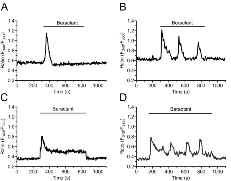
In resting, non stimulated NHLF, the application of 500μg/ml beractant, a concentrationwhich has been shown to exert a nearly maximum apoptotic effect and decrease in collagen accumulation in NHLF [11], caused a Ca2+signal in 96.27% of Fura-2-loaded cells (568 out of 590 cells). Beractant elicited a heterogeneous pattern of Ca2+signals even in cells from the same microscopic field (Fig 1). Accordingly, the intracellular Ca2+signal evoked by beractant consisted in a rapid Ca2+spike (158/568, 27.82%;Fig 1A), that could be followed by Ca2+ oscil-lations (75/568, 13.20%;Fig 1B), a sustained plateau (252/568, 44.37%;Fig 1C), or a plateau overlapped by Ca2+oscillations (plateau + oscillations, 83/568, 14.61%;Fig 1D). Conversely, the IP3-synthesizing autacoid ATP (100μM) induced a biphasic Ca2+response in all the cells
analyzed (n = 28) (S1A Fig).
The pattern and amplitude of the Ca
2+response to beractant depends
on basal [Ca
2+]
iin quiescent lung fibroblasts
By using digital imaging of Fura-2 fluorescence, we have monitored the [Ca2+]isimultaneously
in many individual NHLF from the same population. The mean value of basal [Ca2+]i
mea-sured in all cells studied in the present work was 0.463±0.009 ratio arbitrary units (A.U.) (n = 590). In the majority of cells which had a resting [Ca2+]ihigher than 0.56 ratio A.U (Fig
2A, left panel), beractant evoked a Ca2+signal which displayed either a single spike (n = 158), or repetitive Ca2+oscillations (n = 75) (such as those described inFig 1A and 1B, respectively). When the resting [Ca2+]iwas below 0.4 ratio A.U. (Fig 2A, left panel), beractant evoked Ca2+
signals featured by the appearance of a plateau phase (i.e. plateau, n = 252, and plateau + oscilla-tions, n = 83, as shown inFig 1C and 1D, respectively). In addition, the amplitude of initial Ca2+spike evoked by beractant was seemingly higher in cells that presented high resting [Ca2+]iat rest (i.e. single spike and oscillations), as related to those that had levels of basal
Fig 2A(right panel). However, when the peak fluorescence was normalized to the resting fluo-rescence (ΔR/Ri), the magnitude of the initial Ca2+spike was significantly (p<0.05) higher in
plateauing cells as compared to single-spiking and oscillating cells (Fig 2B). Overall, these results strongly suggest that resting Ca2+levels influence both the magnitude and the pattern of the Ca2+response to beractant.
Beractant elicits a concentration-dependent increase in [Ca
2+]
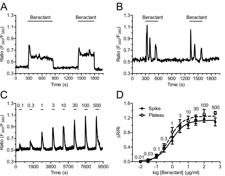
iBeractant effect in NHLF was reversible: the Ca2+signal ceased when the agonist was removed from the bath and a similar Ca2+transient was evoked on beractant restoration. As shown in
Fig 3A, the second application of a supramaximal concentration of beractant (500μg/ml)
pro-duced a Ca2+response with similar kinetics and peak amplitude of that evoked by the first application of beractant (n = 55). The same results were obtained when the Ca2+response to beractant consisted in the onset of repetitive Ca2+oscillations (Fig 3B, n = 45). There was a
Fig 1. Heterogeneity in the Ca2+response elicited by beractant in NHLF.The application of 500μg/ml of beractant, elicited different patterns of Ca2+
signals in cultured NHLF loaded with Fura-2. The intracellular Ca2+signal consisted inA)a rapid Ca2+spike (27.82% of the cells tested) which could be
followed byB)Ca2+oscillations (13.20%),C)sustained plateau (44.37%) or aD)plateau overlapped by Ca2+oscillations (14.61%).
slight reduction in the mean peak amplitude of the second Ca2+transient (13.46%), but the decrease was not statistically significant (Fig 4A, compare Beractant 1stvsBeractant 2nd, p>0.05).
The high reproducibility and lack of desensitization of beractant-induced Ca2+signals enabled us to establish the concentration-response relationship by the repeated administration of the agonist to the same cells. The application of increasing concentrations of beractant (0.03–500μg/ml) to Fura-2 loaded NHLF produced a concentration-dependent increase in
[Ca2+]i.Fig 3Cshows a representative time-course of the Ca2+increases in response to
berac-tant (0.1 to 500μg/ml) in a NHLF that presented a single spike pattern response (seeFig 1A).
Similar results were obtained in NHLF exhibiting the other three patterns of Ca2+response (not shown). The non-cumulative concentration-response curve of beractant-induced eleva-tion in [Ca2+]iis depicted inFig 3Dfor cells that displayed a single spike (closed symbols) and
a plateau response (open symbols). The maximum increase in the peak amplitude was observed at concentrations higher than 100μg/ml (n = 32 cells), whereas raising beractant concentration
up to 500μg/ml did not significantly augment the height of the response (n = 18 cells). Slight
stimulation occurred at 0.1μg/ml (n = 30 cells), while no effect was detectable at
concentra-tions lower than 0.01μg/ml (n = 7). The concentration of beractant required to produce a
half-maximal response (EC50), calculated by fitting the concentration-response curve as described
in Materials and Methods, was 0.82μg/ml. Notably the R2value for the curve fit was 0.9928
Fig 2. The pattern and initial spike-amplitude of the Ca2+response to beractant depend on resting
[Ca2+]i. A)Resting [Ca2+]imeasured in quiescent, non stimulated Fura-2 loaded NHLF that presented the
correspondent Ca2+signal pattern indicated in labels in response to beractant (left). Amplitude of the initial Ca2+spike evoked by beractant (right).B)Initial spike amplitude/basal [Ca2+]
i(ΔR/Ri;ΔR was defined as the
difference between the peak [Ca2+]
iafter stimulation and the value of the resting [Ca2+]i, where Ri is the basal
level of R). Results, expressed as means±SE were analyzed statistically by Student's t test.**p<0.01, ****p<0.0001.
doi:10.1371/journal.pone.0134564.g002
(Fig 3D, closed circles). Similar results were obtained in NHLF cells displaying a long-lasting plateau (Fig 3D, open circles), whose EC50and R2value were 0.95μg/ml and 0.9890,
respectively.
Beractant triggers the Ca
2+response through the PLC/InsP
3signaling
pathway
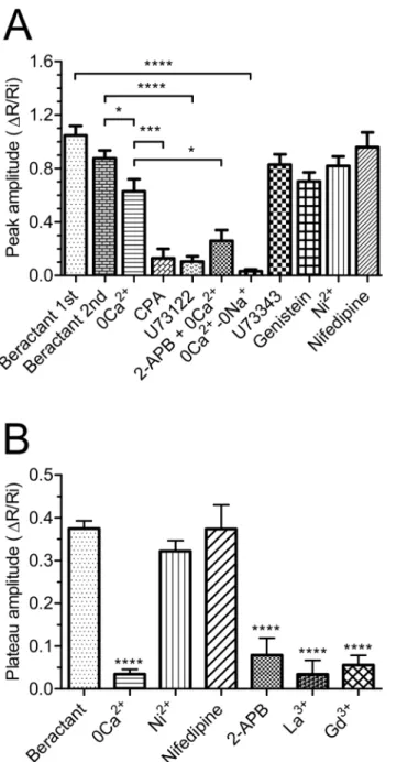
We then sought to dissect the molecular underpinnings of beractant-induced intracellular Ca2+ signals. To assess the contribution of intracellular and extracellular Ca2+stores to the Ca2+ response to 500μg/ml beractant, fibroblasts were exposed to the agonist in the absence of
exter-nal Ca2+(0Ca2+) to prevent Ca2+entry through the plasma membrane. Beractant caused an
Fig 3. Beractant induces a reversible and concentration-dependent Ca2+signal in NHLF.Ca2+
response evoked in a single HNLF cell stimulated repeatedly with the same concentration of beractant (500μg/ml), a reproducible cell-specific pattern of [Ca2+]
isignal is observed in:A)a cell showing a rapid
spike followed by a sustained plateau andB)in a cell showing a rapid spike followed by Ca2+oscillations.
C)A typical trace illustrating the increase in [Ca2+] i
induced by beractant (0.1–500μg/ml).D)Concentration-response relationship. TheΔR/Ri relationship is plotted against the logarithm of beractant concentration. Data points are means±SE, n = 7–39 cells. The continuous curves were obtained by fitting the data toEq 1, which yielded EC50values of 0.8μg/ml and 0.95μg/ ml for cells exhibiting either a single transient (closed symbols) or a sustained plateau (open symbols), respectively. R2value for the curve fits were 0.9928 and
0.9890, respectively. Numbers into the graphics represent de beractant concentration inμg/ml.
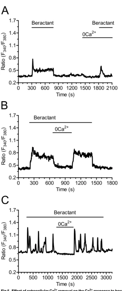
immediate increase in [Ca2+]iin the absence of extracellular Ca2+in 48 out of 50 cells, although
both the Ca2+oscillations and the plateau phase disappeared (Fig 5A). In addition, the mean amplitude of the initial Ca2+spike observed in Ca2+-free solution was significantly reduced by 28.08±10.21% (n = 48; p<0.05) as compared to the Ca2+transient evoked by beractant in
pres-ence of extracellular Ca2+(see statistics inFig 4A: compare 0Ca2+vs Beractant 2nd). These results indicate that the peak response is due to both Ca2+influx and Ca2+release, whereas Ca2+entry sustains both the plateau phase and the following oscillations in [Ca2+]i. This notion
is corroborated by the experiment depicted inFig 5B, where removal of extracellular Ca2+
Fig 4. Effect of beractant on the initial Ca2+spike amplitude and plateau phase amplitude in NHLF. A) ΔR/Ri for the peak response to beractant in presence of designated drugs.B)ΔR/Ri for the plateau phase of beractant-evoked Ca2+increase. See the text for drugs concentrations. Data expressed as means±SE were
analysed statistically by Student’s t-test.*p<0.05,***p<0.001,****p<0.0001.
doi:10.1371/journal.pone.0134564.g004
reversibly inhibited the plateau in 28 of 28 cells (Fig 4B; p<0.0001, n = 28), and inFig 5C,
which shows the abrupt interruption of beractant-induced repetitive Ca2+spikes in 16 out of 16 cells. Both the plateau phase (Fig 5B) and the Ca2+oscillations (Fig 5C) resumed upon Ca2+ restoration to the bath.
No Ca2+signal was ever observed after depletion of the intracellular Ca2+reservoir by cyclo-piazonic acid (CPA, 10μM) (Fig 6A); CPA is an inhibitor of the ER Ca2+-ATPase that prevents
Ca2+reuptake into the stores, thus leading to their depletion [20–22]. In Ca2+-free solution, CPA evoked a transient increase in [Ca2+]idue to passive emptying of the intracellular Ca2+
reservoir through ER leak channels and decreased the Ca2+signal elicited by beractant by 79.39% in 16 of 16 cells (Figs6Aand4A; p<0.001 n = 16). These findings suggest that the
onset of the Ca2+signal evoked by beractant depends on Ca2+mobilization from the intracellu-lar Ca2+pool. Consistently, CPA blocked also the Ca2+response to ATP (n = 10) (S1B Fig).
The involvement of PLC in the transduction pathway leading to beractant-evoked Ca2+ sig-nals was studied by preincubating the cells with U73122 (10μM), a widely employed PLC
inhibitor [23–25]. Accordingly, U73122 (10μM) inhibited the Ca2+response to ATP in 14 out
of 16 NHLF (S1C Fig). Cell pretreatment with U73122 caused a significant reduction in the peak amplitude of beractant-evoked Ca2+transient (87.7± 4.25%, n = 19, p<0.0001) (Figs6B
and4A). Conversely, its inactive structural analogue, U73343 (10μM), did not significantly
affect the Ca2+response to beractant in 19 out of 19 cells (Fig 6C). In the majority of the cells (i.e. 12 out of 19), U73343 (10μM) exerted little or no effect on basal [Ca2+]i, but in a fewer
cells (7 out of 19 cells) it caused a slow rise in intracellular Ca2+levels accompanied by the development of several Ca2+spikes; however, beractant-induced Ca2+elevation was neither prevented nor affected (Figs6Cand4A; p>0.05, n = 19). Taken together, these data suggest
that the initiation of the Ca2+signal by beractant requires the activation of PLC and the release of Ca2+from ER stores, presumably through the IP3-sensitive Ca2+channels. In order
to assess whether PLC activity is triggered following TKR activation, NHLF were preincubated with 100μM genistein, a widely used TKR inhibitor [25–27]. This maneuver did not prevent
or alter the Ca2+response of NHLF to 500μg/ml beractant (Figs6Dand4A; n = 8; p>0.05).
Genistein reduced the amplitude of the initial Ca2+spike evoked by beractant by 19.47±7.5%, however, no statistically relevant difference was found (p>0.05) (Fig 4A, compare Genisteinvs
Beractant 2nd). Therefore, PLCβis the most likely isoform involved in the generation of beractant-induced Ca2+signals. The contribution of IP3-dependent signaling was further
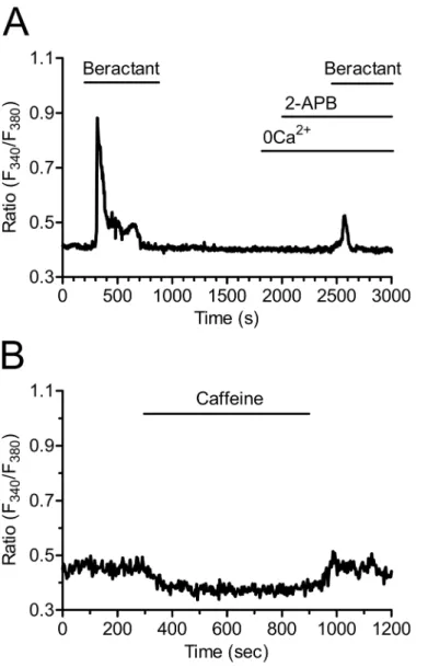
probed by exposing the cells to beractant in the presence of 2-aminoethoxydiphenyl borate (2-APB; 50μM), a widely employed inhibitor of IP3Rs. These experiments were conducted in
the absence of extracellular Ca2+as 2-APB has also been reported to affect SOCs at this concen-tration [28–30]. Accordingly, this treatment dramatically reduced beractant-induced Ca2+ discharge from ER by approximately 58.69% (Figs7Aand4A, p<0.05, n = 18). Moreover,
caf-feine (10 mM), which is a membrane-permeable stimulator of ryanodine receptors (RyRs), failed to increase [Ca2+]iin 16 of 16 NHLF tested (Fig 7B). These results, therefore, hint at
IP3Rs as the main mediators of Ca2+release from ER upon exposition to beractant.
SOCE sustains the Ca
2+response to Beractant
As previously shown, both the prolonged plateau phase (Fig 5B; n = 48) and the oscillations in [Ca2+]ithat may follow the initial Ca2+spike triggered by beractant (Fig 5C) do not occur in
Ca2+-free solution. These findings suggest that Ca2+entry from the extracellular space is essen-tial to sustain the elevation in [Ca2+]iover time, whatever its sub-cellular temporal dynamics,
Fig 5. Effect of extracellular Ca2+removal on the Ca2+response to beractant. A)Ca2+signal elicited by
beractant in the presence and absence of extracellular Ca2+(0Ca2+) in the same single cell. Note that the
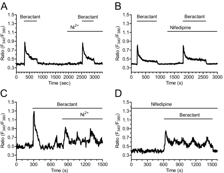
demonstrated that mouse embryonic fibroblasts express voltage-operated Ca2+channels (VOCC) as well. In order to assess the hypothesis that the sustained Ca2+signal evoked by beractant was mediated by VOCC in NHLF, we probed the effects of Ni2+(100μM), a non
spe-cific blocker of VOCC, and nifedipine (1μM), which selectively antagonizes L-type VOCC.
Neither Ni2+(Figs8Aand4B; p>0.05; n = 24) nor nifedipine (Figs8Band4B; p>0.05;
n = 30) inhibited the sustained Ca2+response elicited by beractant. All together, these results rule out the contribution of VOCC to the plateau phase that may follow the initial Ca2+
prolonged decay phase did not occur in absence of extracellular Ca2+.B)Withdrawing extracellular Ca2+ (0Ca2+) during an established response to beractant (500μg/ml) immediately interrupted the Ca2+plateau
andC)repetitive Ca2+oscillations. The Ca2+plateau and Ca2+oscillations resumed upon readmission of
external Ca2+.
doi:10.1371/journal.pone.0134564.g005
Fig 6. Beractant releases Ca2+from intracellular stores by activating PLC.The initial Ca2+increase evoked by beractant was:
A)abolished by depletion of intracellular Ca2+stores with CPA (10μM) in absence of extracellular Ca2+(0Ca2+), andB)by blockage of PLC activity with U73122 (10μM), a widely used PLC blocker,C)but not by its inactive analogue U73343 (10μM),D)and by genistein (100μM), a tyrosine kinase inhibitor.
response to beractant. Similarly, neither Ni2+(Fig 8C) nor nifedipine (Fig 8D) interfered with beractant-induced intracellular Ca2+oscillations.
The most important route for Ca2+inflow into non-excitable cells is represented by SOCE [28,33]. SOCE contribution to beractant-induced Ca2+entry was first assessed by treating the NHLF with 2-APB (50μM) [34]. In addition to IP3Rs, this drug may indeed interfere with
SOCE and prevent Ca2+influx in the presence of extracellular Ca2+[29,35]. 2-APB reduced by 79% (p<0.001) the amplitude of the Ca2+plateau in 18 of 22 cell tested (Figs9Aand4B, p<
0.0001, n = 22). Likewise, 2-APB (50μM) reversibly abolished beractant-induced oscillations
in [Ca2+]iin 10 out of 10 cells (Fig 9B).
As shown by the experiments conducted in the absence of external Ca2+, these results might be explained by the combinatorial inhibition of IP3Rs and SOCE. As a consequence, we Fig 7. Inositol-1,4,5-trisphosphate receptors (IP3Rs) drive the Ca2+response to beractant. A)The Ca2+
signal elicited by beractant is inhibited in the presence of 2-APB (50μM), a well known InsP3R inhibitor.
These experiments were conducted in the absence of extracellular Ca2+(0Ca2+) as 2-APB has also been
reported to affect SOCs at this concentration.B)Caffeine (10 mM), which is a membrane-permeable RyR stimulator, failed to increase [Ca2+]
iin NHLF. doi:10.1371/journal.pone.0134564.g007
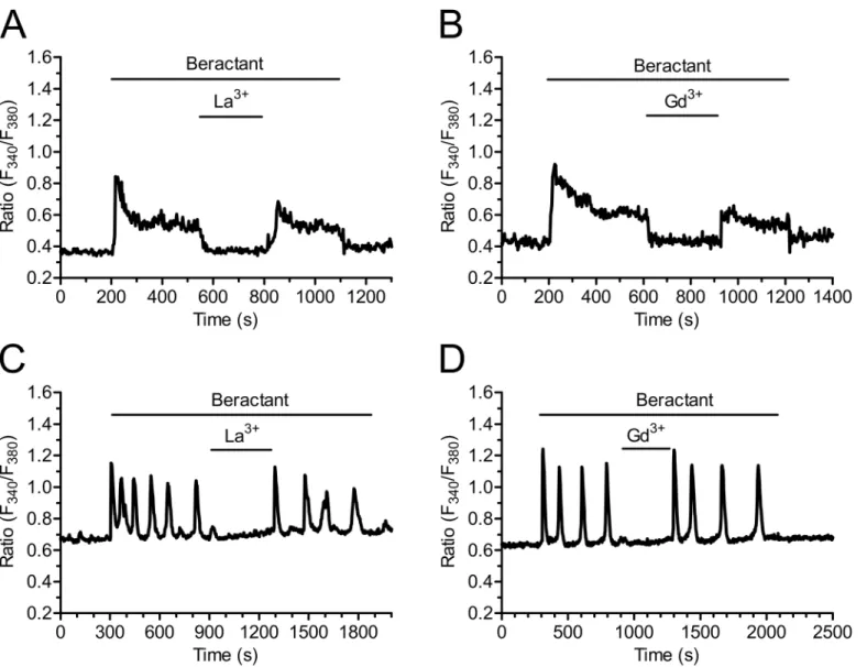
exploited three additional well known inhibitors of SOCs, namely the pyrazole derivative, BTP-2, and the trivalent cations, La3+and Gd3+[28,29,36,37]. Unfortunately, our preliminary experiments revealed that 20μM BTP-2 induced a robust increase in [Ca2+]iin 21 of 21 NHLF
cells and could not be utilized further (S2 Fig). When applied at concentrations ranging from 1 up to 10μM, lanthanides are rather selective towards SOCs and do not affect either
receptor-or second messenger-operated Ca2+channels [28,29,37]. As illustrated inFig 10, both La3+ (10μM) and Gd3+(10μM) reversibly inhibited the plateau phase (Figs10Aand4B, p<0.0001,
n = 18 and Figs10Band4B, p<0.0001, n = 23, respectively), as well as the repetitive oscillations
in [Ca2+]i, that followed the initial Ca2+peak induced by beractant (Fig 10C and 10D,
respec-tively). Overall, these results strongly suggest that SOCE maintains the sustained component of the Ca2+response to beractant in NHLF.
Fig 8. The plateau phase evoked by beractant is not mediated by voltage-operated calcium channels.The plateau phase of beractant-evoked Ca2+ signals was not affectedA) by either Ni2+(100μM) orB)Nifedipine (1μM). Cells were preincubated for 10 min with nifedipine (1μM) before applying beractant (500μg/ml).C)Nifedipine (1μM) andD)Ni2+(100μM) did not inhibit beractant-induced Ca2+oscillations.
The Ca
2+response to beractant is not mimicked by phospholipids, but
requires membrane depolarization
In order to assess which of the single components of beractant trigger the Ca2+response and how they are related to PLCβactivation, we first probed the effect of albumin, dipalmitoylpho-sphatidylcholine (DPPC) and diacylglycerol (DG). Albumin is a protein which is not associated to lung surfactant, and failed to evoke any Ca2+signal in beractant-responsive NHLF (Fig 11A, n = 18). Actually, albumin caused a slight decrease in basal Fura-2 fluorescence, but this did not prevent beractant from elevating [Ca2+]i(Fig 11A). Similarly, DPPC (200μg/ml) and DG
(50μg/ml), which are two phospholipid constituents of beractant, did not elicit any increase
in [Ca2+]i(Fig 11B and 11C, n = 15 and 18, respectively). Overall, these findings strongly
sug-gest that the Ca2+response to beractant is mediated by SAPs. More specifically, beractant con-tains SAP-B and SAP-C, which were recently shown to bring about Ca2+signals through the
Fig 9. 2-APB inhibits beractant-induced Ca2+plateau and Ca2+oscillations in NHLF.2-APB (50μM),
which may also block SOCs, reversibly inhibited:A)the sustained plateau andB)the [Ca2+]
ioscillations
evoked by beractant (500μg/ml) in NHLF.
doi:10.1371/journal.pone.0134564.g009
insertion of monovalent cation channels on the plasma membrane. The resulting depolariza-tion leads to IP3-dependent Ca2+release by a yet to be discovered mechanism [38,39].
There-fore, we then analyzed the triggering mechanism of beractant-induced Ca2+release by stimulating NHLF in the absence of external Na+and Ca2+to prevent membrane depolariza-tion. This procedure reversibly abolished beractant-induced increase in [Ca2+]i(Fig 11D).
SOCE inhibitors prevent beractant-induced apoptosis and collagen
expression downregulation
Finally, we investigated the role of intracellular Ca2+signals in beractant-induced NHLF apo-ptosis and collagen expression. Pre-incubating the cells for 24 hrs with U73122 (10μM), La3+
(10μM) or 2-APB (50μM) prevented beractant-induced apoptosis, as evaluated by monitoring
caspase 3 and 7 activity (Fig 12A; p<0.05). Collectively, these data indicate that SOCE
pro-motes the pro-apoptotic effect of beractant in NHLF. Likewise, pre-treating the cells with Gd3+
Fig 10. La3+and Gd3+block beractant-induced Ca2+plateau and Ca2+oscillations in NHLF.A) Addition of La3+(10μM) and B) Gd3+(10μM) reversibly inhibited the sustained plateau phase of the Ca2+signal induced by beractant (500μg/ml). C) Application of La3+(10μM) and D) Gd3+(10μM) reversibly inhibited beractant-elicited Ca2+oscillations (500μg/ml).
(10μM, 24 hrs) prevented beractant from downregulatingα1(I) procollagen gene expression
(Fig 12B). Unfortunately, 2-APB (50μM) caused a reduction inα1(I) procollagen gene
expres-sionper se(Fig 12B) and did not interfere with the action of beractant. These data, however, strongly suggests that SOCE contributes also to reduce beractant-dependentα1(I) procollagen transcript.
Discussion
Restoration of surfactant activity has been introduced in the routine care of patients affected by respiratory distress syndrome [4], and might be a suitable tool to adverse intraluminal fibrosis in IPF and other interstitial lung diseases. Beractant is a natural bovine extract enriched with phospholipids, neutral lipids, fatty acids, and the hydrophobic proteins SP-B and SP-C, and is widely employed in clinical practice, albeit the underlying signal transduction mechanisms are
Fig 11. The Ca2+response to beractant is not mimicked by albumin, (DPPC) and diacylglycerol, but is inhibited by preventing membrane
depolarization. A)Albumin,B)dipalmitoylphosphatidylcholine (DPPC) (200μg/ml) andC)diacylglucerol (DG) (50μg/ml) did not evoke any detectable increase in [Ca2+]
iin NHLF.D)the Ca2+response to beractant (500μg/ml) was abrogated by replacing extracellular Na+with an equimolar amount of NMDG
in the absence of external Ca2+(0Ca2+-0Na+).
doi:10.1371/journal.pone.0134564.g011
Fig 12. Effect of SOCE inhibitors on Beractant effect in apoptosis and collagen expression.NHLF were incubated for 24 hours with either SOCE inhibitors alone (U73122, 10μM; La3+, 10μM; Gd3+, 10μM or 2-APB, 50μM) or in combination with beractant 500μg/ml in serum-free medium.A)Caspase 3 and 7 activity, the Caspase-Glo assay kit (Promega, Madison, WI) was used to measure the executioner caspases 3 and 7. Cisplatin 20μM was used as positive control. Despite the fact that U73122 and 2-APB exerted a modest, albeit significant, pro-apoptotic effect, they blocked beractant-induced apoptosis.B)Collagen expression. RT-qPCR was used to measure the expression of collagen. All real time PCRs were performed in triplicate at least two times. Results were normalized to human GAPDH using the delta-delta Ct method (2
-ΔΔCt). Results are expressed as means±SE. ANOVA was used witha priorycomparisons of selected pairs.
far from being fully elucidated. Importantly, it shows anti-inflammatory and anti-fibrotic prop-erties [10,11]. Ca2+signaling regulates a myriad of cellular processes, including those elicited by beractant in fibroblasts, i.e. DNA replication, gene expression, apoptosis, and differentiation [13,33,40]. In this context, our results provide the first evidence that beractant elicits an hetero-geneous increase in [Ca2+]iin NHLF, which might be involved in its functional effect on these
cells.
Beractant evoked a complex pattern of elevations in [Ca2+]iin neighboring NHLF, which
displayed at least 4 types of responses upon agonist stimulation: 1) a rapid Ca2+spike which quickly decayed to the baseline; 2) a biphasic Ca2+signal, which comprised an initial Ca2+ spike followed by a prolonged plateau phase of intermediate amplitude; 3) repetitive oscilla-tions in [Ca2+]iand 4) a biphasic elevation in [Ca2+]ifeatured by the superimposition of Ca2+
oscillations on the plateau phase. Studies of the Ca2+responses of a wide variety of cell types at the single cell level have consistently revealed cell-to-cell heterogeneity [23,25,41–44]. Cell cycle heterogeneity as an explanation of the variability in the Ca2+response to beractant is very unlikely. First, all our experiments were carried on in serum-starved NHLF for 48 hours, which arrests cell cycle in Gophase [45]. Second, in spite that cell cycle asynchrony has been proposed
as a source of variability in Ca2+signaling, existing data do not support this idea. Thus, syn-chronization of cultured human foreskin fibroblasts failed to prevent the variety of patterns in [Ca2+]ielevations elicited by bradykinin [46]. Similarly, Ambler and Cols [43] showed that
syn-chronized cycling BC3H-1 cells responded asynchronously to histamine stimulation. Finally, serum-starved rat cardiac coronary microvascular endothelial cells still displayed a heteroge-neous Ca2+response to EGF [41]. Collectively, these considerations lead us to conclude that individual NHLF produce asynchronous changes in [Ca2+]iwhen exposed to beractant and
that this is not due to cell heterogeneity in the cell cycle.
Recently, Ishida and coworkers (2014) showed that cell-to-cell variability in the pattern of Ca2+signals in histamine-stimulated in HeLa cells is due to heterogeneity in the process of IP3
production. Modulation of IP3dynamics by knockdown or overexpression of PLCβ1 and
PLCβ4 resulted in specific changes in the characteristics of Ca2+signals within the range of the cell-to-cell variability found in wild-type cell populations [44]. Moreover, the cell-specific pat-tern of beractant–induced increase in [Ca2+]iin NHLF might invoke single-cell heterogeneity
regarding membrane receptors or elements of the phosphoinositide signaling pathways, such as PLCβ, IP3Rs, and SOCE (see below), as recently suggested in [23,47]. An additional, albeit
not mutually exclusive, explanation for the cell-to-cell variability observed in beractant-stimu-lated NHLF resides in their resting Ca2+levels: The basal [Ca2+]iis higher in cells displaying
either a single Ca2+spike or discrete Ca2+oscillations as compared to those experiencing the plateau phase, with or without the superimposition of sinusoidal oscillations. Similar results were found by Toescu and coworkers [48], who reported about acetylcholine (Ach)-induced intracellular Ca2+waves in cultured mouse pancreatic acinar cells. When the basal Ca2+levels were lower than a threshold concentration of 150 nM, ACh always evoked high frequency short-lasting Ca2+spikes, whereas it elicited less frequent, long-lasting Ca2+transients at [Ca2+]i>150 nM [48]. Future work is required to ascertain how the basal Ca2+concentration
impacts on beractant-induced Ca2+signals. Nevertheless, it is possible to conclude that the pat-tern of the Ca2+response in NHLF is specific to each cell: when a given NHLF is repeatedly stimulated with the same concentration of beractant, a reproducible and cell-specific pattern of [Ca2+]isignal, the so-called Ca2+fingerprint [42], occurs (see, for instance,Fig 3A and 3B).
The EC50of beractant-induced elevation in [Ca2+]iis equal to 0.82μg/ml, while its maximal
effect was achieved at 100–500μg/ml. Likewise, in NHLF, beractant was found to induce
apo-ptosis and reduce collagen deposition at 500μg/ml [11], whereas it interfered with DNA
syn-thesis and secondary inflammatory mediator production at 500–1000μg/ml [10]. Moreover,
beractant was shown to insert plasmalemmal cation monovalent channels and trigger Ca2+ release in human neutrophils in the same concentration range [38,39]. Therefore, we believe that the concentrations of beractant employed in the present investigation are very close to those established by other authors. The following pieces of evidence indicate that beractant-elicited intracellular Ca2+signals in NHLF are patterned by the coordinated interplay between IP3-dependent Ca2+release and SOCE. First, the increase in [Ca2+]iis prevented by U73122, a
widely employed PLC inhibitor, while it is unaffected by its structural analogue, U73343. genis-tein, a broad spectrum protein tyrosin kinase inhibitor, did not interfere with the onset of the Ca2+response to beractant. Therefore, PLC activity is likely to be induced by the activation of GPCRs and to involve theβ-isoform. Second, beractant-induced Ca2+signals are prevented by 2-APB, a membrane-permeable blocker of InsP3-dependent Ca2+release in the absence of
external Ca2+. Third, no Ca2+signal could be detected in response to caffeine, which stimulates endogenous RyRs by sensitizing them to resting Ca2+levels [49]. Fourth, Ni2+and nifedipine, two established VOCC blockers, did not affect beractant-induced increase in [Ca2+]i. Fifth,
lan-thanides reversibly abrogated the sustained component of the Ca2+response to beractant, by interrupting both the prolonged plateau phase and the repetitive Ca2+oscillations. This effect was observed when both La3+and Gd3+were applied at 10μM, a concentration which
selec-tively hinders SOCs [28,37,50]. BTP-2, another well known SOCE inhibitor, could not be probed in the present study due to its ability to increase [Ca2+]iin NHLF. These findings
strongly indicate that, while the single Ca2+transients exclusively derive from IP3-dependent
Ca2+mobilization, the sustained response involve Ca2+entry through plasmalemmal SOCs. This concept is corroborated by the finding that, when 2-APB is administrated in the presence of extracellular Ca2+to interfere with IP3Rs and SOCE, both the prolonged plateau phase and
the repetitive Ca2+spikes rapidly run down. According to the most popular models proposed to describe intracellular Ca2+oscillations [28,51,52], SOCE refills the intracellular Ca2+stores during maintained stimulation and provides IP3Rs with a sufficient amount of intraluminal
Ca2+to sustain their spiking activity. In this scenario, however, Ca2+oscillations do not cease immediately after the removal of extracellular Ca2+, but persist for some time in the absence of Ca2+entry. Conversely, beractant-induced Ca2+transients are instantaneously inhibited by perfusing the cells with a Ca2+-deficient solution, whereas they quickly resume on Ca2+ res-toration to the bath. This observation might be explained by a role for Ca2+entry in governing IP3-dependent Ca2+release. Environmental Ca2+controls IP3-mediated Ca2+mobilization,
whereas surrounding Ca2+stimulates or inhibits IP3gating at [Ca2+] lower and higher than
10μM, respectively [53]. The immediate interruption of Ca2+oscillations in 0Ca2+suggests
that SOCE is required to achieve adequate levels of stimulating Ca2+nearby intracellular IP3Rs,
as observed in spiking HeLa cells stimulated with histamine [54] and exocrine epithelial cells challenged with carbachol [55]. The molecular structure of SOCE may vary depending on the cell type [34]. This pathway is mediated by the interaction between the ER Ca2+sensor, Stim1, and the Ca2+-permeable channels, Orai1 and TRPC1, in normal rat kidney fibroblasts [56]. Nevertheless, Orai1 and TRPC1 have reported to serve as independent SOCs, each activated by Stim1, in human submandibular gland cells [57]. Consistent with these data, Stim1, Orai1 and TRPC1 have been reported in human cardiac fibroblasts [58]; however, the molecular compo-sition of SOCE in NHLF is yet to be elucidated and will require further work.
Our results are consistent with previous investigations, which demonstrated the influence of pulmonary surfactant on intracellular Ca2+homeostasis in both human neutrophils [38] and rat alveolar macrophages [59]. Beractant contains only two hydrophobic low-molecular weight proteins, i.e. SAP-B and SAP-C. Boston et al. [38] demonstrated that beractant causes a tran-sient increase in [Ca2+]iin neutrophils due to G protein-mediated release from intracellular
monovalent cationic channels by SAP-B and SAP-C, which depolarize the cells by causing Na+ influx [38,60]. Likewise, we found that preventing membrane depolarization by exposing NHLF to an external solution devoid of Na+, blocked beractant-induced elevation in [Ca2+]i.
The mechanistic link between the positive shift in membrane potential and PLCβactivation is likely to be provided by voltage-dependent GPCRs, such as P2Y1, 5HT2A, thromboxane (TPα), M1 and M3 receptors [61,62]. For instance, cell depolarization activates P2Y1 receptors in rodent megakaryocytes, thereby leading to InsP3synthesis and InsP3-dependent Ca2+
mobilization, while voltage fails to stimulate TKRs [61,62]. These metabotropic receptors are expressed by human fibroblasts [63] and could mediate the effect of membrane depolarization on PLCβ. Conversely, two abundant phospholipid components of beractant, such as DPPC and DG, do not change intracellular Ca2+levels, just like albumin, which is not contained either in natural surfactant or in beractant.
After lung injury, recovery depends on reestablishment of the air-lung interface through the elimination of intra-alveolar mesenchymal cells. Beractant induces apoptosis and decreases collagen accumulation in NHLF [11]; accordingly, it can be speculated that the use of exoge-nous surfactant may have a beneficial role in avoiding the formation of intraluminal fibrosis, and that the changes in the intracellular Ca2+concentration observed may be implicated in this effect. A prolonged elevation in [Ca2+]i, such as that produced by a sustained plateau, is amid
the most powerful apoptogenic signals [64]. Accordingly, we found that inhibiting SOCE with La3+and 2-APB prevented beractant-induced NHLF apoptosis. On the other hand, intracellu-lar Ca2+oscillations encode the information driving the Ca2+-dependent activation of several transcription factors [34,65]. Intriguingly, Gd3+prevented beractant from suppressing the downregulation ofα1(I) procollagen transcript. 2-APB reducedα1(I) procollagen expression
per seand could not be used further, but we should recall that this drug is far less selective than 10μM Gd3+and interferes also with other Ca2+-permeable pathways [33]. The hypothesis that
the distinct modes of Ca2+signaling induced by beractant in NHLF control different cellular processes is currently under evaluation.
In summary, our results describe for the first time the pattern of Ca2+signals elicited by a natural lung surfactant extract in primary cultures of human lung fibroblasts. The Ca2+ response to beractant is triggered by PLCβrecruitment following the activation of a GPCR. The subsequent cleavage of the membrane phospholipid, PIP2, leads to the generation of IP3,
which releases intraluminally stored Ca2+, thereby activating SOCE. The interplay between IP3-dependent Ca2+mobilization and SOCE results in a variety of Ca2+signals depending on
the resting Ca2+levels. Beractant-induced Ca2+signals might protect against pulmonary struc-tural remodeling in IPF as well as other severe fibrosing respiratory diseases.
Supporting Information
S1 Fig. ATP-induced Ca2+signals in NHLF. A)Ca2+response to ATP (100μM) in NHLF.
ATP-induced Ca2+elevation was abrogated byB)depletion of intracellular Ca2+stores with CPA (10μM) in absence of extracellular Ca2+(0Ca2+), andC)by blockage of PLC activity with
U73122 (10μM).
(TIF)
S2 Fig. Effect of BTP-2 on the resting [Ca2+]iin NHLF.Ca2+signal evoked by the pyrazole derivative, BTP-2 (20μM), in a single NHLF cell.
(TIF)
Author Contributions
Conceived and designed the experiments: RBR LGVL FT FM JTJ. Performed the experiments: AGS CMM AVG MFF ALM. Analyzed the data: AGS CMM RBR FM ALM. Contributed reagents/materials/analysis tools: EPS. Wrote the paper: RBR LGVL FM FT.
References
1. Agassandian M, Mallampalli RK. Surfactant phospholipid metabolism. Biochim Biophys Acta. 2013; 1831: 612–25. doi:10.1016/j.bbalip.2012.09.010PMID:23026158
2. Lopez-Rodriguez E, Pérez-Gil J. Structure-function relationships in pulmonary surfactant membranes: from biophysics to therapy. Biochim Biophys Acta. 2014; 1838: 1568–85. doi:10.1016/j.bbamem.2014. 01.028PMID:24525076
3. Glasser J, Mallampalli R. Surfactant and its role in the pathobiology of pulmonary infection. Microbes Infect. Elsevier Masson SAS; 2012; 14: 17–25. doi:10.1016/j.micinf.2011.08.019PMID:21945366
4. Zuo YY, Veldhuizen RAW, Neumann AW, Petersen NO, Possmayer F. Current perspectives in pulmo-nary surfactant—inhibition, enhancement and evaluation. Biochim Biophys Acta. 2008; 1778: 1947–
77. doi:10.1016/j.bbamem.2008.03.021PMID:18433715
5. Akella A, Deshpande SB. Pulmonary surfactants and their role in pathophysiology of lung disorders. Indian J Exp Biol. 2013; 51: 5–22. PMID:23441475
6. Tzouvelekis A, Bonella F, Spagnolo P. Update on therapeutic management of idiopathic pulmonary fibrosis. Ther Clin Risk Manag. 2015; 11: 359–370. doi:10.2147/TCRM.S69716PMID:25767391
7. King TE, Pardo A, Selman M. Idiopathic pulmonary fibrosis. Lancet. 2011; 378: 1949–61. doi:10.1016/ S0140-6736(11)60052-4PMID:21719092
8. Harari S, Caminati A. IPF: new insight on pathogenesis and treatment. Allergy. 2010; 65: 537–53. doi: 10.1111/j.1398-9995.2009.02305.xPMID:20121758
9. Selman M, King TE, Pardo A. Idiopathic pulmonary fibrosis: prevailing and evolving hypotheses about its pathogenesis and implications for therapy. Ann Intern Med. 2001; 134: 136–51. PMID:11177318
10. Thomassen MJ, Antal JM, Barna BP, Divis LT, Meeker DP, Wiedemann HP. Surfactant downregulates synthesis of DNA and inflammatory mediators in normal human lung fibroblasts. Am J Physiol. 1996; 270: L159–L163. PMID:8772539
11. Vazquez de Lara L, Becerril C, Montano M, Ramos C, Maldonado V, Melendez J, et al. Surfactant com-ponents modulate fibroblast apoptosis and type I collagen and collagenase-1 expression. Am J Physiol Lung Cell Mol Physiol. 2000; 279: L950–957. PMID:11053032
12. Naon D, Scorrano L. At the right distance: ER-mitochondria juxtaposition in cell life and death. Biochim Biophys Acta—Mol Cell Res. 2014; 1843: 2184–2194. doi:10.1016/j.bbamcr.2014.05.011
13. Berridge MJ, Bootman MD, Roderick HL. Calcium signalling: dynamics, homeostasis and remodelling. Nat Rev Mol Cell Biol. Nature Publishing Group; 2003; 4: 517–29. doi:10.1038/nrm1155PMID: 12838335
14. Thurley K, Skupin A, Thul R, Falcke M. Fundamental properties of Ca2+ signals. Biochim Biophys Acta. 2012; 1820: 1185–94. doi:10.1016/j.bbagen.2011.10.007PMID:22040723
15. Streb H, Irvine R, Berridge M, Schulz I. Release of Ca2+ from a nonmitochondrial intracellular store in pancreatic acinar cells by inositol-1, 4, 5-trisphosphate. Nature. Nature Publishing Group; 1983; 306: 67–69. doi:10.1038/306067a0
16. Fukami K, Inanobe S. Phospholipase C is a key enzyme regulating intracellular calcium and modulating the phosphoinositide balance. Prog Lipid Res. Elsevier Ltd; 2010; 49: 429–37. doi:10.1016/j.plipres. 2010.06.001PMID:20553968
17. Maeda N, Kawasaki T, Nakade S, Yokota N, Taguchi T, Kasai M, et al. Structural and functional charac-terization of inositol 1,4,5-trisphosphate receptor channel from mouse cerebellum. J Biol Chem. 1991; 266: 1109–16. PMID:1845986
18. Berra-Romani R, Raqeeb A, Avelino-Cruz JE, Moccia F, Oldani A, Speroni F, et al. Ca2+ signaling in injured in situ endothelium of rat aorta. Cell Calcium. 2008; 44: 298–309. doi:10.1016/j.ceca.2007.12. 007PMID:18276005
19. Berridge MJ. Cell Signalling Biology: Module 2—Cell signalling pathways. In: Biochemical Journal [Internet]. 10 Apr 2012 [cited 22 May 2013] pp. 1–130. doi:10.1042/csb0001002
primary myelofibrosis. PLoS One. 2014; 9: e91099. doi:10.1371/journal.pone.0091099PMID: 24603752
21. Berra-Romani R, Mazzocco-Spezzia A, Pulina M V, Golovina VA. Ca2+ handling is altered when arte-rial myocytes progress from a contractile to a proliferative phenotype in culture. Am J Physiol Cell Phy-siol. 2008; 295: C779–90. doi:10.1152/ajpcell.00173.2008PMID:18596214
22. Seidler NW, Jona I, Vegh M, Martonosi A. Cyclopiazonic acid is a specific inhibitor of the Ca2+-ATPase of sarcoplasmic reticulum. J Biol Chem. 1989; 264: 17816–23. PMID:2530215
23. Berra-Romani R, Raqeeb A, Torres-Jácome J, Guzman-Silva A, Guerra G, Tanzi F, et al. The mecha-nism of injury-induced intracellular calcium concentration oscillations in the endothelium of excised rat aorta. J Vasc Res. Karger Publishers; 2012; 49: 65–76. doi:10.1159/000329618PMID:21997119
24. Mukherjee S, Duan F, Kolb MRJ, Janssen LJ. Platelet derived growth factor-evoked Ca(2+) wave and matrix gene expression through phospholipase C in human pulmonary fibroblast. Int J Biochem Cell Biol. Elsevier Ltd; 2013; 45: 1516–1524. doi:10.1016/j.biocel.2013.04.018PMID:23618877
25. Dragoni S, Laforenza U, Bonetti E, Lodola F, Bottino C, Berra-Romani R, et al. Vascular endothelial growth factor stimulates endothelial colony forming cells proliferation and tubulogenesis by inducing oscillations in intracellular Ca2+ concentration. Stem Cells. 2011; 29: 1898–907. doi:10.1002/stem. 734PMID:21905169
26. Potenza DM, Guerra G, Avanzato D, Poletto V, Pareek S, Guido D, et al. Hydrogen sulphide triggers VEGF-induced intracellular Ca2+signals in human endothelial cells but not in their immature progeni-tors. Cell Calcium. 2014; 56: 225–34. doi:10.1016/j.ceca.2014.07.010PMID:25113159
27. Moccia F, Berra-Romani R, Tritto S, Signorelli S, Taglietti V, Tanzi F. Epidermal growth factor induces intracellular Ca2+ oscillations in microvascular endothelial cells. J Cell Physiol. 2003; 194: 139–50. doi: 10.1002/jcp.10198PMID:12494452
28. Moccia F, Berra-Romani R, Tanzi F. Update on vascular endothelial Ca(2+) signalling: A tale of ion channels, pumps and transporters. World J Biol Chem. 2012; 3: 127–58. doi:10.4331/wjbc.v3.i7.127 PMID:22905291
29. Parekh AB. Store-operated CRAC channels: function in health and disease. Nat Rev Drug Discov. Nature Publishing Group; 2010; 9: 399–410. doi:10.1038/nrd3136PMID:20395953
30. Dragoni S, Laforenza U, Bonetti E, Lodola F, Bottino C, Guerra G, et al. Canonical Transient Receptor Potential 3 channel triggers VEGF-induced intracellular ca2+ oscillations in endothelial progenitor cells isolated from umbilical cord blood. Stem Cells Dev. Mary Ann Liebert, Inc. 140 Huguenot Street, 3rd Floor New Rochelle, NY 10801 USA; 2013; 22: 2561–2580. doi:10.1089/scd.2013.0032PMID: 23682725
31. Catterall W. Voltage-gated calcium channels. Cold Spring Harb Perspect Biol. 2011; 3: a003947. doi: 10.1101/cshperspect.a003947PMID:21746798
32. Yang S, Huang X-Y. Ca2+ influx through L-type Ca2+ channels controls the trailing tail contraction in growth factor-induced fibroblast cell migration. J Biol Chem. 2005; 280: 27130–7. doi:10.1074/jbc. M501625200PMID:15911622
33. Moccia F, Dragoni S, Lodola F, Bonetti E, Bottino C, Guerra G, et al. Store-dependent Ca(2+) entry in endothelial progenitor cells as a perspective tool to enhance cell-based therapy and adverse tumour vascularization. Curr Med Chem. 2012; 19: 5802–18. PMID:22963562
34. Parekh AB, Putney JW. Store-operated calcium channels. Physiol Rev. 2005; 85: 757–810. doi:10. 1152/physrev.00057.2003PMID:15788710
35. Moccia F, Bonetti E, Dragoni S, Fontana J, Lodola F, Berra Romani R, et al. Hematopoietic Progenitor and Stem Cells Circulate by Surfing on Intracellular Ca2+ Waves: A Novel Target for Cell-based Ther-apy and Anti-cancer Treatment? Curr Signal Transduct Ther. Bentham Science Publishers; 2012; 7: 161–176. doi:10.2174/157436212800376672
36. Berra-Romani R, Avelino-Cruz JE, Raqeeb A, Della Corte A, Cinelli M, Montagnani S, et al. Ca2 +-dependent nitric oxide release in the injured endothelium of excised rat aorta: a promising mecha-nism applying in vascular prosthetic devices in aging patients. BMC Surg. 2013; 13 Suppl 2: S40. doi: 10.1186/1471-2482-13-S2-S40PMID:24266895
37. Moccia F, Dragoni S, Poletto V. Orai1 and Transient Receptor Potential Channels as Novel Molecular Targets to Impair Tumor Neovascularisation in Renal Cell Carcinoma and other Malignancies. Antican-cer agents Med Chem. 2014; 14: 296–312. PMID:23869775
38. Boston ME, Frech GC, Chacon-Cruz E, Buescher ES, Oelberg DG. Surfactant releases internal cal-cium stores in neutrophils by G protein-activated pathway. Exp Biol Med (Maywood). SAGE Publica-tions; 2004; 229: 99–107.
39. Oelberg DG, Xu F. Pulmonary surfactant proteins insert cation-permeable channels in planar bilayers. Mol Genet Metab. 2000; 70: 295–300. doi:10.1006/mgme.2000.3022PMID:10993716
40. Misquitta CM, Ghosh P, Mwanjewe J, Grover AK. Role of cis-acting elements in the control of SER-CA2b Ca2+ pump mRNA decay by nuclear proteins. Biochem J. 2005; 388: 291–7. doi:10.1042/ BJ20041568PMID:15656788
41. Moccia F, Berra-Romani R, Tritto S, Signorelli S, Taglietti V, Tanzi F. Epidermal growth factor induces intracellular Ca2+ oscillations in microvascular endothelial cells. J Cell Physiol. 2003; 194: 139–50. doi: 10.1002/jcp.10198PMID:12494452
42. Prentki M, Glennon M, Thomas A. Cell-specific patterns of oscillating free Ca2+ in carbamylcholine-stimulated insulinoma cells. J Biol Chem. 1988; 263: 11044–7. PMID:2841314
43. Ambler S, Poenie M, Tsien R, Taylor P. Agonist-stimulated oscillations and cycling of intracellular free calcium in individual cultured muscle cells. J Biol Chem. 1988; 263: 1952–9. PMID:2892835
44. Ishida S, Matsu-Ura T, Fukami K, Michikawa T, Mikoshiba K. Phospholipase C-β1 andβ4 contribute to non-genetic cell-to-cell variability in histamine-induced calcium signals in HeLa cells. PLoS One. 2014; 9: e86410. doi:10.1371/journal.pone.0086410PMID:24475116
45. Iyer VR, Eisen MB, Ross DT, Schuler G, Moore T, Lee JC, et al. The transcriptional program in the response of human fibroblasts to serum. Science. 1999; 283: 83–7. PMID:9872747
46. Byron KL, Villereal ML. Mitogen-induced [Ca2+]i changes in individual human fibroblasts. Image analy-sis reveals asynchronous responses which are characteristic for different mitogens. J Biol Chem. 1989; 264: 18234–9. PMID:2808375
47. Diambra L, Marchant JS. Localization and socialization: experimental insights into the functional archi-tecture of IP3 receptors. Chaos. 2009; 19: 037103. doi:10.1063/1.3147425PMID:19792028
48. Toescu EC, Lawrie AM, Gallacher DV, Petersen OH. The pattern of agonist-evoked cytosolic Ca2+ oscillations depends on the resting intracellular Ca2+ concentration. J Biol Chem. 1993; 268: 18654–8. PMID:8360161
49. Guerreiro S, Marien M, Michel P. Methylxanthines and ryanodine receptor channels. In: Fredholm BB, editor. Methylxantines. Berlin, Heidelberg: Springer Berlin Heidelberg; 2011. pp. 135–150. doi:10. 1007/978-3-642-13443-2
50. Putney JW, Bird GS. Cytoplasmic calcium oscillations and store-operated calcium influx. J Physiol. 2008; 586: 3055–9. doi:10.1113/jphysiol.2008.153221PMID:18388136
51. Berridge MJ. Inositol trisphosphate and calcium signalling mechanisms. Biochim Biophys Acta. 2009; 1793: 933–40. doi:10.1016/j.bbamcr.2008.10.005PMID:19010359
52. Berridge MJ. Inositol trisphosphate and calcium oscillations. Biochem Soc Symp. 2007; 74: 1–7. doi: 10.1042/BSS0740001
53. Foskett JK, White C, Cheung K-H, Mak D-OD. Inositol trisphosphate receptor Ca2+ release channels. Physiol Rev. 2007; 87: 593–658. doi:10.1152/physrev.00035.2006PMID:17429043
54. Bootman MD, Berridge MJ. Subcellular Ca2+ signals underlying waves and graded responses in HeLa cells. Curr Biol. 1996; 6: 855–865. PMID:8805305
55. Shuttleworth TJ, Thompson JL. Ca2+ entry modulates oscillation frequency by triggering Ca2+ release. Biochem J. 1996; 313 (Pt 3: 815–9.
56. Almirza WHM, Peters PHJ, van Zoelen EJJ, Theuvenet APR. Role of Trpc channels, Stim1 and Orai1 in PGF2α-induced calcium signaling in NRK fibroblasts. Cell Calcium. 2012; 51: 12–21. doi:10.1016/j. ceca.2011.10.001PMID:22050845
57. Ong HL, Jang S-I, Ambudkar IS. Distinct contributions of Orai1 and TRPC1 to agonist-induced [Ca(2+)] (i) signals determine specificity of Ca(2+)-dependent gene expression. PLoS One. 2012; 7: e47146. doi:10.1371/journal.pone.0047146PMID:23115638
58. Chen J-B, Tao R, Sun H-Y, Tse H-F, Lau C-P, Li G-R. Multiple Ca2+ signaling pathways regulate intra-cellular Ca2+ activity in human cardiac fibroblasts. J Cell Physiol. 2010; 223: 68–75. doi:10.1002/jcp. 22010PMID:20039269
59. Castro R, Sun XH, Liu X-B, Martinez JR, Zhang GH. Inhibition of Ca2+ influx by surfactant in NR8383 alveolar macrophages. Inflamm Res. 2008; 57: 489–96. doi:10.1007/s00011-008-6214-yPMID: 18827971
60. Chacon-Cruz E, Buescher E, Oelberg D. Surfactant modulates calcium response of neutrophils to physiologic stimulation via cell membrane depolarization. Pediatr Res. 2000; 47: 405–13. PMID: 10709743
61. Mahaut-Smith MP, Hussain JF, Mason MJ. Depolarization-evoked Ca2+ release in a non-excitable cell, the rat megakaryocyte. J Physiol. 1999; 515 (Pt 2: 385–90.
63. Janssen LJ, Mukherjee S, Ask K. Calcium-homeostasis and Ionic Mechanisms in Pulmonary Fibro-blasts. Am J Respir Cell Mol Biol. 2015; doi:10.1165/rcmb.2014-0269TR
64. Pinton P, Giorgi C, Siviero R, Zecchini E, Rizzuto R. Calcium and apoptosis: ER-mitochondria Ca2+ transfer in the control of apoptosis. Oncogene. 2008; 27: 6407–18. doi:10.1038/onc.2008.308PMID: 18955969
65. Lewis R. Calcium oscillations in T-cells: mechanisms and consequences for gene expression. Portland Press Ltd.; 2003; 31: 925–929.
![Fig 2. The pattern and initial spike-amplitude of the Ca 2+ response to beractant depend on resting [Ca 2+ ] i](https://thumb-eu.123doks.com/thumbv2/123dok_br/18441952.363294/7.918.298.717.113.538/pattern-initial-spike-amplitude-response-beractant-depend-resting.webp)
![Fig 9. 2-APB inhibits beractant-induced Ca 2+ plateau and Ca 2+ oscillations in NHLF. 2-APB (50 μM), which may also block SOCs, reversibly inhibited: A) the sustained plateau and B) the [Ca 2+ ] i oscillations evoked by beractant (500 μg/ml) in NHLF.](https://thumb-eu.123doks.com/thumbv2/123dok_br/18441952.363294/15.918.297.677.113.723/inhibits-beractant-oscillations-reversibly-inhibited-sustained-oscillations-beractant.webp)