29 2929 2929 Mem Inst Oswaldo Cruz, Rio de Janeiro, Vol. 102(1): 29-33, February 2007
Slime production and antibiotic susceptibility in staphylococci
isolated from clinical samples
Seza Arslan
+, Fatma Özkardes,
Department of Biology, Faculty of Arts and Sciences, Abant Izzet Baysal University, 14280, Gölköy/Bolu, Turkey
A total of 187 isolates from several clinical specimens were identified to species level as 129 Staphylococcus aureus strains and 58 coagulase-negative staphylococci (CNS) strains by the API Staph System (Biomerieux). Slime production was detected both by the conventional Christensen’s method as well as by the Congo red agar method. Seventy-two strains of staphylococci isolates (38.5%) were found to be slime producers by Christensen’s test tube method whereas 58 strains (31%) were slime positive with Congo red agar method. There was no statistically significant difference between the two methods for the detection of slime production (P > 0.05). Susceptibility of isolates against antimicrobial agents was tested by the disk diffusion method. Staphylococcal species had resistance to one or more antibiotics. Among the various antimicrobial agents, oxacillin (71.1%) and erythromycin (47.1%) showed higher resistance than most of the agents used against all isolates. Oxacillin resistant S. aureus (ORSA) and oxacillin resistant coagulase-negative staphylococci (ORCNS), 97 (75.2%) and 36 (62.1%) respectively were frequently observed in strains isolated from clinical materials. Among the ORSA strains, two strains were resistant to vancomycin. Moreover, 96 (74.4%) of 129 S. aureus strains were positive for β-lactamase enzyme. However, 78 (81.25%) of 96 β-lactamase positive S. aureus strains were β-lactamase positive ORSA isolates, but none of them had vancomycin resistance.
Key words: Staphylococcus sp. - slime - antimicrobial susceptibility - β-lactamase - clinical isolates
Slime production is considered to be a significant virulence factor for some strains of staphylococci (Christensen et al. 1982, Davenport et al. 1986, Kleeman et al. 1993, Ammendolia et al. 1999, Mack et al. 2000). In coagulase-negative staphylococci (CNS), a loosely bound exopolysaccharides layer (slime) has been found in addition to capsule, and it has been associated with sepsis, including intravenous-catheter-related bacter-emia and other prosthetic device infections (Ishak et al. 1985, Diaz-Mitoma et al. 1987, Etienne et al. 1988, Rupp & Archer 1994).
Similarly, Staphylococcus aureus strains have bac-terial capsules, which are closely associated with the bacterial cell wall. These strains may also have an extra-capsular and labile extrapolysaccharidic structure (Caputy & Costerton 1982). Formerly slime production of S. aureus has never been considered as a virulence factor. Recently, some investigators reported that slime-producing S. aureus strains had a higher colonization capacity than its non-slime-producing variants did. There-fore, S. aureus slime may play a role in the establish-ment of infection (Baselga et al. 1993, Ammendolia et al. 1999).
The importance of the role played by slime is further increased by its frequent association to reduced antibi-otic susceptibility (Kloos & Bannerman 1994). The dif-ficulty in eradicating a chronic infection associated with slime formation has been reported, and
slime-produc-ing bacteria has been shown to resist higher antibiotic concentrations than non-slime-producing bacteria (Gristina et al. 1987). Moreover, detection of resistance to oxacillin in staphylococci is important to guide the therapy and prevent the patient from being unnecessar-ily treated with vancomycin, which is an antimicrobial agent that presents therapeutic complications, high costs, and may lead to the selection of resistant mutants (Marshall et al. 1999).
In this study, we wanted to evaluate the occurrence of slime production among clinical isolates of both CNS and S. aureus by comparing different methods. To as-sess the relationship between slime and pathogenicity, we investigated the susceptibility to certain antimicro-bial agents, particularly oxacillin.
MATERIALS AND METHODS
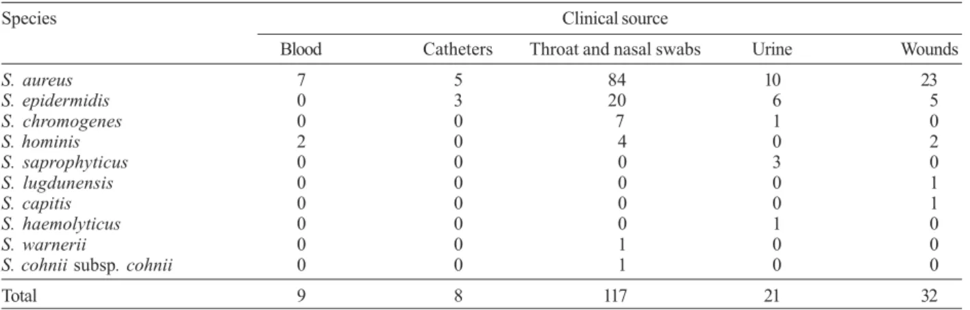
Bacterial isolates - One hundred eighty seven sta-phylococcal isolates, provided by hospital laboratory, were obtained from culture of several specimens; 117 isolated from throat and nasal swabs, 32 from wounds, 21 from urine, 9 from blood, and 8 from catheters. Table I shows distribution of species and clinical samples in the 187 staphylococcal strains. These staphylococcal strains, specifically 115 S. aureus strains, 34 S. epidermidis strains, eight S. chromogenes strains, eight S. hominis strains, three S. saprophyticus strains, one S. lugdunensis strain, one S. capitis strain, one S. haemolyticus strain, one S. warneri strain,one S. cohnii subsp. cohnii strain, isolated from diverse clinical sources were studied. Isolates were characterized at the species level by the API Staph system (Biomerieux, France) according to the instructions of the manufac-turers. The organims were stored in Trypticase soy broth (TSB), to which 15% sterile glycerol was added, at –20°C.
+Corresponding author: [email protected]
Slime production - Slime production of all isolates was evaluated by two different methods, Christensen method (Tube adherence) and Congo red agar method.
Briefly, a loop of isolates from a blood agar plate was inoculated into a glass tube containing 5 ml of TSB and incubated at 37°C for 48 h. The contents of the tubes were removed and then stained with 0.25% safranin. An adherent film on the surface of the glass tube was taken as evidence of slime formation. The absence of a film or the mere presence of a ring at the liquid-air interface was interpreted as a negative result (–). In the study, posi-tive results were recorded as strong (+++), moderate (++), weak (+). Each test was interpreted by two differ-ent observers (Christensen et al. 1982).
In Congo red test, the medium was prepared with 37 g/l brain heart infusion broth, 50 g/l sucrose, 10 g/l agar, and 0.8 g/l Congo red. Congo red stain was prepared as a concentrated aqueous solution and autoclaved at 121°C for 15 min and separately from the other medium con-stituents, and was then added when the agar had cooled to 55°C. Plates were inoculated and incubated at 37°C for 24 h. A positive result was indicated by black colo-nies on the surface. Non-slime producing strains devel-oped red colonies. Each plate was interpreted by two different observers (Freeman et al. 1989).
Antibiotic susceptibility - Susceptibility to antibiot-ics was determined by the disk diffusion method on Mueller-Hinton agar plates (NCCLS 1997). Nine anti-biotics were chosen for the study according to their com-mon use in research, especially human medicine. They belonged to the following groups: penicillins (oxacil-lin), cephalosporins (cefepime), beta-lactamase inhibi-tors (amoxicillin-clavulanic acid), aminoglycosides (gen-tamicin), macrolides (erythromycin), quinolones (ciprofloxacin), glycopeptides (vancomycin), and mis-cellaneous (clindamycin and trimethoprim-sulfa-methoxazole).
β-lactamase test- β-lactamase strips (Fluka, Ger-many) for the acidimetric detection of the β-lactamase activity of all S. aureus strains were used. The test strips
were inserted into test tube with prepared bacterial sus-pension in saline. A positive result was indicated by the appearance of a yellow color. Negative reaction remained red color.
Statistical analysis - Differences between two meth-ods used for the detection of the slime production were evaluated by the Chi-square analysis. P values of less than 0.05 were considered significant.
RESULTS
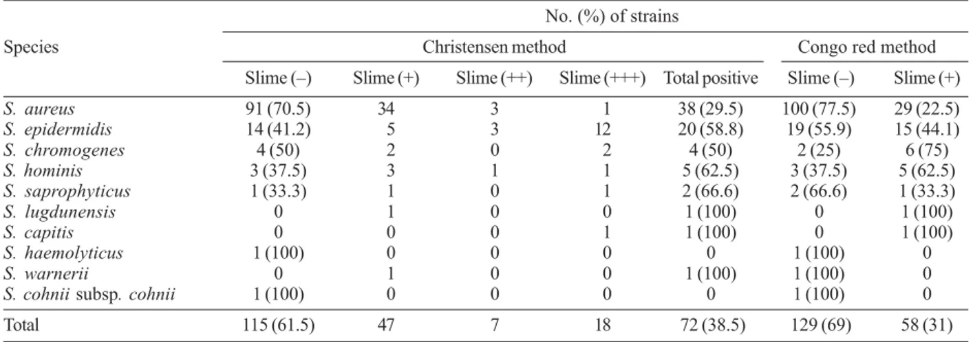
Slime production was detected by Christensen method and Congo red agar method. Table II shows the results of slime production by different staphylococcal species by both methods in details. In Christensen method, slime production was found in 72 (38.5%) of 187 staphylococcal isolates whereas 115 (61.5%) strains of staphylococcal isolates were not slime producers. All staphylococcal species isolated from clinical samples had one or more of slime producer strains according to tube adherence test, except S. haemolyticus and S. cohnii subsp. cohnii. Fifty eight (31%) of staphylococcal strains were slime positive and 129 (69%) strains were slime negative by Congo red agar method. In this test, species of S. haemolyticus, S. warnerii, and S. cohnii subsp. cohnii had no any slime producer strains. Posi-tive cases in the tube adherence test in staphylococ-cal isolates were more frequently observed in the Christensen’s method than in the Congo red agar method, but there was no statistically significant difference be-tween two methods used for detection of the slime pro-duction (P > 0.05).
Table III shows the correlation between the results ofantibiotic susceptibility assays for detection of anti-biotic resistance and distribution among them staphylo-cocci. Resistance to antibacterial agents has increased among many species of bacterial pathogens. All the iso-lates were susceptible to gentamicin and vancomycin, except for two strains of S. aureus and one strain of S. epidermidis. Among the various antimicrobial agents, oxacillin (71.1%) and erythromycin (47.1%) had higher
TABLE I
Distribution in the 187 staphylococcal strains isolated from clinical materials
Species Clinical source
Blood Catheters Throat and nasal swabs Urine Wounds
S. aureus 7 5 84 10 23
S. epidermidis 0 3 20 6 5
S. chromogenes 0 0 7 1 0
S. hominis 2 0 4 0 2
S. saprophyticus 0 0 0 3 0
S. lugdunensis 0 0 0 0 1
S. capitis 0 0 0 0 1
S. haemolyticus 0 0 0 1 0
S. warnerii 0 0 1 0 0
S. cohnii subsp. cohnii 0 0 1 0 0
Total 9 8 117 21 32
3 1 3 13 1 3 1 3 1 Mem Inst Oswaldo Cruz, Rio de Janeiro, Vol. 102(1), February 2007
resistance than most of the agents used against all iso-lates. Oxacillin resistant S. aureus (ORSA) and oxacil-lin resistant coagulase-negative staphylococci (ORCNS), 97 (75.2%) and 36 (62.1%) respectively were frequently observed in strains isolated from clinical materials. Among the ORSA strains, two strains were resistant to vancomycin.
β-lactamase was produced by 96 (74.4%) of 129 S. aureus strains with statistically significant difference among the strains from each clinical source (P < 0.05).
β-lactamase positive strains and their clinical sources are shown in Table IV. However, 78 (81.25%) of 96 ORSA strains were β-lactamase positive ORSA isolates, but none of them had vancomycin resistant.
DISCUSSION
In recent years, the most common infectious agents have been Staphylococcus sp. They are frequently iso-lated from clinical specimens, where they may be only a contaminant or the cause of infections. S. aureus is
TABLE II
Results of slime production in staphylococcal strains according to Christensen method and Congo red agar method No. (%) of strains
Species Christensen method Congo red method Slime (–) Slime (+) Slime (++) Slime(+++) Total positive Slime (–) Slime (+)
S. aureus 91 (70.5) 34 3 1 38 (29.5) 100 (77.5) 29 (22.5)
S. epidermidis 14 (41.2) 5 3 12 20 (58.8) 19 (55.9) 15 (44.1)
S. chromogenes 4 (50) 2 0 2 4 (50) 2 (25) 6 (75)
S. hominis 3 (37.5) 3 1 1 5 (62.5) 3 (37.5) 5 (62.5)
S. saprophyticus 1 (33.3) 1 0 1 2 (66.6) 2 (66.6) 1 (33.3)
S. lugdunensis 0 1 0 0 1 (100) 0 1 (100)
S. capitis 0 0 0 1 1 (100) 0 1 (100)
S. haemolyticus 1 (100) 0 0 0 0 1 (100) 0
S. warnerii 0 1 0 0 1 (100) 1 (100) 0
S. cohnii subsp. cohnii 1 (100) 0 0 0 0 1 (100) 0 Total 115 (61.5) 47 7 18 72 (38.5) 129 (69) 58 (31) strong (+++), moderate (++), weak (+) for Christensen method; S: Staphylococcus.
TABLE III
Susceptibility of clinical staphylococcal isolates to various antimicrobial agents Staphylococcal species % resistant
and no. of isolates CN SXT AMC VA FEP DA E OX CIP
S. aureus, 129 0 4.7 22.5 1.6 24 20.9 41.1 75.2 5.4
S. epidermidis, 34 2.9 38.2 2.9 0 20.6 20.6 70.6 61.8 17.6
S. chromogenes, 8 0 37.5 0 0 12.5 25 50 50 12.5
S. hominis, 8 0 12.5 0 0 25 12.5 37.5 62.5 25
S. saprophyticus, 3 0 0 0 0 0 0 33.3 100 0
S. lugdunensis, 1 0 0 0 0 100 0 0 100 0
S. capitis,1 0 0 0 0 0 100 100 100 0
S. haemolyticus, 1 0 100 0 0 100 0 0 100 0
S. warnerii, 1 0 100 0 0 0 0 100 0 0
S. cohnii subsp. cohnii, 1 0 100 0 0 0 100 100 0 0 CN, gentamicin; SXT, trimethoprim-sulfamethoxazole; AMC, amoxicillin-clavulanic acid; VA, vancomycin; FEP, cefepime; DA, clindamycin; E, erythromycin; OX, oxacillin; CIP, ciprofloxacin; S: Staphylococcus.
TABLE IV
β- lactamase of Staphylococcus aureus strains with respect to clinical source
β-lactamase (%) Clinical source (+) (–) Blood 3 (42.9) 4 (57.1) Catheters 2 (40) 3 (60) Throat and nasal swabs 67 (79.8) 17 (20.2) Urine 4 (40) 6 (60) Wounds 20 (87) 3 (13) Total 96 (74.4) 33 (25.6)
(Gemmel 1986, Khatib et al. 1995, Fux et al. 2005). In this study, we isolated several staphylococcal strains from different clinical samples. The staphylococcal clinical species most commonly isolated were S. aureus, S. epidermidis, and S. hominis, a distribution similar to those found by other authors (Ammendolia et al. 1999, Urdez-Hernandez et al. 1999). S. lugdunensis, S. capi-tis, S. warnerii, and S. cohnii subsp. cohnii were less frequent although these strains have also been isolated from several clinical specimens (Kleeman et al. 1993, Urdez-Hernandez et al. 1999). S. epidermidis was the most prevalent species isolated among clinical strains of CNS. This finding confirms that of a recent study (Ammendolia et al. 1999).
On the other hand, slime production plays an impor-tant role in the pathogenesis of infections caused by dif-ferent microorganisms, especially staphylococci. Except for S. haemolyticus and S. cohnii subsp. cohnii, slime production was by both methods in all staphylococci species (Table II). The data reported here indicate an important role of slime production as a virulence marker for clinically significant S. epidermidis isolates. Its oc-currence among a majority of clinical CNS isolates and its association with the strains’ ability to produce thicker biofilms may suggest a role of slime in pathogenesis (Table II). The results are similar to those reported by other authors (Ishak et al. 1985, Muller et al. 1993, Rupp & Archer1994, Ammendolia et al. 1999), who found that S. epidermidis frequently causes nosocomial septice-mia, and infects indwelling medical devices like intra-vascular catheters and prosthetic valves. However, the findings of the present study showed that slime forma-tion was not more prominent in S. aureus strains iso-lated from various clinical samples than in clinical CNS strains isolated (Table II).
Among the resistant pathogens, methicillin-(oxacil-lin) resistant S. aureus (MRSA) is one of the most im-portant causes of nosocomial infections worldwide and can cause outbreaks that are difficult to control (Boyce 1990, Gold & Moellering 1996, Salmenlinna 2002). Hsueh et al. (2004) reported that a rapid emergence of nosocomical methicillin-resistant S. aureus infection from 26.3% in 1986 to 77% in 2001 was found at a hos-pital. According to our results, S. aureus strains isolated from clinical materials showed the highest resistance to antibiotic oxacillin in all antimicrobials. Of the 129 S. aureus isolates, 97 isolates (75.2%) was ORSA strains. In general, multidrug resistance (resistance to three or more antibiotics) was observed in S. aureus clinical strains against antimicrobial agents (Table III).
CNS tend to be more resistant to antimicrobial agents than S. aureus, especially to methicillin (Hussain et al. 2000). Currently, more than 70% of the CNS isolates worldwide are resistant to methicillin or oxacillin. In addition, those CNS strains acquired in hospitals have become resistant to various other antimicrobial agents (Diekema et al. 2001). In the present study, we found that CNS clinical isolates were resistant to oxacillin in 62.1%. Surprisingly CNS had somewhat high resistance against erythromycin (60.3%). However, resistance of
CNS to other antimicrobial agents tested were frequently observed (Table III).
In addition, all clinical staphylococcal S. aureus strains were tested for β-lactamase production. β -lac-tamase was produced by 96 (74.4%) of 129 S. aureus strains with statistically significant difference among the strains from each clinical source (P < 0.05). The results are shown in Table IV. However, 78 (81.25%) of 96 β -lactamase positive S. aureus strains were β-lactamase positive ORSA isolates, but none of them had vancomy-cin resistance. Some authors reported that clinical in-fections of both community and nosocomial origins caused by ORSA strains continue to be major therapeu-tic and infection control challenges (Sorrell et al. 1982, Hackbarth & Chambers 1989). Recently, the vast major-ity of ORSA have been shown to produceβ-lactamases (Massanari et al. 1988, Montanari et al. 1990), and van-comycin has been generally thought to be the agent of choice for invasive ORSA infections (Sorrell et al. 1982). Our results are consistent with data obtained from previous studies.
In conclusion, we found that clinical CNS isolates had a high frequency of slime production and drug resis-tance, particularly S. epidermidis strains. Also, all sta-phylococcal strains isolated from clinical samples showed resistance against various antimicrobial agents (one or more agents), especially human medicine used. The findings of present study may be advantageous in the appropriate use of antibiotics for the successful treat-ment of infections caused by staphylococci.
REFERENCES
Ammendolia MG, Di rosa R, Montanaro R, Arciola CR, Baldassarri L 1999. Slime production and expression of the slime-associated antigen by staphylococal clinical isolates.
J Clin Microbiol 37: 3235-3238.
Baselga R, Albizu L, De La Cruz M, Del Cacho E, Barberan M, Amorena B 1993. Phase variation of slime production in Sta-phylococcus aureus: implications in colonization and viru-lence. Infect Immun 61: 4857-4862.
Boyce JM 1990. Increasing prevalence of methicillin-resistant
Staphylococcus aureus in the United States. Infect Con-trol Hosp Epidemiol 11: 639-642.
Caputy GG, Costerton JW 1982. Morphological examination of the glycocalyses of Staphylococcus aureus strains Wiley and Smith. Infect Immun 36: 759-767.
Christensen GD, Simpson WA, Bisno AL, Beachey EH 1982. Adherence of slime producing strains of Staphylococcus epidermidis to smooth surfaces. Infect Immun 37: 318-326. Davenport DS, Massanari RM, Pfaller MA, Bale MJ, Streed SA, Hierholzer WJ 1986. Usefulness of a test for slime pro-duction as a marker for clinically significant infections with coagulase negative staphylococci. J Infect Dis 153: 332-339.
3 3 3 33 3 3 3 3 3 Mem Inst Oswaldo Cruz, Rio de Janeiro, Vol. 102(1), February 2007
RN, Beach M and the SENTRY Participants Group 2001. Survey of infections due to Staphylococcus species: fre-quency of occurrence and antimicrobial susceptibility of iso-lates collected in the United States, Canada, Latin America, Europe, and the Western Pasific region for the SENTRY Antimicrobial Surveillance Program, 1997-1999. Clin Infect Dis 32: S114-S132.
Etienne J, Brun Y, Solh NE, Delorme V, Mouren C, Bes M, Fleurette J 1988. Characterization of clinically significant iso-lates of Staphylococcus epidermidis from patients with endocardatis. J Clin Microbiol 26: 613-617.
Freeman DJ, Falkiner FR, Keane CT 1989. New method for detecting slime production by coagulase negative staphylo-cocci. J Clin Pathol 42: 872-874.
Fux CA, Uehlinger D, Bodmer T, Droz S, Zellweger C, Muhlemann K2005. Dynamics of hemodialysis catheter colo-nization bycoagulase-negativestaphylococci.Infect Cont Hosp Ep 26: 567-574.
Gemmel CG 1986. Coagulase-negative staphylococci. J Med Microbiol 22: 285-295.
Gold HS, Moellering RC 1996. Drug therapy: antimicrobial-drug resistance. New Engl J Med 335: 1445-1453.
Gristina AG, Hobgood CD, Webb LX, Myrvik QN 1987. Adhe-sive colonization of biomaterials and antibiotic resistance.
Biomaterials 8: 423-426.
Hackbarth CJ, Chambers HF 1989. Methicillin-resistant staphy-lococci: detection methods and treatment of infections.
Antimicrob Agents Chemother 33: 995-999.
Hsueh PR, Teng LJ, Chen WH, Pan HJ, Chen ML, Chang SC, Luh KT, Lin FY 2004. Increasing prevalence of methicillin-resistant Staphylococcus aureus causing nosocomial infec-tions at a university hospital in Taiwan from 1986 to 2001.
Antimicrob Agents Chemother 48: 1361-1364.
Hussain Z, Stoakes L, Garrow S, Longo S, Fitzgerald V, Lannigan R 2000. Rapid detection of mecA-positive and mecA -nega-tive coagulase-nega-nega-tive staphylococci by an anti-penicillin binding protein 2a slide latex agglutination test. J Clin Microbiol 38: 2051-2054.
Ishak MA, Groschel DH, Mandell GL, Wenzel RP 1985. Asso-ciation of slime with pathogenicity of coagulase-negative sta-phylococci causing nosocomial septicemia. J Clin Microbiol 22: 1025-1029.
Khatib R, Riederer KM, Clark JA, Khatib S, Briski LE, Wilson FM 1995. Coagulase-negative staphylococci in multiple blood cultures: strain relatedness and determinants of same strain bacteremia. J Med Microbiol 33: 816-820.
Kleeman KT, Bannerman TL, Kloos WE 1993. Species distribu-tion of coagulase-negative staphylococcal isolates at a com-munity hospital and implications for selection of staphylococ-cal identification procedures. J Clin Microbiol 31: 1318-1321.
Kloss WE, Bannerman TL 1994. Update on clinical significance of coagulase-negative staphylococci. Clin Microbiol Rev 7: 117-140.
Kluytmans JAJW, Wertheim HFL 2005. Nasal carriage of Sta-phylococcus aureus and prevention of nosocomial infec-tions. Infection 33: 3-8.
Mack KD, Bartscht S, Dobinsky MA, Horskotte H, Kiel K-M, Knobloch A, Schafer P 2000. Staphylococcal factors involved in adhesion and biofilm formation on biometarials. In YH An, RJ Friedman (eds), Handbook of Bacterial Adhesion. Prin-ciples Methods and Applications, Humana Press, Totowa, NJ, p. 307-330.
Marshall SA, Pfaller MA, Jones RN 1999. Ability of the modi-fied vitek card to detect coagulase-negative staphylococci with mecA and oxacillin resistant phenotypes. J Clin Microbiol 37: 2122-2123.
Massanari RM, Pfaller MA, Wakesfield DS, Hammons GT, McNut LA, Woolson RF, Helms CM 1988. Implications of acquired oxacillin resistance in the management and control of Staphylococcus aureus infections. J Infect Dis 158: 702-709.
Montanari MP, Tonin E, Biavasco F, Varaldo PE 1990. Further Characterization of borderline methicillin-resistant Staphy-lococcus aureus and analysis of penicillin-binding protein.
Antimicrob Agents Chemother 34: 911-913.
Muller E, Takeda S, Shiro H, Goldmann DA, Pier GB 1993. Oc-currence of capsular polysaccharide/adhesin among clinical isolates of coagulase-negative staphylococci. J Infect Dis 168: 1211-1218.
NCCLS-National Committee for Clinical Laboratory Standards 1997. Performance standards for antimicrobial disk suscep-tibility tests. Approved Standard, M2-A6, Wayne, PA. Rupp ME, Archer GD 1994. Coagulase-negative staphylococci:
pathogens associated with medical progress. Clin Infect Dis 19: 231-245.
Salmenlinna S, Lyytikainen O, Vuopio-Varkila J 2002. Commu-nity acquired methicillin-resistant Staphylococcus aureus, Finland. Emerg Infect Dis 8: 602-607.
Sorrell TC, Packham DR, Shanker S, Foldes M, Munro R 1982. Vancomycin therapy for methicillin-resistant Staphylococ-cus aureus. Ann Intern Med 97: 344-350.
Tenover FC, Gaynes RP 2000. The epidemiology of Staphylo-coccus infections. In VA Fishetti, RP Novick, JJ Ferretti, DA Portnoy, JI Rood (eds), Gram-positive Pathogens, ASM Press, Washington DC, p. 414-421.