e80
Correspondência: Humberto Villacorta •
Hospital Quinta D’Or - Rua Almirante Baltazar 435 – 7º andar – CEP 20941-150 – Rio de Janeiro, RJ - Brasil
Email: hvillacorta@quintador.com.br
Artigo recebido em 28/11/08; revisado recebido em 24/03/09; aceito em 06/07/09
The non-compacted myocardium is characterized by excessive trabeculation and ventricular recesses (usually of the left ventricle) due to the interruption of the myocardial fiber compaction during embryogenesis. This abnormality usually courses with heart failure, thromboembolic phenomena and cardiac arrhythmia, predicting a bad prognosis. We report the case of a 26-year-old male individual with refractory heart failure due to isolated left ventricular non-compacted myocardium that needed a heart transplant.
Refractory Heart Failure in a Patient with Non-compacted
Myocardium
Humberto Villacorta
1,2, Jacqueline Miranda Sampaio
1,
Fernanda Beatriz Amador Dos Santos
1, Valdo Carrera
1,
Carlos Cleverson Pereira
1,
Evandro Tinoco Mesquita
2Hospital Quinta D’Or 1; Universidade Federal Fluminense2, Rio de Janeiro, RJ - Brasil
Introduction
The ventricular myocardium non-compaction (VMNC) is characterized by excessive trabeculation and recesses of the ventricles (usually the left ventricle), due to the interruption of the process of myocardial fiber compaction during embryogenesis1,2. This structural abnormality frequently
courses with heart failure, thromboembolic phenomena and cardiac arrhythmia, presenting a poor prognosis3. In some
cases, the patients develop refractory heart failure and need a heart transplant.
The VMNC was initially described associated with other congenital heart anomalies4 and also associated with
neuromuscular disorders5. In the 1990s, the first cases of its
isolated form were described6. Although it is categorized as
a cardiomyopathy that is not classified by the World Health Organization due to its unique characteristics, some authors describe it as a distinct cardiomyopathy3,7.
Case Report
A 26-year-old patient, with a history of dilated cardiomyopathy for 8 years, presented symptom worsening in the last few months, when a new assistant physician was assigned to him. At that time, the suspected diagnosis of VMNC affecting the left ventricle was attained, based on
Key Words
Heart Failure; Cardiomyopathies; Heart Transplantation.
echocardiographic parameters. In January, 2007, the patient was admitted at the hospital with pain and abdominal distension, diarrhea and fatigue. He was initially admitted at the Internal Medicine Clinic, for assessment of the digestive symptoms. He received diuretics and vasodilators with no symptom improvement. He was referred to the Cardiology Service 48 hours after admission. After the digestive causes had been ruled out, he was diagnosed with decompensated heart failure, with signs of low cardiac output and the digestive symptoms were attributed to splenic ischemia.
At this point, the patient was slight dyspneic, had normal skin color, blood pressure (BP) of 100x50 mmHg and heart rate (HR) of 110 bpm, presented pulmonary rales at the base and oxygen saturation of 92% in ambient air and 99% with oxygen mask. The cardiac auscultation showed the presence of third and fourth heart sounds and systolic murmur in mitral focus with an intensity of ++++/6+.
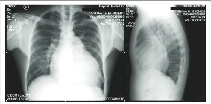
The electrocardiogram showed sinus rhythm and left chamber overload. The laboratory assessment showed hemoglobin = 13 g/dL, leucocytes = 6600 (3 rods), urea = 34 g/dL, creatinine = 1.4 g/dL, serum sodium = 137 mEq/L, serum potassium = 5.1 mEq/L and prothrombin activity time = 62%. Liver and pancreatic function tests were normal. The chest x-ray showed a large increase in the cardiac area, with little pulmonary congestion (Figure 1). He was submitted to a bi-dimensional echocardiogram, which showed a marked left ventricle (LV) enlargement, with an end-diastolic diameter of 8.2 cm and EF of 20%. Moreover, there were echocardiographic criteria for the diagnosis of VMNC affecting the LV, with extensive trabeculation of this chamber (Figure 2). The criteria used for the diagnosis on non-compaction were the classic ones described in the literature2,6, i.e.: a) numerous
and prominent trabeculations and deep intertrabecular recesses in the ventricular wall; b) communication of the intertrabecular recesses with the ventricular cavity, through the demonstration of flow in these recesses; and c) non-compacted/compacted myocardium ratio > 2, measured in telesystole. All these criteria were found in our patient. The right ventricle was preserved.
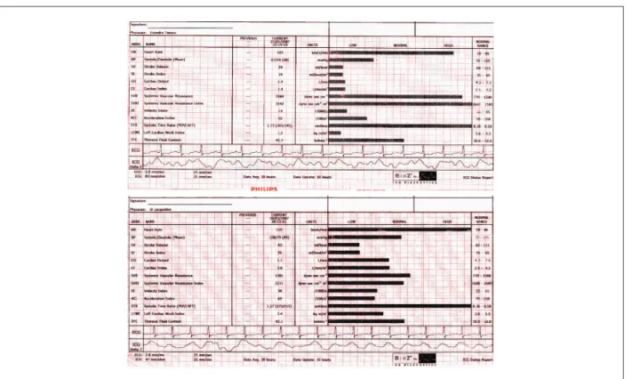
The patient was submitted to non-invasive assessment of hemodynamic parameters through electrical bioimpedance, which showed a decreased cardiac index (1.4 L/min/m2) and
increased peripheral artery resistance (3542 dinas.sec.cm-5/m2)
and normal chest fluid content (Figure 3A). The diuretic was withdrawn at this moment and the patient started to receive levosimendan, at an initial dose of 0.05 µ/Kg/min, increased up to 1 µ/Kg/min. After 24 hours of drug infusion, the clinical picture improved. The hemodynamic parameters showed improvement in the cardiac index (CI), which increased to 3 L/min/m2, and
Figure 1 – Chest X-ray in PA and proile showing a large increase in the cardiac area.
2A
2B
LV
LA
Figure 2 – Echocardiogram in the longitudinal parasternal view, disclosing the presence of trabeculations (white arrows) and left ventricular recesses (2A), with marked dilatation of this chamber (2B); LV = left ventricle; LA= left atrium.
the peripheral artery resistance, which decreased to 2211 dinas. sec.cm-5/m2 (Figure 3B). The patient was then submitted to a
heart MRI, which confirmed the diagnosis of LV myocardium non-compaction. The criteria used for the diagnosis through the MRI were the same employed for the echocardiography. Holter showed 353 ventricular extrasystoles, isolated and polymorphic, 18 coupled ones and 13 episodes of nonsustained ventricular tachycardia.
The patient was submitted to an implantable cardiac defibrillator implant, due to the high risk of sudden death. He was discharged and put in a waiting list for heart transplant.
Three days after the discharge he was hospitalized again with the same picture and his condition deteriorated rapidly,
thus necessitating the use of inotropic drugs. A new assessment by transthoracic bioimpedance showed a return of the CI to 1.4 L/min/m2. There was no possibility of hospital discharge
and during this period, the patient was dependent on vasoactive amines, alternating infusions of levosimendan and dobutamine. Finally, a month later, the patient was submitted to a heart transplant. The patient’s explanted heart presented extensive trabeculation of the LV, confirming the diagnosis of LV myocardium non-compaction, as shown in Figure 4. He is currently well, presents NYHA Functional Class I, after 21 months of follow-up. As the disease has a genetic trait, a screening of the family members was carried out through MRI and the mother and a younger sibling showed to have the disease, with both being asymptomatic.
Arq Bras Cardiol 2009; 93(6) : e80-e83
Villacorta et al Heart Failure and Non-compacted Myocardium
e81
Figure 3 – Hemodynamic measurements obtained by transthoracic bioimpedance before (3A) and after (3B) Levosimendan. Note the improvement in the cardiac index from 1.4 to 3.0 L/min/m2 and the decrease in the indexed peripheral artery resistance from 3542 to 2211 dinas.sec.cm-5/m2, 24 h after the drug administration (red rectangle).
Figure 4 - Explanted heart showing left ventricle with extensive trabeculation and the intertrabecular recess communication with the ventricular cavity, typical
indings in non-compaction of the myocardium.
Discussion
We present a case of VMNC in a young patient, with refractory HF. In our country, there have been previous reports of the disease, but the presentation in these cases was as ventricular arrhythmias and not HF8,9. This case is special as
it describes a case of refractory HF, which, due to the fact that the patient was submitted to a heart transplant, allowed us to directly observe the typical findings of the disease in the explanted heart.
VMNC is a relatively rare entity, which affects mainly the male sex, representing 56% to 82% of the cases in the four major series1. The initial series of the description of
the isolated form of the disease consisted mainly of young individuals (aged between 11 months and 22 years)6, but
later, the disease was also described in adults, including elderly individuals1. Its prevalence, in patients referred to
echocardiography for HF investigation, has been estimated at 0.014%3. The prevalence in the general population,
however, has not been established and can be higher than the estimated one, due to missed diagnoses.
Ina recent study, Kohli et al10, assessing 200 patients referred
to HF investigation, using the current echocardiographic criteria, found that almost one fourth of the individuals met the criteria for the diagnosis of myocardium non-compaction10,11.
The VMNC can be found in asymptomatic individuals or can present as HF, thromboembolic phenomena or cardiac arrhythmias. The HF can manifest from asymptomatic LV
systolic dysfunction to cases of severe HF, as presented by our patient. In one of the largest isolated series, 2/3 of the patients presented symptomatic HF3. In the series by Chin et al6, 63%
of the patients presented decreased LVEF. Subendocardial hypoperfusion to alterations in microcirculation can be involved in the genesis of systolic dysfunction and arrhythmogenesis6. Diastolic dysfunction can also be found
and results from the abnormal relaxation or restricted filling due to the numerous trabeculations1.
Arq Bras Cardiol 2009; 93(6) : e80-e83
Villacorta et al
Heart Failure and Non-compacted Myocardium
e82
References
1. Engberding R, Yelbuz TM, Breithardt G. Isolated noncompaction of the left ventricular myocardium: a review of the literature two decades after the initial case description. Clin Res Cardiol. 2007; 96: 481-8.
2. Sá MI, Reis H, Cabral S, Fernandes P, Oliveira F, Torres S, et al. Não compactação do miocárdio ventricular. Rev Port Cardiol. 2006; 25: 835-44.
3. Oechslin EN, Jattenhofer Jost CH, Rojas JR, Kaufmann PA, Jenni R. Long-term follow-up of 34 adults with isolated left ventricular noncompaction: a distinct cardiomyopathy with poor prognosis. J Am Coll Cardiol. 2000; 36: 493-500.
4. Dusek J, Ostadal B, Duskova M. Postnatal persistence of spongy myocardium with embrionic blood supply. Arch Pathol. 1975; 99: 312-7.
5. Stollberg C, Finsterer J, Blazek G. Left ventricular hypertrabeculation/ noncompaction and association with additional cardiac abnormalities and neuromuscular disorders. Am J Cardiol. 2002; 90: 899-902.
6. Chin TK, Perloff JK, Williams RG, Jue K, Mohrmann R. Isolated noncompaction of the left ventricular myocardium: a study of eight cases. Circulation. 1990; 82: 507-13.
7. Conraads V, Paelinck B, Vorlat A, Goethals M, Jacobs W, Vrints C. Isolated non-compaction of the left ventricle: a rare indication for transplantation. J Heart Lung Transplant. 2001; 20 (8): 904-7.
8. Elias J, Valadão W, Kuniyoshi R, Queiroz A, Peixoto CA. Isolated noncompaction of the myocardium. Arq Bras Cardiol. 2000; 74: 253-7.
9. de Oliveira DC, Malta MM, Pinheiro JA, Piegas LS. Isolated noncompaction of the myocardium. Arq Bras Cardiol. 2007; 88 (2): e36-9.
10. Kohli SK, Pantazis AA, Shah JS, Adeyemi B, Jackson G, McKenna WJ, et al. Diagnosis of left-ventricular non-compaction in patients with left-ventricular systolic dysfunction: time for reappraisel of diagnostic criteria? Eur Heart J. 2008; 29: 89-95.
11. Anderson RH. Ventricular non-compaction: a frequently ignored finding? Eur Heart J. 2008; 29: 10-1.
12. Klaassen S, Probst S, Oechslin E, Gerull B, Krings G, Schuler P, et al. Mutations in sarcomere protein genes in left ventricular noncompaction. Circulation. 2008; 117: 2893-901.
Cardiac arrhythmias are frequent findings, with ventricular tachyarrhythmias being particularly common, observed in around 47% of the cases and atrial fibrillation, described in more than 25% of the patients1,3. Sudden death was
responsible for around 50% of the deaths in the largest series of isolated VMNC1,3,6.
The diagnosis of VMNC can be attained by echocardiography in most cases, according to pre-defined criteria2,6. In
inconclusive cases, the heart MRI is an extremely valuable tool. Although the diagnosis is relatively easy in the typical forms, the more subtle forms require a differential diagnosis with other diseases, such as apical hypertrophic cardiomyopathy, dilated cardiomyopathy, right ventricular arrhythmogenic dysplasia, endocardial fibroelastosis, normal myocardial hypertrabeculation and LV apical thrombus.
It is worth mentioning that the disease presents a genetic trait. Mutations have been described in the heavy chain of myosin, actin and troponin T in individuals with VMNC12.
Therefore, as demonstrated in our case, the patient’s family members can present the asymptomatic forms of the disease and must be screened.
In conclusion, we present a case of VMNC in a young male individual that developed refractory heart failure and was submitted to a heart transplant, with good evolution to date, after 21 months of follow-up.
Acknowledgements
We thank Ana Karla Palis, Clerio Azevedo, Marcelo Hadlich, Plinio Resende and Walter Omena for their participation in the management of the case.
Arq Bras Cardiol 2009; 93(6) : e80-e83
Villacorta et al Heart Failure and Non-compacted Myocardium

